

 PDF
PDF ePub
ePub Citation
 Print
Print


Abstract
Objective
We wanted to present the clinical manifestation and imaging findings of a rare case of acute sciatic nerve palsy with a foot drop similar to lumbar disc herniation developed after sleeping for 8 hours in a sitting position in inebriated condition.
Summary of Literature Review
Sciatic nerve palsy as a complication from being operated in a sitting position have been reported, but here have not been any reported cases of after-sleep sciatic nerve palsy.
Study Subject and Methods
Sixty eight year old male admitted to hospital due to acute onset of right foot drop, subsequent walking difficulty, and numbness of the right calf and foot. Symptoms began after 8 hours of sleeping in a sitting position. Pelvic MRI exam revealed sciatic neuropathy, and also electrophysiological exam revealed sciatic nerve palsy.
REFERENCES
1. Baima J, Krivickas L. Evaluation and treatment of peroneal neuropathy. Curr Rev Musculoskelet Med. 2008; 1:147–53.

2. Benson ER, Schutzer SF. Posttraumatic piriformis syndrome: diagnosis and results of operative treatment. J Bone Joint Surg Am. 1999; 81:941–9.
3. El-Rubaidi OA, Horcajadas-Almansa A, Rodriguez-Rubio D, Galicia-Bulnes JM. Sciatic nerve compression as a complication of the sitting position. Neurocirugia. 2003; 14:426–30.
4. Gozal Y, Pomeranz S. Sciatic nerve palsy as a complication after acoustic neurinoma resection in the sitting position. J Neurosurg Anesthesiol. 1994; 6:40–2.
5. Henson JT, Roberts CS, Giannoudis PV. Gluteal compartment syndrome. Acta Orthop Belg. 2009; 75:147–52.
6. Manigoda M, Dujmovic-Basuroski I, Trikic R, Drulovic J. Acute sciatic neuropathy “post-Saturday palsy”. Srp Arh Celok Lek. 2005; 133:58–61.
7. Tacconi P, Manca D, Tamburini G, Cannas A, Giagheddu M. Bed footboard peroneal and tibial neuropathy. A furtherunusual type of Saturday night palsy. J Peripher Nerv Syst. 2004; 9:54–6.
Figures and Tables%
Fig. 1.
Preoperative (A) and postoperative (B) image of T2 sagittal MR show decompression state of lumbar spinal canal.
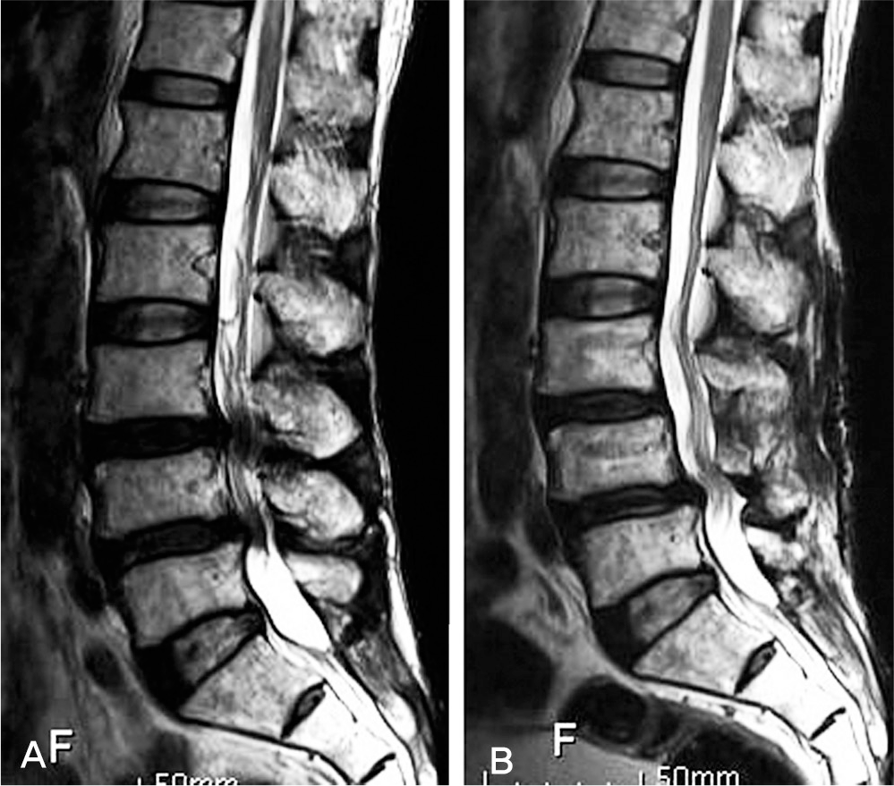
Fig. 2.
(A) Pelvis T2 weighted coronal MR image showing sciatic nerve (arrow), bilateral gluteus muscle, obturator muscle and right pyriformis muscle swelling and marked enhancement (B). Axial image of T2 weighted (C) and T1 weighted enhance MR (D) show sciatic nerve and adjacent soft tissue edema.
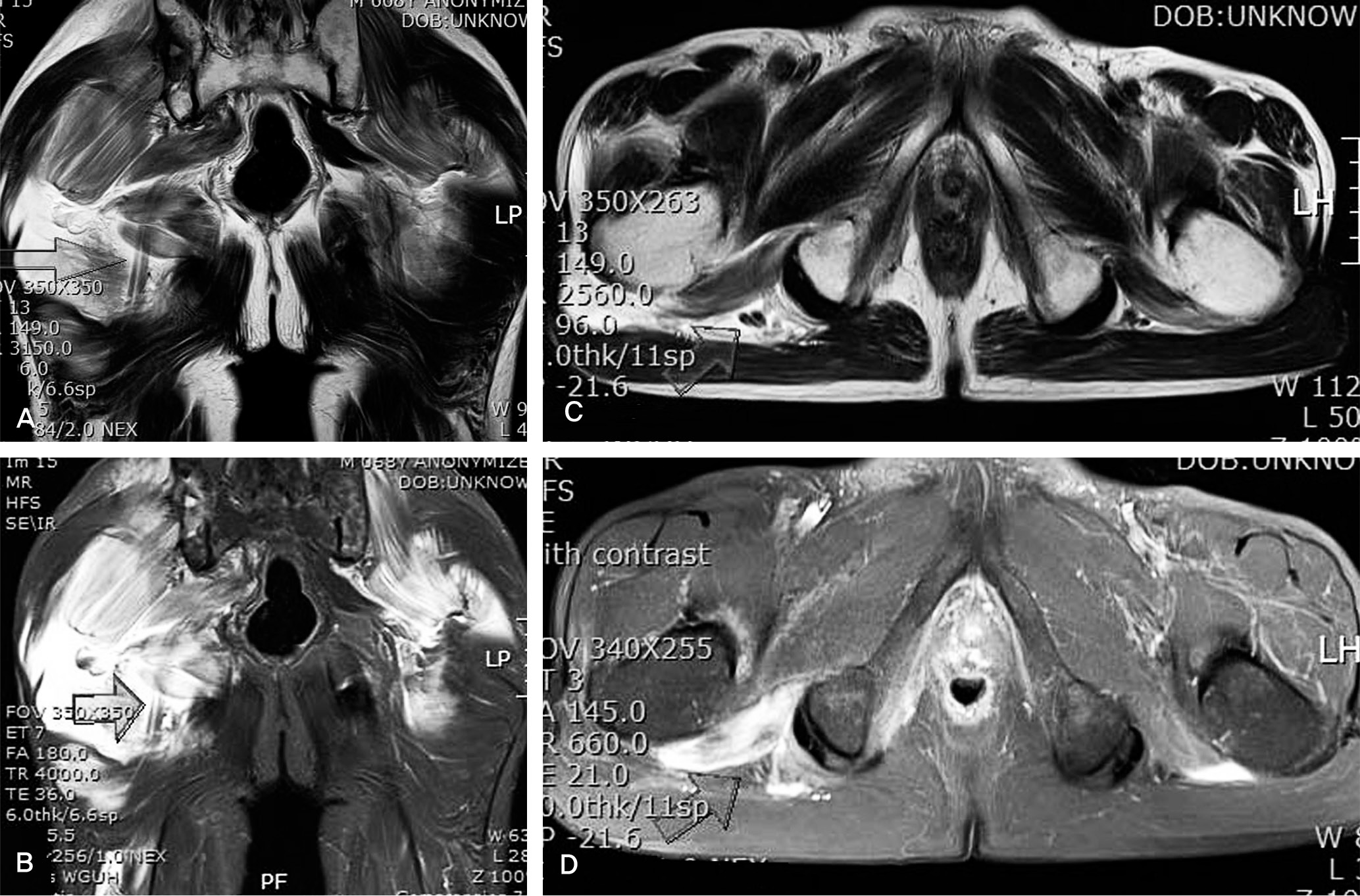
Table 1.
Findings of Electromyography
 XML Download