Abstract
Objectives
This case report presents a child who was treated conservatively after having being diagnosed with cervical intervertebral disc calcification.
Summary of Literature Review
Cervical intervertebral disc calcification is considered as a degenerative change of spine. It is common in adults and in most cases, no symptoms are observed. In children, by contrast, it is a rare condition and frequently accompanies symptoms such as severe neck pain and dysphagia.
Materials and Methods
A 7-year-old male patient who suffered from neck pain and torticollis without trauma had been diagnosed with cervical intervertebral disc calcification and was treated conservatively. He was discharged after symptom relief, and has been followed up and observed in our outpatient department.
Conclusions
Cervical intervertebral disc calcification shows similar symptoms to laryngopharyngeal abscess, traumatic injury and infective spondylitis, but through careful physical examination and radiologic evaluation, differential diagnosis is possible. After diagnosis, conservative treatment alone is sufficient. Antibiotic usage and surgical treatment are avoidable.
REFERENCES
2. Gerlach R, Zimmermann M, Kellermann S, Lietz R, Raabe A, Seifert V. Intervertebral disc calcification in childhood— a case report and review of the literature. Acta Neurochir (Wien). 2001; 143:89–93.
3. Calderone M, Severino M, Pluchinotta FR, Zangardi T, Martini G. Idiopathic intervertebral disc calcification in childhood. Arch Dis Child. 2009; 94:233–4.

4. Dai LY, Ye H, Qian QR. The natural history of cervical disc calcification in children. J Bone Joint Surg Am. 2004; 86:1467–72.

5. Girodias JB, Azouz EM, Marton D. Intervertebral disk space calcification. A report of 51 children with a review of the literature. Pediatr Radiol. 1991; 21:541–6.
6. Swischuk LE, Jubang M, Jadhav SP. Calcific discitis in children: vertebral body involvement (possible insight into etiology). Emerg Radiol. 2008; 15:427–30.

7. Sonnabend DH, Taylor TK, Chapman GK. Intervertebral disc calcification syndromes in children. J Bone Joint Surg Br. 1982; 64:25–31.

8. Bagatur AE, Zorer G, Centel T. Natural history of paediatric intervertebral disc calcification. Arch Orthop Trauma Surg. 2001; 121:601–3.

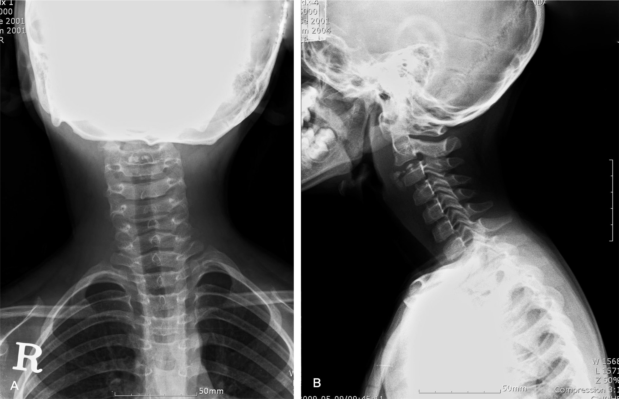
Fig. 1.
Initial X-ray. Intervertebral disc calcification at C2-3 with right anterior hernication with inferior migration and associated retropharyngeal widening (A) AP view (B) Lateral view

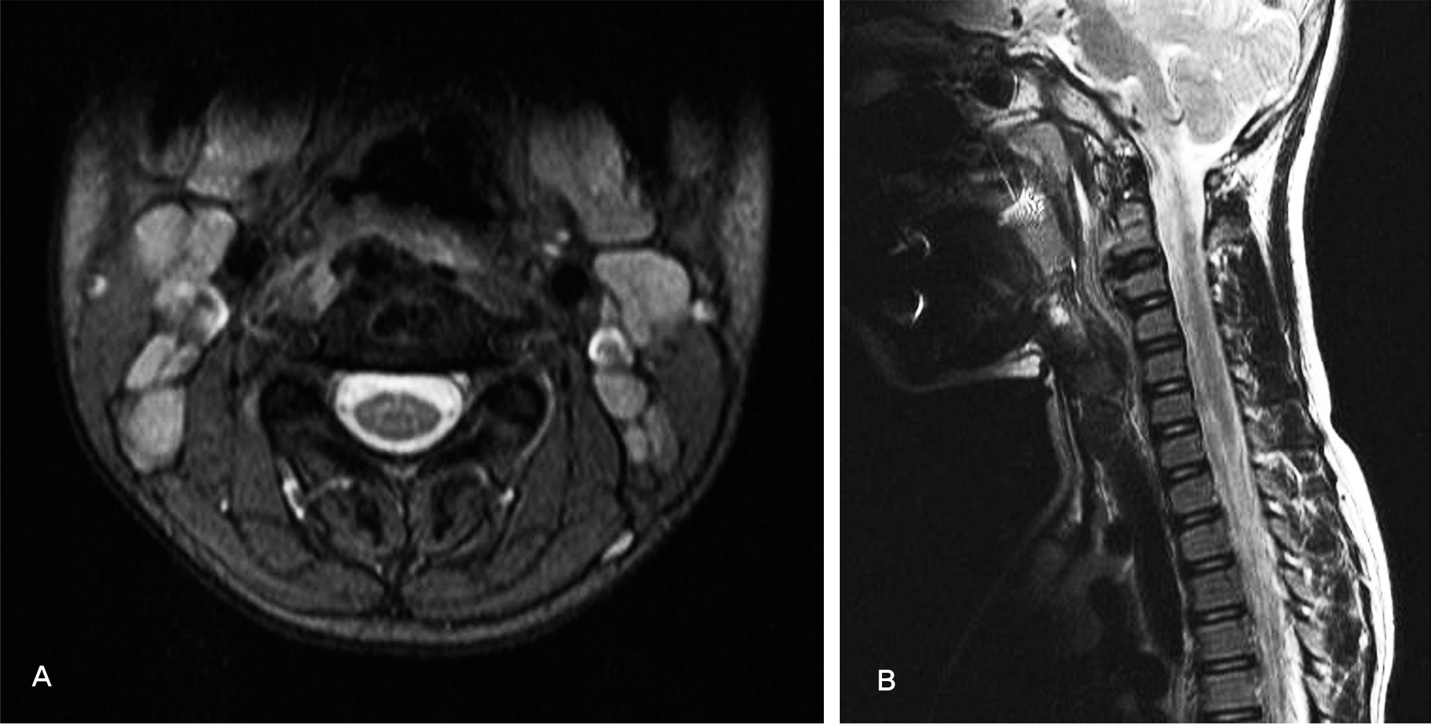
Fig. 2.
Initial MRI. Acute symptomatic intervertebral disc calcification at C2-3 with right sided prevertebral hermiation of the calcification migrating inferiorly (A) T2WI Axial view (B) T1WI Sagittal view


 PDF
PDF ePub
ePub Citation
 Print
Print


 XML Download