

 PDF
PDF ePub
ePub Citation
 Print
Print


Abstract
Objectives
To evaluate the clinical and radiological outcomes of anterior cervical fusion within Harms cage versus an iliac bone block graft.
Summary of Literature Review
There is no current consensus regarding the optimal material for anterior cervical fusion.
Materials and Methods
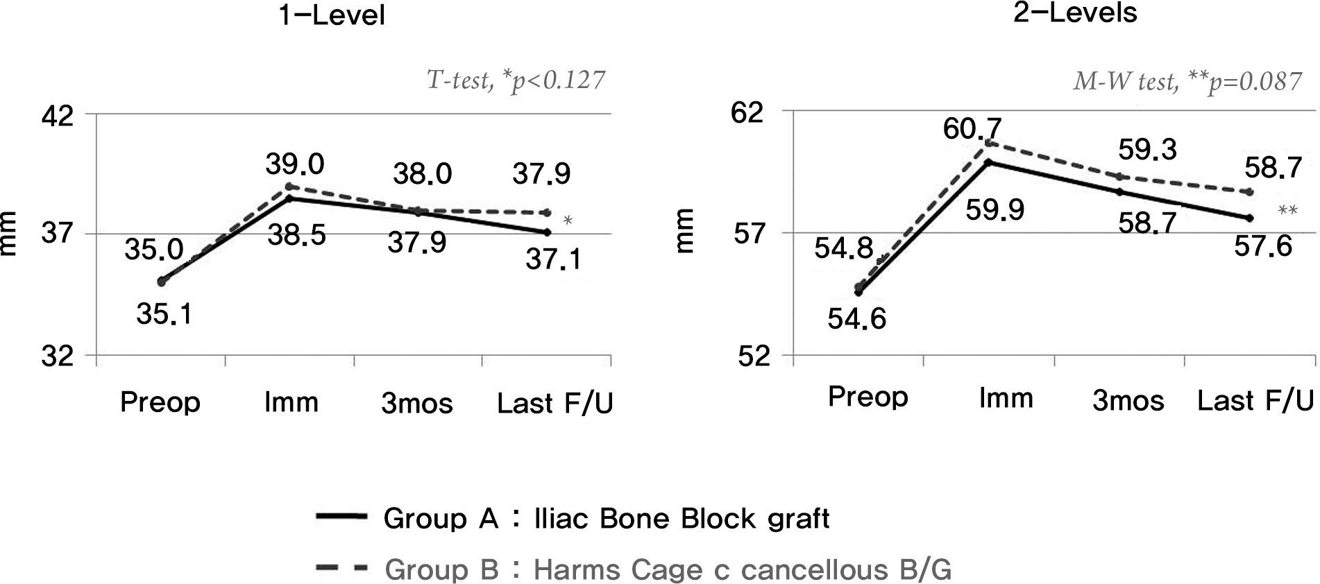
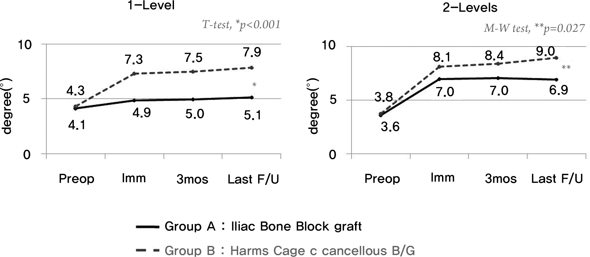
This was a single-center study of 107 patients who either underwent anterior cervical fusion with an iliac bone block graft (n=56; group A) or a cancellous bone graft within the cervical Harms titanium cage (n=51; group B). Anterior plating occurred in all cases. Clinical outcomes and complications were evaluated using Visual Analogue Scale (VAS) scores and Odom's Criteria. Radiological outcomes were evaluated by the height of vertebral bodies, sagittal lordosis, the rate of bony union, and the subsidence of cage.
Results
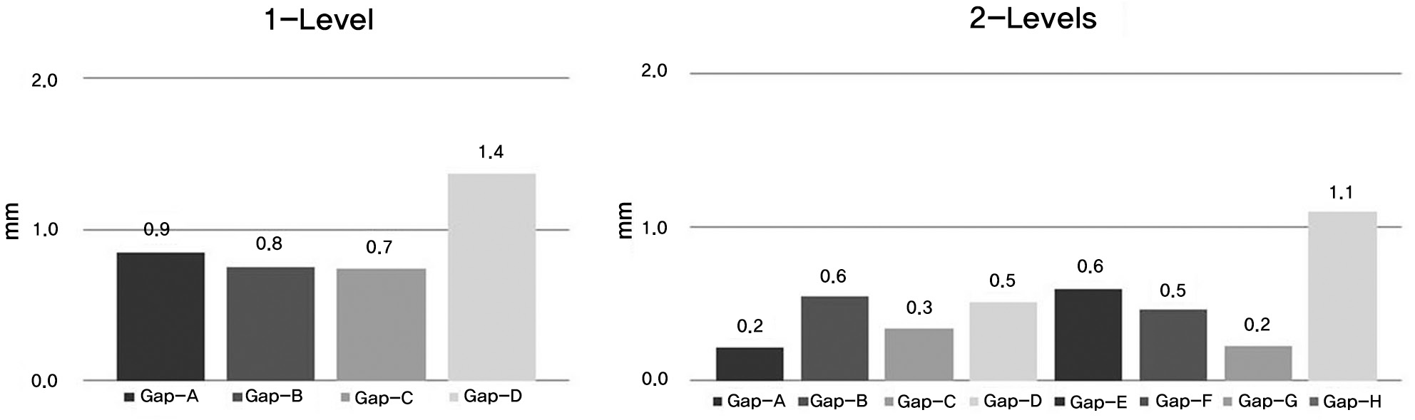
The VAS of donor site pain was significantly higher in group A than in group B at the final followup. Sagittal lordosis was increased in both groups, but was significantly higher in group B than group A. The rate of bony union was 95% and 91% for both groups 6 months after surgery and reached 100% for both groups at the final followup. In terms of cage subsidence, the highest point of subsidence was at the inferior and posterior aspect of the cage and the average amount of subsidence was approximately 1.3 mm at final followup.
Go to : 
REFERENCES
1. Cloward RB. The anterior approach for removal of ruptured cervical disks. J Neurosurg. 1958; 15:602–17.

2. Smith GW, Robinson RA. The treatment of certain cervical-spine disorders by anterior removal of the intervertebral disc and interbody fusion. J Bone Joint Surg Am. 1958; 40:607–24.
3. Coric D, Branch CL Jr, Jenkins JD. Revision of anterior cervical pseudoarthrosis with anterior allograft fusion and plating. J Neurosurg. 1997; 86:969–74.
4. Wang JC, McDonough PW, Endow KK, Delamarter RB. Increased fusion rates with cervical plating for two-level anterior cervical discectomy and fusion. Spine (Philla Pa 1976). 2000; 25:41–5.
5. Abd-Alrahman N, Dokmak AS, Abou-Madawi A. Anterior cervical discectomy (ACD) versus anterior cervical fusion (ACF), clinical and radiological outcome study. Acta Neurochir. 1999; 141:1089–92.
6. Bishop RC, Moore KA, Hadley MN. Anterior cervical interbody fusion using autogeneic and allogeneic bone graft substrate: a prospective comparative analysis. J Neurosurg. 1996; 85:206–10.
7. Hacker RJ, Cauthen JC, Gilbert TJ, Griffith SL. A prospective randomized multicenter clinical evaluation of an anterior cervical fusion cage. Spine (Philla Pa 1976). 2000; 25:2646–54.
8. Kulkarni AG, Hee HT, Wong HK. Solis cage (PEEK) for anterior cervical fusion: preliminary radiological results with emphasis on fusion and subsidence. Spine J. 2007; 7:205–9.
9. Savolainen S, Rinne J, Hernesniemi J. A prospective randomized study of anterior single-level cervical disc operations with longterm followup: surgical fusion is unnecessary. Neurosurgery. 1998; 43:51–5.
10. Shapiro S. Banked fibula and the locking anterior cervical plate in anterior cervical fusions following cervical discectomy. J Neurosurg. 1996; 84:161–5.
11. Wilke HJ, Kettler A, Goetz C, Claes L. Subsidence resulting from simulated postoperative neck movements: an in vitro investigation with a new cervical fusion cage. Spine (Philla Pa 1976). 2000; 25:2762–70.
12. Park HJ, Shim YJ, Yang JH. Anterior decompression and fusion in the treatment of single-level cercival disc herniation. J Kor Soc Spine Surg. 2008; 15:140–8.
13. Gercek E, Arlet V, Delisle J, Marchesi D. Subsidence of stand-alone cervical cages in anterior interbody fusion: warning. Eur Spine J. 2003; 12:513–6.
14. Kandziora F, Schollmeier G, Scholz M, et al. Influence of cage design on interbody fusion in a sheep cervical spine model. J Neurosurg. 2002; 96:321–2.
15. Majd ME, Vadhva M, Holt RT. Anterior cervical reconstruction using titanium cages with anterior plating. Spine (Philla Pa 1976). 1999; 24:1604–10.
16. Odom GL, Finney W, Woodhall B. Cervical disk lesions. J Am Med Assoc. 1958; 166:23–8.
17. Sawin PD, Traynelis VC, Menezes AH. A comparative analysis of fusion rates and donor-site morbidity for autogeneic rib and iliac crest bone grafts in posterior cervical fusions. J Neurosurg. 1998; 88:255–65.
18. Cauthen JC, Kinard RE, Vogler JB, et al. Outcome analysis of noninstrumented anterior cervical discectomy and interbody fusion in 348 patients. Spine (Philla Pa 1976). 1998; 23:188–92.
19. Greene DL, Crawford NR, Chamberlain RH, Park SC, Crandall D. Biomechanical comparison of cervical interbody cage versus structural bone graft. Spine J. 2003; 3:262–9.
20. Emery SE, Bolesta MJ, Banks MA, Jones PK. Robinson anterior cervical fusion comparison of the standard and modified techniques. Spine (Philla Pa 1976). 1994; 19:660–3.
21. Wang JC, McDonough PW, Kanim LE, Endow KK, Delamarter RB. Increased fusion rates with cervical plating for three-level anterior cervical discectomy and fusion. Spine (Philla Pa 1976). 2001; 26:643–7.
22. Papadopoulos EC, Huang RC, Girardi FP, Synnott K, Cammisa FP Jr. Three-level anterior cervical discectomy and fusion with plate fixation: radiographic and clinical results. Spine (Philla Pa 1976). 2006; 31:897–902.
23. Lee SH, Lee ST, Lee YK, et al. Three-level anterior cervical discectomy and fusion with cervical plate. J Korean Soc Spine Surg. 2002; 9:238–44.
24. Thome C, Leheta O, Krauss JK, Zevgaridis D. A prospective randomized comparison of rectangular titanium cage fusion and iliac crest autograft fusion in patients undergoing anterior cervical discectomy. J Neurosurg Spine. 2006; 4:1–9.
25. Zaveri GR, Ford M. Cervical spondylosis: the role of anterior instrumentation after decompression and fusion. J Spinal Disord. 2001; 14:10–6.
26. Fujibayashi S, Neo M, Nakamura T. Stand-alone interbody cage versus anterior cervical plate for treatment of cervical disc herniation: sequential changes in cage subsidence. J Clin Neurosci. 2008; 15:1017–22.
27. van Jonbergen HP, Spruit M, Anderson PG, Pavlov PW. Anterior cervical interbody fusion with a titanium box cage: early radiological assessment of fusion and subsidence. Spine J. 2005; 5:645–9.
28. Schmieder K, Wolzik-Grossmann M, Pechlivanis I, Engelhardt M, Scholz M, Harders A. Subsidence of the wing titanium cage after anterior cervical interbody fusion: 2-year followup study. J Neurosurg Spine. 2006; 4:447–53.
Go to :
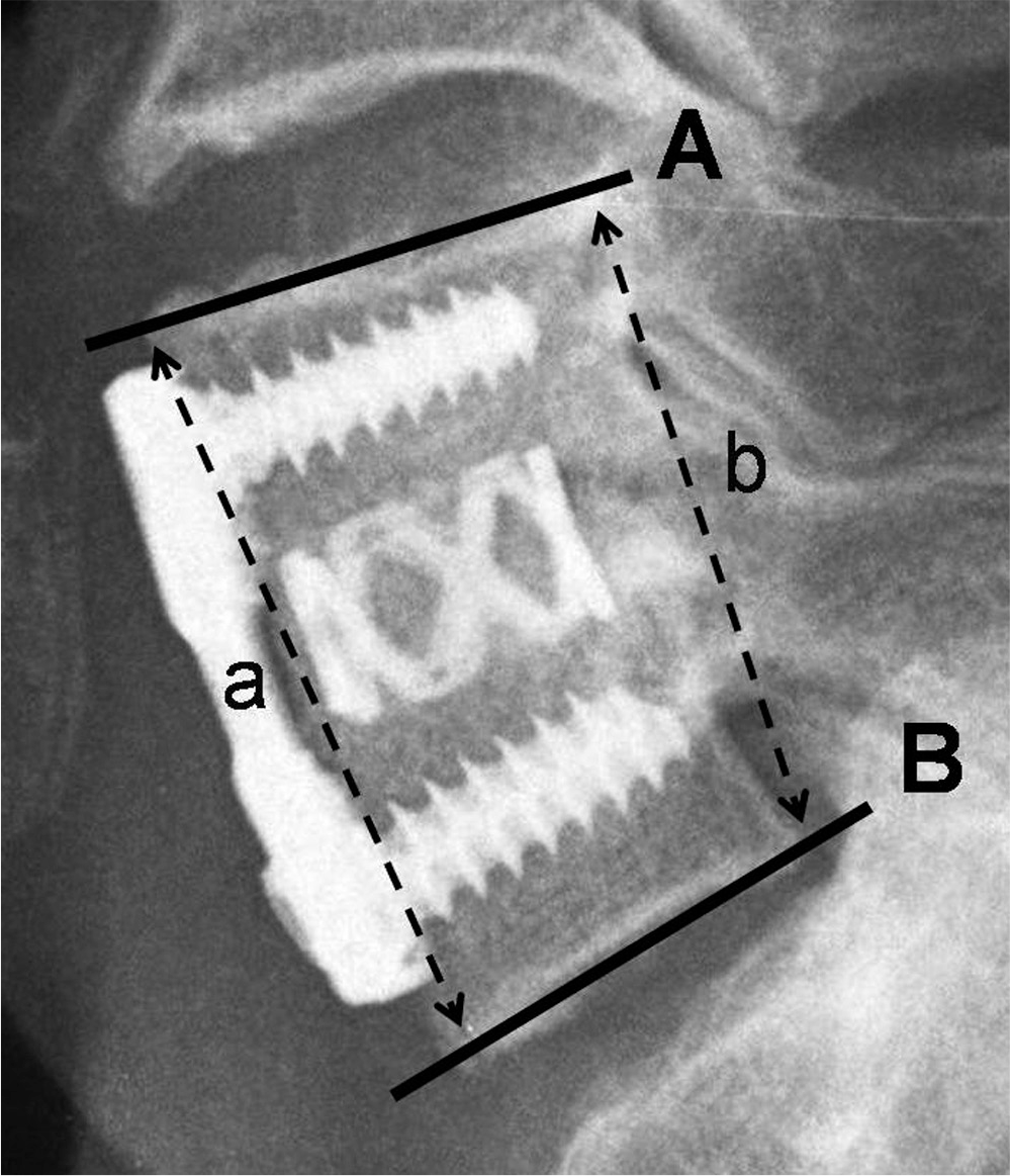 | Fig. 2.Lateral radiograph of cervical spine showing vertebral height measuring method of operation segment (a+b / 2) and segmental alignment measuring method (Cobb's angle of A & B). |
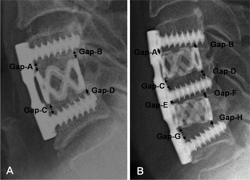 | Fig. 3.Lateral radiograph of cervical spine showing cage subsidence measuring method which is checked a difference of gap that right angle distance from each edge of cage to screw for one level (A) and two levels (B). |
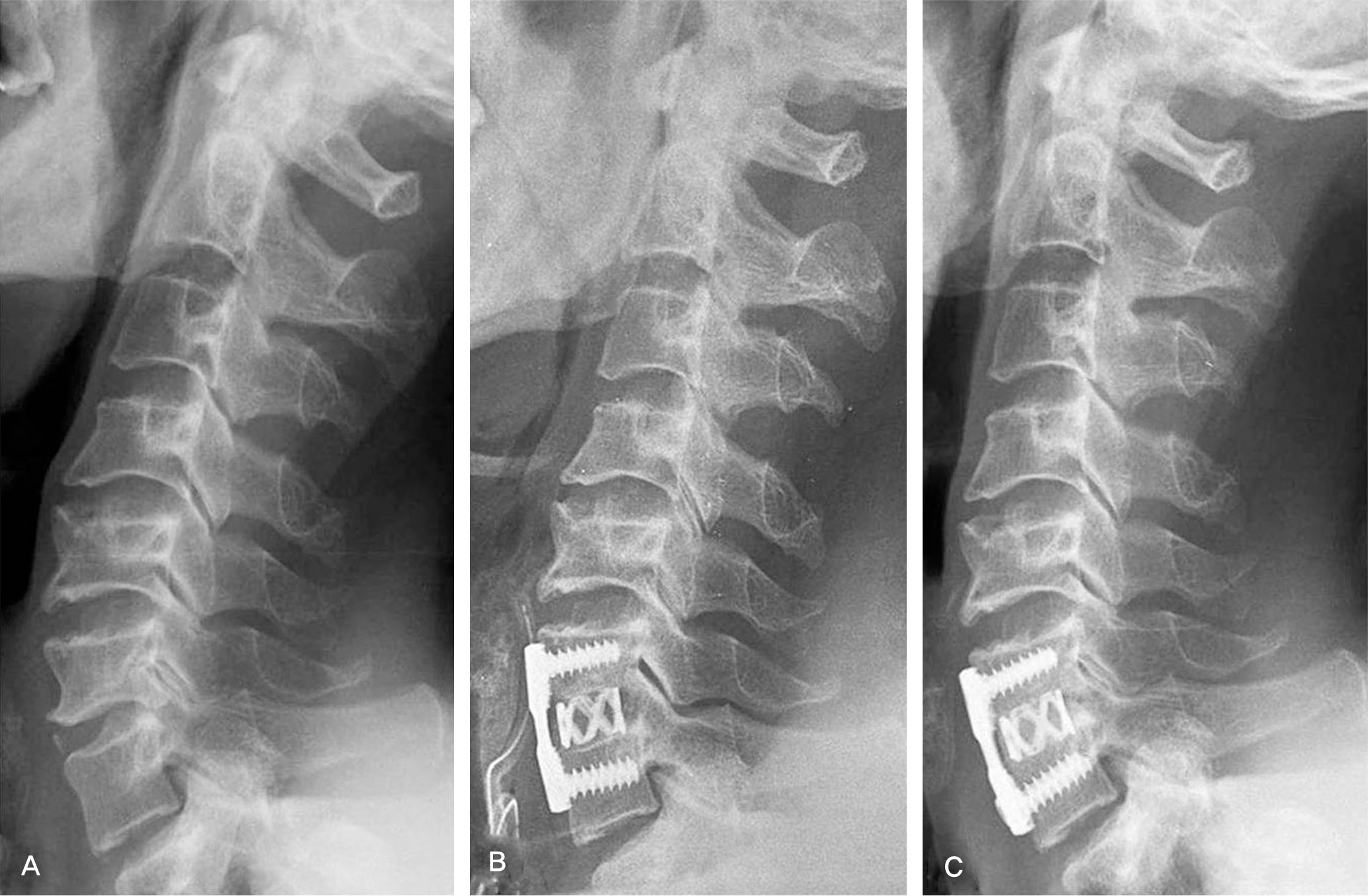 | Fig. 4.Serial radiographs of 57-year-old male patient underwent C6/7 anterior cervical fusion using Harms cage with anterior plating. Preoperative (A) and immediate postoperative radiograph (B) show increased disc height in C6/7. Follow radiographs (C) at 6 months showing solid union. |
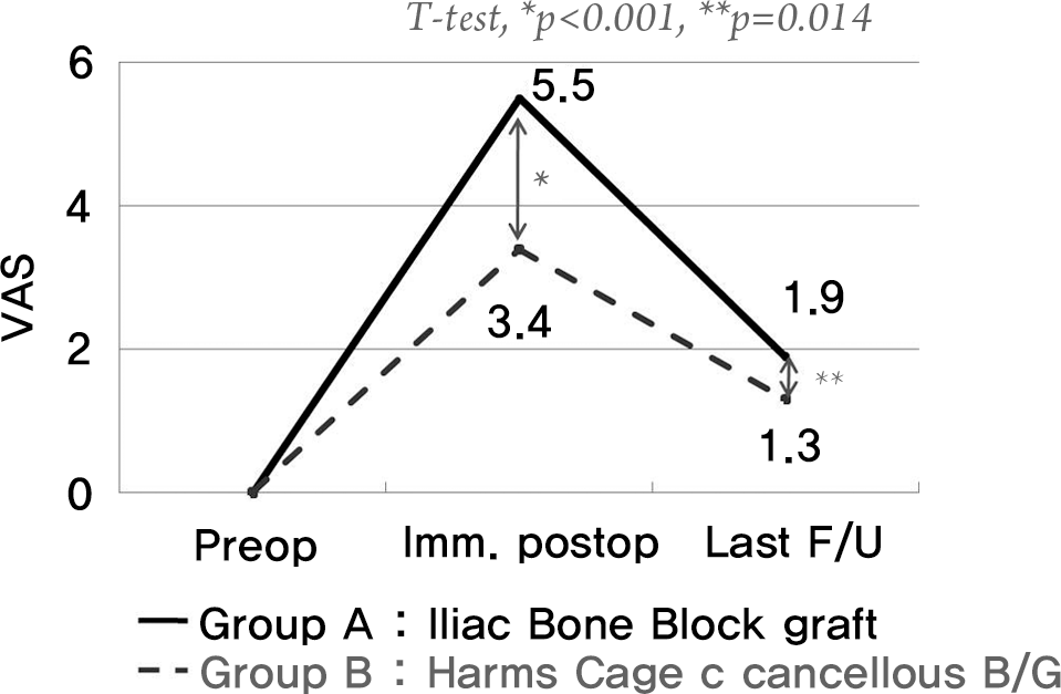 | Fig. 5.Graph showing the clinical outcome analysis with Visual Analogue Scale(VAS) for donor site pain. |
 XML Download