

 PDF
PDF ePub
ePub Citation
 Print
Print


Abstract
Objectives
To evaluated the clinical and radiological effectiveness of sacral alar screws for augmentation of S1 pedicle screws in long-level fusion including L5-S1 segment.
Summary of Literature Review
The fusion rates of lumbosacral junction in long-level fusion are various when S1 pedicle screws are used without augmentation. But, reports of sacral alar screw augmentation are rare.
Material and Methods
From 1996 to 2005, 63 patients performed more than two-level fusion including lumbosacral junction were reviewed. 47 patients underwent lumbosacral fusion with S1 pedicle screws only (S1 group), and 16 patients with sacral alar screws augmentation in addition to S1 pedicle screws (S1-2 group). Radiologically, bony union, halo sign, and breakage of implants were evaluated. Clinically, complications associated with screw placement and general complications were evaluated.
Results
Bony union was obtained in 56 cases(89%) at postoperative 4.3 months. Nonunion was observed in 7 cases(11%, S1 group:5, S1-2 group:2). Loosening of S1 pedicle screw was observed in 32 cases(89%) of S1 group and in 4 cases(25%) of S1-2 group. It showed statistical significance between two groups. Sacral alar screw loosening occurred in 8 cases(50%) of S1-2 group. Metal breakage was developed in 2 cases of S1 group without nonunion or loosening. Postoperative infection occurred in 7 cases(11%, S1 group:5, S1-2 group:2).
Go to : 
REFERENCES
1. Cunningham BW, Lewis SJ, Long J, Dmitriev AE, Linville DA, Bridwell KH. Biomechanical evaluation of lumbosacral reconstruction techniques for spondylolisthesis: an in vitro porcine model. Spine (Phila Pa 1976). 2002; 27:2321–7.
2. Grobler LJ, Frymoyer JW, Robertson PA, Novotny JE. Biomechanics of lumbar spine surgery. Semin Spine Surg. 1993; 5:59–72.
3. Grubb SA, Lipscomb HJ. Results of lumbosacral fusion for degenerative disc disease with and without instrumentation. Two-to five-year followup. Spine (Phila Pa 1976). 1992; 17:349–55.
4. Kornblatt MD, Casey MP, Jacobs RR. Internal fixation in lumbosacral spine fusion. A biomechanical and clinical study. Clin Orthop Relat Res. 1986; 203:141–50.

5. Lonstein JE. The Galveston technique using Luque or Cotrel-Dubousset rods. Orthop Clin North Am. 1994; 25:311–20.
6. Johnston CE 2nd, Ashman RB, Baird AM, Allard RN. Effect of spinal construct stiffness on early fusion mass incorporation. Experimental study. Spine (Phila Pa 1976). 1990; 15:908–12.
7. Lee CS, Chung SS, Choi SW, Yu JW, Sohn MS. Critical Length of Fusion Requiring Additional Fixation to Prevent Nonunion of the Lumbosacral Junction. Spine (Phila Pa 1976). 2010; 6:E206–11.
8. Ashman RB, Birch JG, Bone LB, et al. Mechanical testing of spinal instrumentation. Clin Orthop Relat Res. 1988; 227:113–25.
9. Ashman RB. Mechanical testing of spinal implants. Semin Spine Surg. 1993; l5:73–9.
10. Bernhardt M, Swartz DE, Clothiaux PL, Crowell RR, White AA 3rd. Posterolateral lumbar and lumbosacral fusion with and without pedicle screw internal fixation. Clin Orthop Relat Res. 1992; 284:109–15.
11. Carlson GD, Abitbol JJ, Anderson DR, et al. Screw fixation in the human sacrum. An in vitro study of the biomechanics of fixation. Spine (Phila Pa 1976). 1992; 17(6 Suppl):S196–203.
12. Jackson R. Intrasacral fixation and segmental corrections with adjustable contoured translating axes. Spine State Art Rev. 1994; 8:307–41.
13. Lenke LG, Bridwell KH, Bullis D, Betz RR, Baldus C, Schoenecker PL. Results of in situ fusion for isthmic spondylolisthesis. J Spinal Disord. 1992; 5:433–42.
14. Hambly MF, Wiltse LL, Raghavan N, Schneiderman G, Koenig C. The transition zone above a lumbosacral fusion. Spine (Phila Pa 1976). 1998; 23:1785–92.
15. Esses SI, Botsford DJ, Huler RJ, Rauschning W. Surgical anatomy of the sacrum. A guide for rational screw fixation. Spine (Phila Pa 1976). 1991; 16(6 Suppl):S283–8.
16. Ogilvie JW, Schendel M. Comparison of lumbosacral fixation devices. Clin Orthop Relat Res. 1986; 203:120–5.
17. Camp JF, Caudle R, Ashmun RD, Roach J. Immediate complications of Cotrel-Dubousset instrumentation to the sacro-pelvis. A clinical and biomechanical study. Spine (Phila Pa 1976). 1990; 15:932–41.
18. Devlin VJ, Boachie-Adjei O, Bradford DS, Ogilvie JW, Transfeldt EE. Treatment of adult spinal deformity with fusion to the sacrum using CD instrumentation. J Spinal Disord. 1991; 4:1–14.
19. Perra JH. Techniques of instrumentation in long fusions to the sacrum. Orthop Clin North Am. 1994; 25:287–99.
20. McKinley TO, McLain RF, Yerby SA, Sharkey NA, Sarigul-Klijn N, Smith TS. Characteristics of pedicle screw loading. Effect of surgical technique on intravertebral and intrapedicular bending moments. Spine (Phila Pa 1976). 1999; 24:18–24.
21. Ogon M, Haid C, Krismer M, Sterzinger W, Bauer R. Comparison between single-screw and triangulated, double-screw fixation in anterior spine surgery. A biomechanical test. Spine (Phila Pa 1976). 1996; 21:2728–34.
22. Leong JC, Lu WW, Zheng Y, Zhu Q, Zhong S. Comparison of the strengths of lumbosacral fixation achieved with techniques using one and two triangulated sacral screws. Spine (Phila Pa 1976). 1998; 23:2289–94.
23. Horton WC, Holt RT, Muldowny DS. Controversy. Fusion of L5-S1 in adult scoliosis. Spine (Phila Pa 1976). 1996; 21:2520–2.
24. Shin BJ, Kim KJ, Kim ST, Kim YI. Survivorship analysis of pedicle screw fixation. J Korean Soc Spine Surg. 1999; 6:355–61.
25. Shin BJ, Kim KJ, Cho YB, Kim YI. Radiologic results of posterior lumbosacral fixation according to sacral fixation methods-single screw vs double screws. J Korean Soc Spine Surg. 2000; 7:15–21.
26. Kim EH, Kim HJ. Long Segment Fusion to L5 Vertebra and Sacral Vertebra in Degenerative Lumbar Spine. J Korean Soc Spine Surg. 2002; 9:216–22.
27. Kuklo TR, Bridwell KH, Lewis SJ, et al. Minimum 2-year analysis of sacropelvic fixation and L5-S1 fusion using S1 and iliac screws. Spine (Phila Pa 1976). 2001; 26:1976–83.
28. Smith SA, Abitbol JJ, Carlson GD, Anderson DR, Taggart KW, Garfin SR. The effects of depth of penetration, screw orientation, and bone density on sacral screw fixation. Spine (Phila Pa 1976). 1993; 18:1006–10.
29. Halvorson TL, Kelley LA, Thomas KA, Whitecloud TS 3rd, Cook SD. Effects of bone mineral density on pedicle screw fixation. Spine (Phila Pa 1976). 1994; 19:2415–20.
30. Kim JH, Kim BJ, Choo SK, Cho JH, Kim YJ. Deep Infection following Instrumented Posterior Fusion. J Korean Orthop Assoc. 2006; 41:617–22.
Go to :
Figures and Tables%
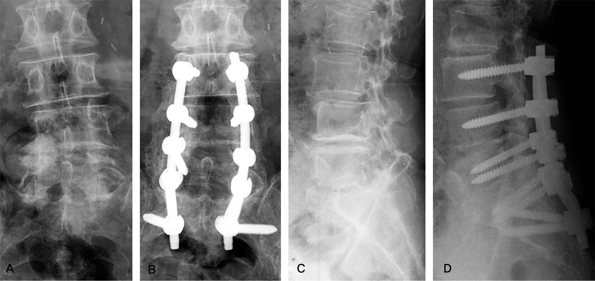 | Fig 1.(A, B) Preoperative anteroposterio(AP) and lateral views of a 59 year-old female show degenerative spondylolisthesis, L3-4 and narrowing of L4-5-S1 disc spaces. (C, D) 2 years followup AP and lateral views fixed with S1 pedicle sacral screw and augmentation with sacral alar screw show solid union and no evidence of metal failure. |
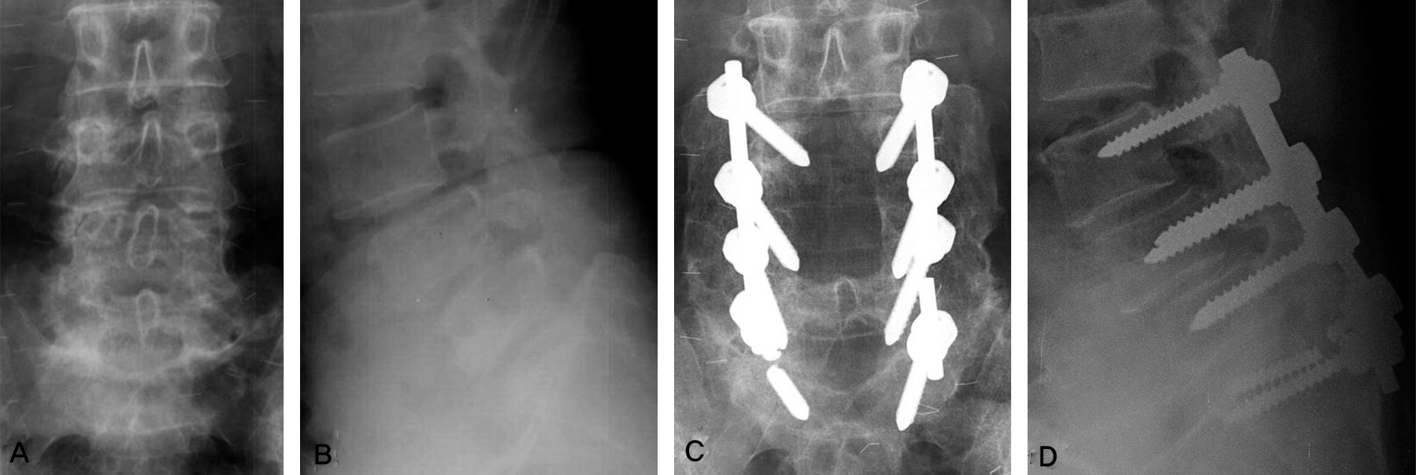 | Fig 2.(A, B)Preoperative AP and lateral views of a 53 year-old female show isthmic spondylolisthesis L3-S1. (C, D) 5 years 10months followup anteroposterior and lateral views fixed with single sacral screw show union of posterolateral fusion but breakages of one sacral screw and one opposite rod. |
Table 1.
Fusion Grades (by Lenke Classification)
Table 2.
Demographic data of S1 and S1-2 group.
 XML Download