Abstract
Objectives
To investigate the clinical results of conservative treatment for mid-to-large lumbar disc herniation diagnosed via magnetic resonance imaging (MRI) and the factors influencing treatment.
Summary of Literature Review
There is limited information regarding the clinical results of conservative treatment for lumbar disc herniation. The recent studies using MRI have suggested favorable treatment results.
Materials and Methods
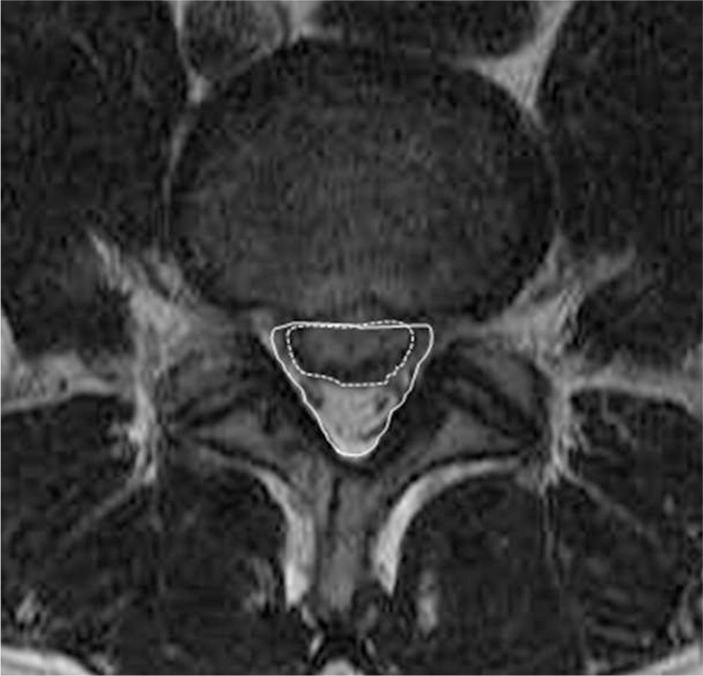
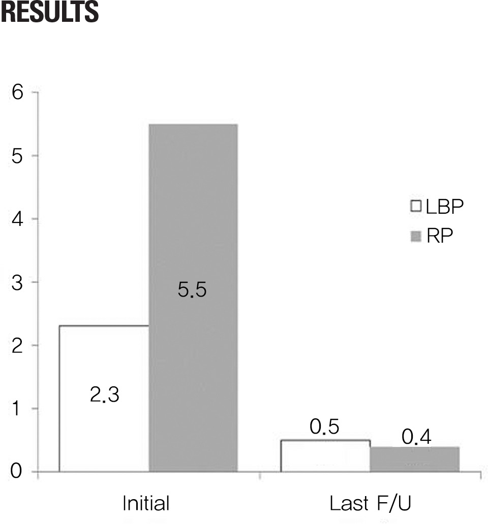
The study subjects were 39 cases of herniated disc patients with over a 1/3 spinal canal encroachment – based on MRI – that were followed up for at least 1 year. The average age was 42.6-years-old (range of 12-76 years-old), and the average followup period was 28 months. The neurological deficit and the visual analogue scale (VAS) of back pain and radiating pain at the time of initial diagnoses and final followups were compared, and the clinical results were evaluated based Kim & Kim's criteria.
Results
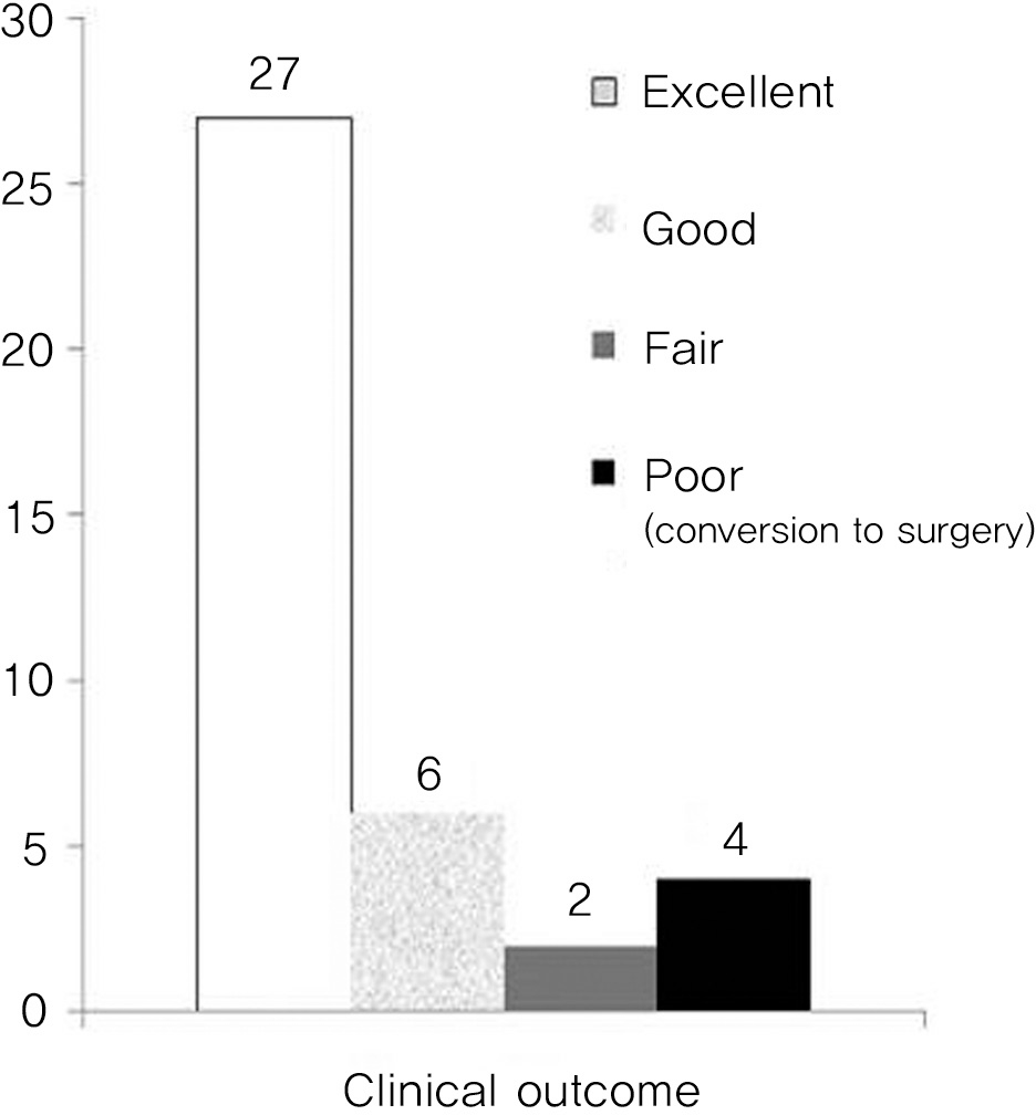
Although 4 of the 39 patients needed to undergo surgery during the followup period, 33 of the remaining 35 patients showed satisfactory (excellent and good ratings) results: 27 excellent, 6 good, 2 fair, i.e., a 85% (33 out of 39) satisfactory results. Of the 14 cases that had neurological defect at the initial diagnosis, only 1 case needed surgery, thereby resulting in a 93% (13 out of 14) satisfactory result. There were no statistically significant correlations among the degree of spinal canal encroachment and other factors such as age, sex, herniation type, and neurological deficit at initial diagnosis, and the clinical results at the final followup, conversion to surgery during followup, and remaining pains.
Go to : 
REFERENCES
1. Deyo RA, Tsui-Wu YJ. Descriptive epidemiology of low-back pain and its related medical care in the United States. Spine (Phila Pa 1976). 1987; 12:264–8.

2. Taylor VM, Deyo RA, Cherkin DC, Kreuter W. Low back pain hospitalization. Recent United States trends and regional variations. Spine (Phila Pa 1976). 1994; 19:1207–12.
3. Hakelius A. Prognosis in sciatica. A clinical followup of surgical and non-surgical treatment. Acta Orthop Scand Suppl. 1970; 129:1–76.

4. Costello RF, Beall DP. Nomenclature and standard reporting terminology of intervertebral disk herniation. Magn Reson Imaging Clin N Am. 2007; 15:167–74.

5. Saal JA, Saal JS. Nonoperative treatment of herniated lumbar intervertebral disc with radiculopathy. An outcome study. Spine (Phila Pa 1976). 1989; 14:431–7.
6. Saal JA, Saal JS, Herzog RJ. The natural history of lumbar intervertebral disc extrusions treated nonoperatively. Spine (Phila Pa 1976). 1990; 15:683–6.

7. Weber H. Lumbar disc herniation. A controlled, prospective study with ten years of observation. Spine (Phila Pa 1976). 1983; 8:131–40.

8. Atlas SJ, Deyo RA, Keller RB, et al. The Maine Lumbar Spine Study, Part II. 1-year outcomes of surgical and nonsurgical management of sciatica. Spine (Phila Pa 1976). 1996; 21:1777–86.
9. Atlas SJ, Keller RB, Chang Y, Deyo RA, Singer DE. Surgical and nonsurgical management of sciatica secondary to a lumbar disc herniation: five-year outcomes from the Maine Lumbar Spine Study. Spine (Phila Pa 1976). 2001; 26:1179–87.
10. Atlas SJ, Keller RB, Wu YA, Deyo RA, Singer DE. Longterm outcomes of surgical and nonsurgical management of sciatica secondary to a lumbar disc herniation: 10 year results from the maine lumbar spine study. Spine (Phila Pa 1976). 2005; 30:927–35.

11. Alaranta H, Hurme M, Einola S, et al. A prospective study of patients with sciatica. A comparison between conservatively treated patients and patients who have undergone operation, Part II: Results after one year followup. Spine (Phila Pa 1976). 1990; 15:1345–9.
12. Hurme M, Alaranta H, Einola S, et al. A prospective study of patients with sciatica. A comparison between conservatively treated patients and patients who have undergone operation, Part I: Patient characteristics and differences between groups. Spine (Phila Pa 1976). 1990; 15:1340–4.
13. Masui T, Yukawa Y, Nakamura S, et al. Natural history of patients with lumbar disc herniation observed by magnetic resonance imaging for minimum 7 years. J Spinal Disord Tech. 2005; 18:121–6.

14. Carlisle E, Luna M, Tsou PM, Wang JC. Percent spinal canal compromise on MRI utilized for predicting the need for surgical treatment in single-level lumbar intervertebral disc herniation. Spine J. 2005; 5:608–14.

15. Cribb GL, Jaffray DC, Cassar-Pullicino VN. Observations on the natural history of massive lumbar disc herniation. J Bone Joint Surg Br. 2007; 89:782–4.

16. Benson RT, Tavares SP, Robertson SC, Sharp R, Marshall RW. Conservatively treated massive prolapsed discs: a 7-year followup. Ann R Coll Surg Engl. 2010; 92:147–53.

17. Dubourg G, Rozenberg S, Fautrel B, et al. A pilot study on the recovery from paresis after lumbar disc herniation. Spine (Phila Pa 1976). 2002; 27:1426–31.

18. Saal JA. Natural history and nonoperative treatment of lumbar disc herniation. Spine (Phila Pa 1976). 1996; 21:1877–83.
19. Takada E, Takahashi M, Shimada K. Natural history of lumbar disc hernia with radicular leg pain: Spontaneous MRI changes of the herniated mass and correlation with clinical outcome. J Orthop Surg (Hong Kong). 2001; 9:1–7.

20. Autio RA, Karppinen J, Niinimä ki J, et al. Determinants of spontaneous resorption of intervertebral disc herniations. Spine (Phila Pa 1976). 2006; 31:1247–52.

21. Jensen TS, Albert HB, Soerensen JS, Manniche C, Leboeuf-Yde C. Natural course of disc morphology in patients with sciatica: an MRI study using a standardized qualitative classification system. Spine (Phila Pa 1976). 2006; 31:1605–12.
Go to : 
Figures and Tables%
 | Fig. 3.Clinical outcome at the final followup (four patients who converted to surgery were defined as “poor”) |
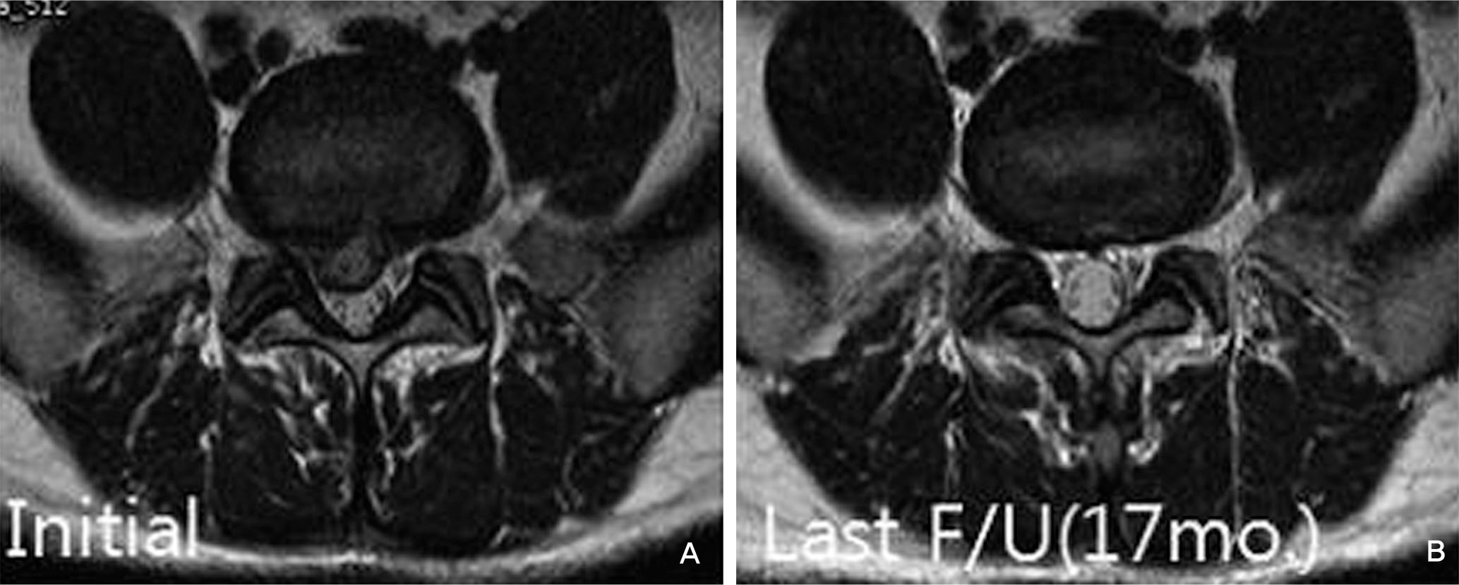
 | Fig. 4.Thirty five year old male patient suffering from bilateral buttock and right leg pain showed canal encroachment about 53% on MRI axial image at the first visit(A). After conservative treatment for 17 months, pain subsided and herniated disc material was absorbed at the final follow up(B). |
Table. 1.
Kim & Kim's criteria of clinical outcome
Table 2.
Summary of cases


 PDF
PDF ePub
ePub Citation
 Print
Print


 XML Download