Abstract
Objectives
The aim of this study was to examine the usefulness of axial and sagittal-reconstructed CT images in the evaluation of spinal canal encroachment by thoracolumbar burst fractures.
Summary of Literature Review
The dimensions of spinal canal encroachment by burst fractures have been described using axial CT images in the thoracolumbar region and sagittal-reconstructed images in the lower cervical region. However, the validity and reliability, depending on the measuring method, have not been fully evaluated.
Materials and Methods
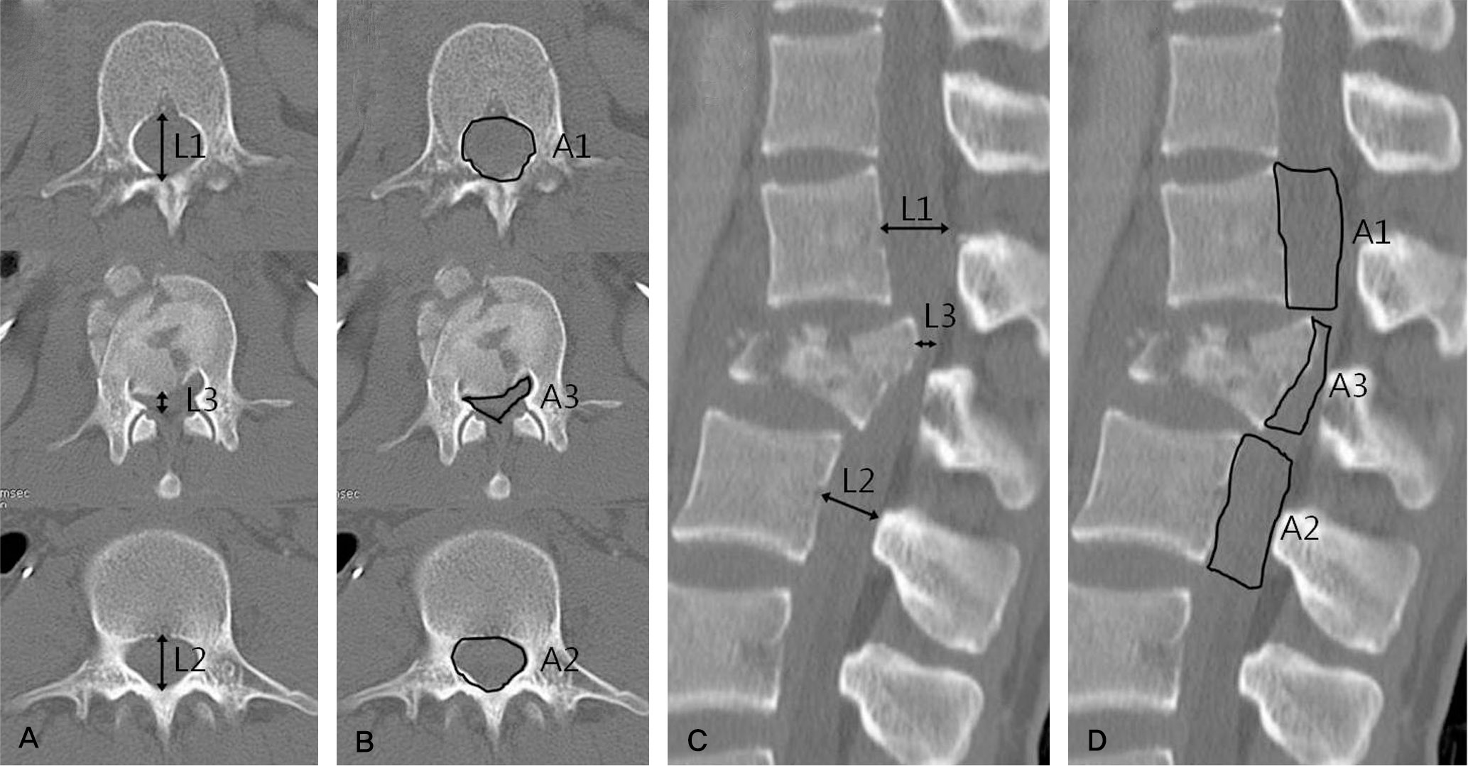
A hundred and ninety-nine patients, who had diagnosed as a thoracolumbar burst fracture, were included in this study. Three orthopedic surgeons independently measured the canal encroachment of the burst fragment in the axial CT images and the sagittal-reconstructed images using the ratio of spinal length (method 1) and the ratio of area (method 2). The validity for the evaluation of the deformity and fracture stability was evaluated. In addition, the reliability of each method was assessed.
Results
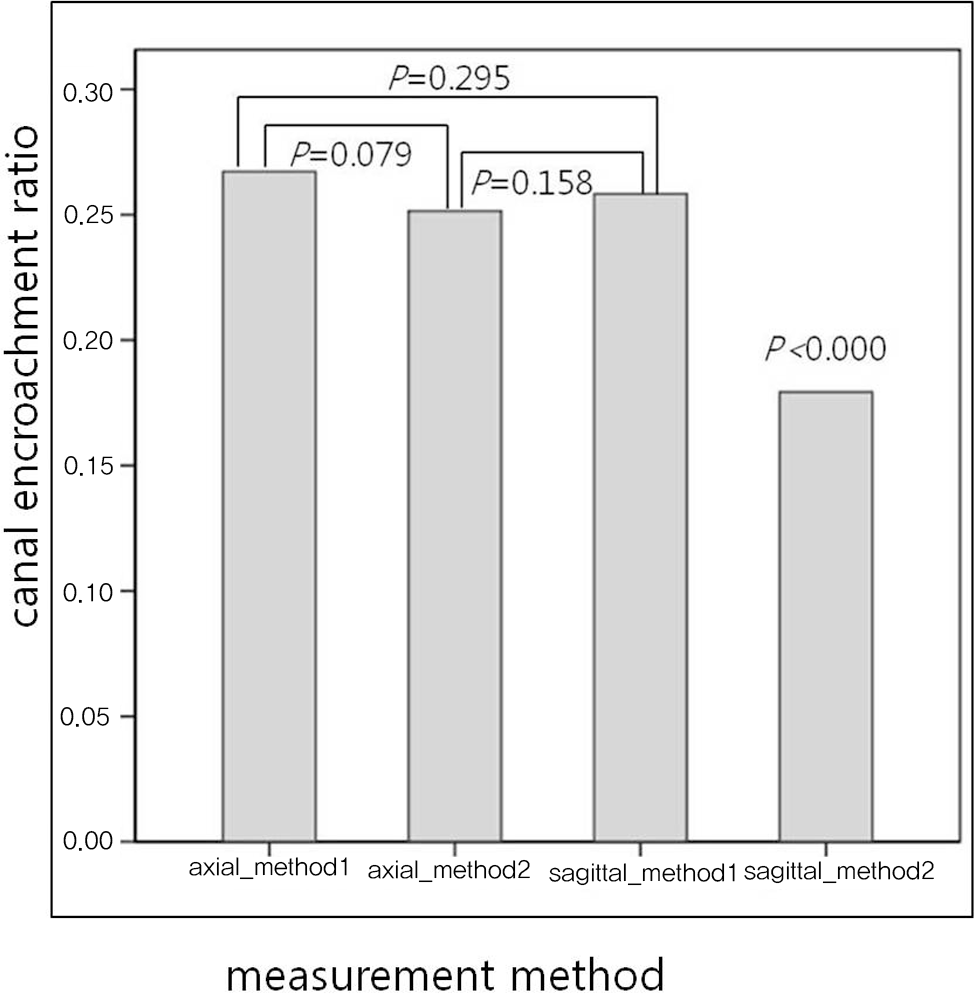
Sixty-seven stable burst fractures and 132 unstable burst fractures were assessed. The mean kyphotic angle of stable and unstable burst fracture were 11.89 ± 8.49°and 15.90 ± 9.63°(P=0.005). The mean canal encroachment ratios of stable fracture were 17.21 ± 15.82 % (axial-method 1), 16.71 ±16.49 % (axial-method 2), 19.54 ± 17.03 % (sagittal reconstructed-method 1), and 11.75 ± 12.33 % (sagittal reconstructed-method 2). The mean canal encroachment ratios of unstable fracture were 31.54 ± 17.10 % (axial-method 1), 29.67 ± 18.47 % (axial-method 2), 28.53 ± 18.60 % (sagittal reconstructed-method 1), and 21.20 ± 15.11 % (sagittal reconstructed-method 2). There was no relationship between the fracture deformity and the canal encroachment ratio in all 4 methods. All ratios in the 4 method showed significant differences in the evaluation of fracture stability. All methods except method 1 in the sagittal-reconstructed images showed significant differences in the assessment of neurologic compromise.
Go to : 
REFERENCES
1. Denis F. The three column spine and its significance in the classification of acute thoracolumbar spinal injuries. Spine (Phila Pa 1976). 1983; 8:817–31.

2. McAfee PC, Yuan HA, Fredrickson BE, Lubicky JP. The value of computed tomography in thoracolumbar fractures. An analysis of one hundred consecutive cases and a new classification. J Bone Joint Surg Am. 1983; 65:461–73.

3. Vaccaro AR, Nachwalter RS, Klein GR, Sewards JM, Albert TJ, Garfin SR. The significance of thoracolumbar spinal canal size in spinal cord injury patients. Spine (Phila Pa 1976). 2001; 26:371–6.

4. Dai LY, Wang XY, Jiang LS. Neurologic recovery from thoracolumbar burst fractures: is it predicted by the amount of initial canal encroachment and kyphotic deformity? Surg Neurol. 2007; 67:232–7.

5. Meves R, Avanzi O. Correlation between neurological deficit and spinal canal compromise in 198 patients with thoracolumbar and lumbar fractures. Spine (Phila Pa 1976). 2005; 30:787–91.

6. Dall BE, Stauffer ES. Neurologic injury and recovery patterns in burst fractures at the T12 or L1 motion segment. Clin Orthop Relat Res. 1988. 171–6.

7. Eberl R, Kaminski A, Muller EJ, Muhr G. Importance of the crosssectional area of the spinal canal in thoracolumbar and lumbar fractures. Is there any correlation between the degree of stenosis and neurological deficit? Orthopade. 2003; 32:859–64.
8. Herndon WA, Galloway D. Neurologic return versus crosssectional canal area in incomplete thoracolumbar spinal cord injuries. J Trauma. 1988; 28:680–3.
9. Keene JS, Fischer SP, Vanderby R Jr, Drummond DS, Turski PA. Significance of acute posttraumatic bony encroachment of the neural canal. Spine (Phila Pa 1976). 1989; 14:799–802.

10. Mohanty SP, Venkatram N. Does neurological recovery in thoracolumbar and lumbar burst fractures depend on the extent of canal compromise? Spinal Cord. 2002; 40:295–9.

11. Zhao X, Fang XQ, Zhao FD, Fan SW. Traumatic canal stenosis should not be an indication for surgical decompression in thoracolumbar burst fracture. Med Hypotheses. 2010; 75:550–2.

12. Bono CM, Vaccaro AR, Fehlings M, et al. Measurement techniques for lower cervical spine injuries: consensus statement of the Spine Trauma Study Group. Spine (Phila Pa 1976). 2006; 31:603–9.
13. Keynan O, Fisher CG, Vaccaro A, et al. Radiographic measurement parameters in thoracolumbar fractures: a systematic review and consensus statement of the spine trauma study group. Spine (Phila Pa 1976). 2006; 31:E156–65.

14. Tran NT, Watson NA, Tencer AF, Ching RP, Anderson PA. Mechanism of the burst fracture in the thoracolumbar spine. The effect of loading rate. Spine (Phila Pa 1976). 1995; 20:1984–8.
15. Willen J, Lindahl S, Nordwall A. Unstable thoracolumbar fractures. A comparative clinical study of conservative treatment and Harrington instrumentation. Spine (Phila Pa 1976). 1985; 10:111–22.
16. Krompinger WJ, Fredrickson BE, Mino DE, Yuan HA. Conservative treatment of fractures of the thoracic and lumbar spine. Orthop Clin North Am. 1986; 17:161–70.

17. Gertzbein SD. Scoliosis Research Society. Multicenter spine fracture study. Spine (Phila Pa 1976). 1992; 17:582–40.
18. Petersilge CA, Pathria MN, Emery SE, Masaryk TJ. Thoracolumbar burst fractures: evaluation with MR imaging. Radiology. 1995; 194:49–54.

19. Oner FC, van Gils AP, Faber JA, Dhert WJ, Verbout AJ. Some complications of common treatment schemes of thoracolumbar spine fractures can be predicted with magnetic resonance imaging: prospective study of 53 patients with 71 fractures. Spine (Phila Pa 1976). 2002; 27:629–36.
20. Haba H, Taneichi H, Kotani Y, et al. Diagnostic accuracy of magnetic resonance imaging for detecting posterior ligamentous complex injury associated with thoracic and lumbar fractures. J Neurosurg. 2003; 99:20–6.

21. Oner FC, van Gils AP, Dhert WJ, Verbout AJ. MRI findings of thoracolumbar spine fractures: a categorisation based on MRI examinations of 100 fractures. Skeletal Radiol. 1999; 28:433–43.

22. Vaccaro AR, Zeiller SC, Hulbert RJ, et al. The thoracolumbar injury severity score: a proposed treatment algorithm. J Spinal Disord Tech. 2005; 18:209–15.
23. Vaccaro AR, Baron EM, Sanfilippo J, et al. Reliability of a novel classification system for thoracolumbar injuries: the Thoracolumbar Injury Severity Score. Spine (Phila Pa 1976). 2006; 31(11 Suppl):S62–9.
Go to : 
Figures and Tables%
 | Fig. 2.Canal encroachment ratio was calculated by the upper and lower adjacent levels (A) using the length ratio on the axial images, (B) using the area ratio on the axial images, (C) using the length ratio on the sagittal-reconstructed images, (D) using the area ratio on the sagittal-reconstructed images |
 | Fig. 3.Paired t-tests revealed that the canal encroachment ratio using the area ratio on the sagittal-reconstructed images was different from those of other methods. |
Table 1.
Patient Demographics
Table 2.
Radiologic measurement


 PDF
PDF ePub
ePub Citation
 Print
Print


 XML Download