Abstract
Objectives
To evaluate the safety and usefulness of implant removal based on fusion by radiological change analyses and non-fused segment motion angle after open reduction, multi-segment fixation, and single segment fusion.
Summary of Literature Review
There have been reports that discuss possible fracture of fixator, loss of reduction, or failure of fixation in certain cases of single segment fixation consistent with thoracolumbar fracture.
Materials and Methods
We analyzed 83 patients who had undergone treatment by fixation of the top 2 segments and the bottom segment. The posterolateral fusions were performed for the top segment for thoracolumbar fractures. The mean followup was 21.3 months. Wedge and local kyphotic angles, anterior, and posterior heights of the vertebral body were measured on plain radiograph. The range of motion of each segment was recorded by flexion-extension lateral radiographs at 6 month after the removal of implants.
Results
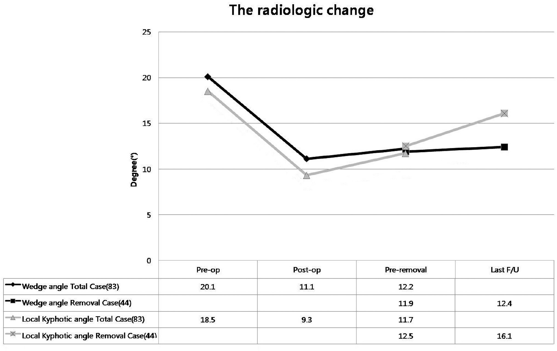
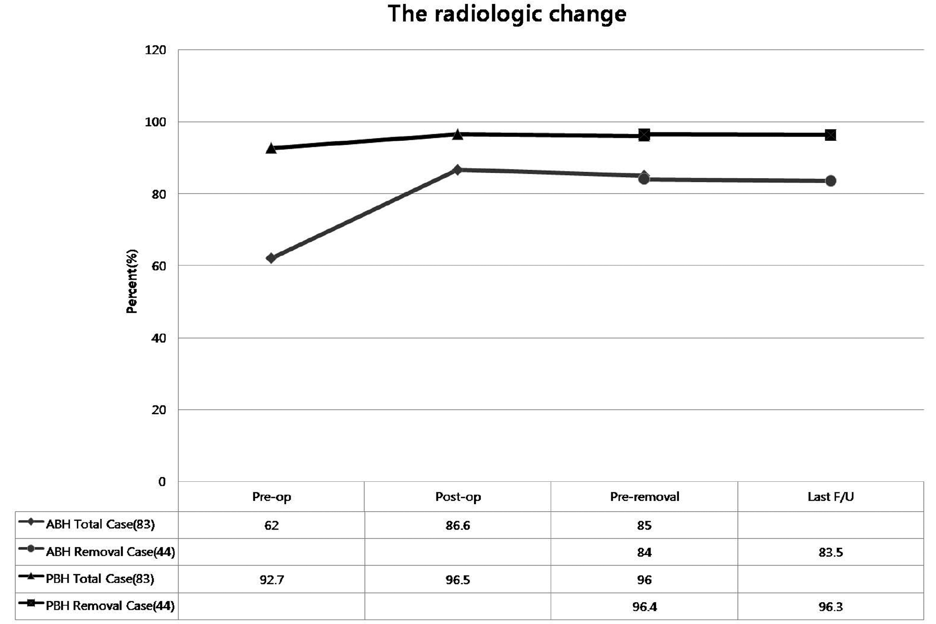
Radiologic assessments performed on 83 patients demonstrated preoperative mean wedge angle, kyphotic angle, mean anterior body height of 20.1°, 18.5° and 62.0%, respectively, and, postoperatively, these were corrected by 9.0°, 9.3° and 24.6%, respectively. In the 44 cases that had the implants removed, the correction losses were 0.4°(P=0.258) and 3.7°(P=0.000), 0.5 % (P=0.756), and at the last followup, compared to measurements prior to the removal. There was no statistical significance in wedge angle or anterior body height. The range of motion measured on the non-fused segment was 3.9°on average at 6-months after the hardware removal.
Conclusions
The multi-segments fixation and single-segment fusion for the thoracolumbar fracture can preserve correction and the motion of non-fusion segment. Although the implant removal after union can sustain motion, further studies regarding degenerative change of the non-fused segment are necessary.
Go to : 
REFERENCES
1. Huler RJ. Thoracolumbar Spine Fracture. in John WF ed. The adult spine-principles and practice. 2nd ed.Philadelphia, Lippincott-Raven: 1997;1473.
2. Ahn JS, Lee JK, Hwang DS, Kim YM, Kim WJ, Byun KH. The Change of Kyphotic Angle and Anterior Vertebral Height after Posterior or Posterolateral Fusion with Transpedicular Screws for Thoracolumbar Bursting Fractures. J Korean Soc Fract. 1999; 12:379–87.

3. Kramer DL, Rodgers WB, Mansfield FL. Transpedicular instrumentation and short-segment fusion of thoracolumbar fractures: a prospective study using a single instrumentation system. J Orthop Trauma. 1995; 9:499–506.

4. Katonis PG, Kontakis GM, Loupasis GA, Aligizakis AC, Christoforakis JI, Velivassakis EG. Treatment of unstable thoracolumbar and lumbar spine injuries using Cotrel-Dubousset instrumentation. Spine (Phila Pa 1976). 1999; 24:2352–7.

5. Lindsey RW, Dick W. The fixateur interne in the reduction and stabilization of thoracolumbar spine fractures in patients with neurologic deficit. Spine (Phila Pa 1976). 1991; 16:140–5.

6. Denis F. The three column spine and its significance in the classification of acute thoracolumbar spinal injuries. Spine (Phila Pa 1976). 1983; 8:817–31.

7. Yue JJ, Sossan A, Selgrath C, et al. The treatment of unstable thoracic spine fractures with transpedicular screw instrumentation: a 3-year consecutive series. Spine (Phila Pa 1976). 2002; 27:2782–7.
8. Farcy JP, Weidenbaum M, Glassman SD. Sagittal index in management of thoracolumbar burst fractures. Spine (Phila Pa 1976). 1990; 15:958–65.

9. Dai LY, Jiang SD, Wang XY, Jiang LS. A review of the management of thoracolumbar burst fractures. Surg Neurol. 2007; 67:221–31.

10. Mikles MR, Stchur RP, Graziano GP. Posterior instrumentation for thoracolumbar fractures. J Am Acad Orthop Surg. 2004; 12:424–35.

11. Inamasu J, Guiot BH, Nakatsukasa M. Posterior instrumentation surgery for thoracolumbar junction injury causing neurologic deficit. Neurol Med Chir. 2008; 48:15–21.

12. Scholl BM, Theiss SM, Kirkpatrick JS. Short segment fixation of thoracolumbar burst fractures. Orthopedics. 2006; 29:703–8.

13. Shen WJ, Liu TJ, Shen YS. Nonoperative treatment versus posterior fixation for thoracolumbar junction burst fractures without neurologic deficit. Spine (Phila Pa 1976). 2001; 26:1038–45.

14. Lee JY, Kim GL. Posterior Instrumentation of Thoracolumbar Fracture. J Korean Soc Spine Surg. 2001; 8:423–7.

15. Defino HL, Scarparo P. Fractures of thoracolumbar spine: monosegmental fixation. Injury. 2005; 36:90–7.

16. Junge A, Gotzen L, von Garrel T, Ziring E, Giannadakis K. Monosegmental internal fixator instrumentation and fusion in treatment of fractures of the thoracolumbar spine. Indications, technique and results. Unfallchirurg. 1997; 100:880–7.
17. Alanay A, Acaroglu E, Yazici M, Oznur A, Surat A. Short-segment pedicle instrumentation of thoracolumbar burst fractures: does transpedicular intracorporeal grafting prevent early failure? Spine (Phila Pa 1976). 2001; 26:213–7.
18. Lee C, Choi JS, Kim YC, et al. Survival Analysis of Posterior Short Fusion in Thoracolumbar Fracture: Significance of Load-Sharing Score and Bone Mineral Density-Changseop. J Korean Soc Spine Surg. 2001; 8:113–20.
19. McLain RF, Sparling E, Benson DR. Early failure of short-segment pedicle instrumentation for thoracolumbar fractures. A preliminary report. J Bone Joint Surg Am. 1993; 75:162–7.

20. Benson DR, Burkus JK, Montesano PX, Sutherland TB, McLain RF. Unstable thoracolumbar and lumbar burst fractures treated with the AO fixateur interne. J Spinal Disord. 1992; 5:335–43.

21. Ebelke DK, Asher MA, Neff JR, Kraker DP. Survivorship analysis of VSP spine instrumentation in the treatment of thoracolumbar and lumbar burst fractures. Spine (Phila Pa 1976). 1991; 16:428–32.

22. Krag MH. Biomechanics of thoracolumbar spinal fixation. A review. Spine (Phila Pa 1976). 1991; 16:84–99.
23. Krag MH, Beynnon BD, Pope MH, Frymoyer JW, Haugh LD, Weaver DL. An internal fixator for posterior application to short segments of the thoracic, lumbar, or lumbosacral spine. Design and testing. Clin Orthop Relat Res. 1986; 203:75–98.

Go to : 
Figures and Tables%
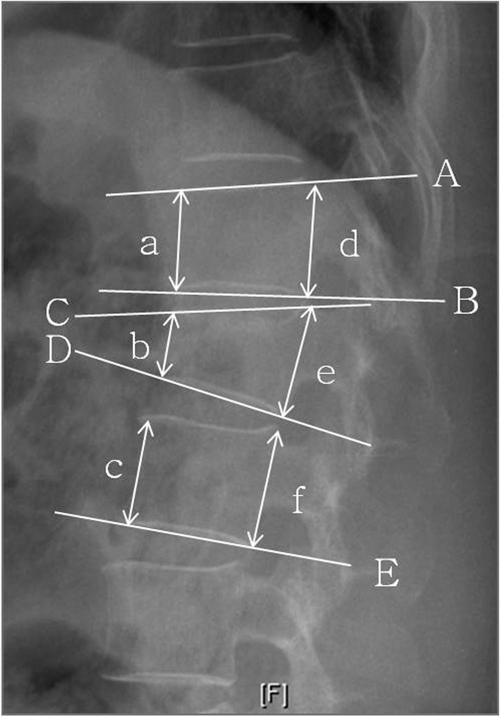
 | Fig. 1.Radiography showing linear and angular measurement. : Wedge angle(∠ CD°), Local kyphotic angle(∠ AE°), Sagittal index(∠ BD°: if T11,∠ BD-5 and if L2,∠ BD+10), Anterior body height(200ⅹ b/(a+c) %), Posterior body height(200× e/(d+f) %) |
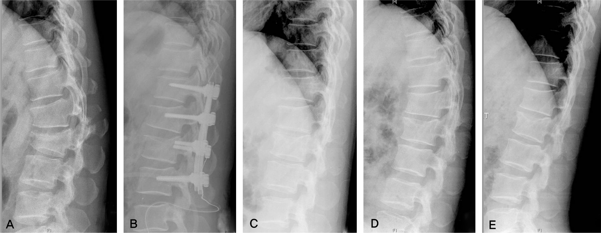
 | Fig. 2.A 41-year old male with flexion-distraction injury on L2. (A) Preoperative lateral roentgenogram shows fracture of L1 spinous process (B) Lateral radiograph, immediately after surgery, shows anatomical reduction. (C,D,E) Lateral roentgenogram of neutral/flexion/extension views that show range of motion of no fusion segment |
Table 1.
The radiologic change e of linear and angular measu urement


 PDF
PDF ePub
ePub Citation
 Print
Print


 XML Download