Abstract
Objectives
We analyzed the risk factors and the surgical results for adjacent segment disease after lumbar fusion.
Summary of Literature Review
Many studies have been performed about the risk factors for adjacent segment disease, but the findings are still controversial.
Materials and Methods
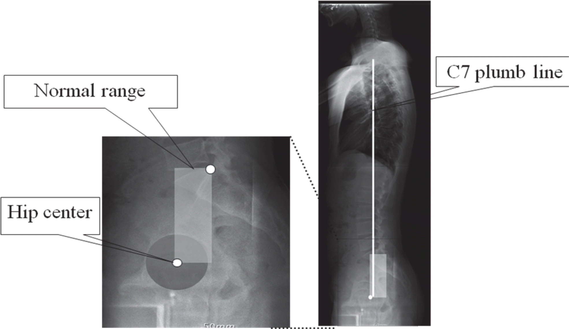
This study was carried out on 35 (13 men, 22 women) of 50 patients who underwent lumbar fusion due to adjacent segment disease with a minimum of 2 year follow-up period from July 1999 to July 2006. The differences of the interval to revision (IR) were statistically analyzed by the examining preexisting degenerative change in the adjacent segments on MRI, the number of fused segments, the lumbar lordosis and the sagittal balance. The surgical outcomes of reoperation were assessed by Brodsky's criteria.
Results
Junctional stenosis as adjacent segment disease was seen in 21 cases (60%) and instability was seen in 14 cases (40%), including 2 iatrogenic flat backs and 2 cases of lumbar degenerative kyphosis. The average IR was 93 months for the cases that had less than 2 segment fusion (20 cases) and 62 months in those with more than 3 segment fusion (15 cases). As for lumbar lordosis, 25 cases (71%) had a normal range of angle as well as 101 months until the IR and 10 cases (29%) had an abnormal range of angle as well as 64 months until IR. Six cases were beyond the normal range of sagittal balance (17%) and their average IR value was 59 months. Otherwise, the cases with a normal range of sagittal balance had 109 months for the IR. The clinical outcome was excellent in 6 cases (17%) and good in 15 cases (43%).
Go to : 
REFERENCES
1.Frymoyer JW., Hanley EN Jr., Howe J., Kuhlmann D., Matteri RE. A comparison of radiographic findings in fusion and non-fusion patients ten or more years following lumbar disc surgery. Spine. 1979. 4:435–40.

2.Kim HT., Kang DW., Yoo CH., Jung JH., Jang SA. Late Changes at the Adjacent Segments to Lumbar Fusions. J Korean Soc Spine Surg. 1996. 3:1–10.
3.Kim WJ., Kang JW., Kim KH, et al. Analysis of Correction Loss after Pedicle Subtraction Osteotomy in Patients with Sagittal Imbalance: Radiologic Aspects. J Korean Orthop Assoc. 2004. 39:629–35.

4.Etebar S., Cahill DW. Risk factors for adjacent segment failure following lumbar fixation with rigid instrumentation for degenerative instability. J Neurosurg. 1999. 90:163–9.
5.Pfirmann CW., Metzdorf A., Zanetti M., Hodler J., Boos N. Magnetic resonance classification of lumbar intervertebral disc degeneration. Spine. 2001. 26:1873–8.
6.Kim DS., Kim YM., Choi ES, et al. Shape and Motion of Each Lumbar Segment in Normal Korean Adults. J Korean Orthop Assoc. 2008. 43:595–600.

7.Jackson RP., McManus AC. Radiographic analysis of sagittal plane alignment and balance in standing volunteers and patients with low back pain matched for age, sex, and size: a prospective controlled clinical study. Spine. 1994. 19:1611–8.
8.Peterson MD., Jackson RP., McManus AC. Standing sagittal spinal balance, alignments and lumbopelvic relationships: Part I. A study of adult volunteers. Presented at the annual meeting of the Scoliosis Research Society, Asheville, North Carolina,. 1995 Sep. 13–17.
9.Aota Y., Kumano K., Hirabayashi S. Postfusion instability at the adjacent segments after rigid pedicle screw fixation for degenerative lumbar spinal disorders. J Spinal Disord. 1995. 8:464–73.

10.Brodsky AE., Hendricks RL., Khalil MA, et al. Segmental lumbar spinal fusions. Spine. 1989. 14:447–50.
11.Harris RI., Wiley JJ. Acquired spondylolysis as a sequel to spine fusion. J Bone Joint Surg Am. 1963. 45:1159–70.

12.Ha KY., Kim YH., Kang KS. Surgery for Adjacent Segment Changes after Lumbosacral Fusion. J Korean Soc Spine Surg. 2002. 9:332–40.

13.Lee CK., Langrana NA. Lumbosacral spine fusion. A biomechanical study. Spine. 1984. 9:574–81.
14.Lehmann TR., Spratt KF., Tozzi JE, et al. Long-term follow-up of lower lumbar fusion patients. Spine. 1987. 12:97–104.

15.Park P., Garton HJ., Gala VC., Hoff JT., McGillicuddy JE. Adjacent segment disease after lumbar or lumbosacral fusion: review of the literature. Spine. 2004. 29:1938–44.

16.Ahn DG., Lee S., Jeong KW., Park JS., Cha SK., Park HS. Adjacent Segment Failure after Lumbar Spine Fusion: Controlled Study for Risk Factors. J Korean Orthop Assoc. 2005. 40:203–8.

17.Gillet P. The fate of the adjacent motion segment after lumbar fusion. J Spinal Disord Tech. 2003. 16:338–45.
18.Kim WJ., Yeom JS., Kang JW., Kim KH., Oh JU., Choy WS. The Changes of Adjacent Segment after Fusions(above 3-levels to L5) in Degenerative Lumbar Disorders. J Korean Soc Spine Surg. 2002. 9:305–12.
19.Cho JL., Park YS., Han JH., Lee CH., Roh WI. The Changes of Adjacent Segments after Spinal Fusion: Follow-up more than Three Years after Spinal Fusion. J Korean Soc Spine Surg. 1998. 5:239–46.
20.Rohlmann A., Neller S., Bergmann G., Graichen F., Claes L., Wilke HJ. Effect of an internal fixator and a bone graft on intersegmental spinal motion and intradiscal pressure in the adjacent regions. Eur Spine J. 2001. 10:301–8.

21.Vialle R., Levassor N., Rillardon L., Templier A., Skalli W., Guigui P. Radiographic analysis of The sagittal alignment and balance of the spine in asymptomatic subjects. J Bone Joint Surg Am. 2005. 87:260–7.

22.Grouw AV., Nadel CI., Weierman RJ., Lowell HA. Long-term follow-up of patients with idiopathic scoliosis treated surgically. Clin Orthop Relat Res. 1976. 117:197–201.
23.Chung JY., Seo HY., Jung JW. Surgical Treatment of Adjacent Degenerative Segment after Lumbar Fusion. J Korean Soc Spine Surg. 2000. 7:264–70.
24.Herkowitz HN., Kurz LT. Degenerative lumbar spondylolisthesis with spinal stenosis: A prospective study comparing decompression with decompression and intertransverse process arthrodesis. J Bone Joint Surg Am. 1991. 73:802–8.

25.Kim WJ., Kim KH., Yeom JS., Choy WS. Clinical Analysis of Failed Lumbar Disc Surgery. J Korean Orthop Assoc. 2001. 36:587–92.

26.Kim WJ., Yeom JS., Kang JW, et al. The Relationship between Sagittal Spinal Aignment and Surgical Results in Degenerative Lumbar Scoliosis with Spinal Stenosis. J Korean Soc Spine Surg. 2002. 9:133–42.
27.Schlegel JD., Smith JA., Schleusener RL. Lumbar motion segment pathology adjacent to thoracolumbar, lumbar and lumbosacral fusion. Spine. 1996. 21:970–81.
28.Kim WJ., Kang JW., Yeom JS, et al. A Comparative Analysis of Sagittal Balance in 100 Asymptomatic Young and Older Aged Volunteers. J Korearn Soc Spine Surg. 2003. 10:327–34.
Go to : 
 | Fig. 2.A 66-year-old woman with 2 segments fusion (A) and a 69-year-old man with 3 segments fusion (B) within normal range of lumbar curvature had a reoperation after several years. But, a 61-year-old woman underwent 2 segments fusion with abnormal range of lumbar curvature had earlier reoperation than previous two patients (C). All three patients had revision surgery due to junctional stenosis. LL: lumbar lordosis |
 | Fig. 3.A 65-year-old woman suffered from stenosis L3-4-5 and spondylolisthesis L3 on L4 (A) was treated with posterior decompression and posterior instrumentation from L3 to L5 (B). Several years later the patient suffered from pain due to junctional stenosis at L3-4, loosening of the most proximal screws and iatrogenic flat back (C) . We treated with implant removal, pedicle subtraction osteotomy(PSO) at L3 vertebral body and posterolateral fusion from T11 to S1 (D). The figure (E) shows follow-up radiograph after several years. SVA: sagittal vertical axis, LL: lumbar lordosis |
Table 1.
Pfirmann classification of disc degeneration
Table 2.
Brodsky's criteria
Table 3.
Lumbar lordosis
| Pre-op(°) | Post-op(°) | IR(months)∗ | ||
|---|---|---|---|---|
| Lumbar lordosis (cases) | Normal(25) | 37.5.±4.9 | 42.0±5.1 | 101(85-125) |
| Abnormal(10) | 15.0±3.7 | 24.5±3.1 | 64(43-90) | |
Table 4.
Sagittal balance
| Pre-op(SVA, cm) | Post-op(SVA, cm) | IR(months)∗ | ||
|---|---|---|---|---|
| Sagittal balance (cases) | Normal(29cases) | +3.4±1.2 | +2.9±2.2 | 109(83-125) |
| Abnormal(6cases) | +5.6±3.2 | +4.7±2.5 | 59(43-80) | |


 PDF
PDF ePub
ePub Citation
 Print
Print


 XML Download