

 PDF
PDF ePub
ePub Citation
 Print
Print


Abstract
Summary of Literature Review
Although numerous studies in the literature have investigated cervical spine injuries, patients with multiple-level cervical fractures have not been commonly described. Multiple non-contiguous cervical fractures are distinctively unusual.
Go to : 
REFERENCES
1.Calenoff L., Chessare JW., Rodgers LF., Toerge J., Rosen JS. Multiple level spinal injuries: importance of early recognition. AJR. 1978. 130:665–9.

2.Kewalramani LS., Taylor RG. Multiple noncontiguous injuries to the spine. Acta Ortho Scand. 1976. 47:52–8.
3.Gupta A., El Masri WS. Multilevel spinal injuries. Incidence, distribution and neurological patterns. J Bone Joint Surg Br. 1989. 71:692–5.
4.Vaccaro AR., An HS., Lin S., Sun S., Balderston RA., Cotler JM. Noncontiguous injuries of the spine. Journal of Spinal Disorders. 1992. 5:320–9.
5.Shear P., Hugenholtz H., Richard MT, et al. Multiple noncontiguous fractures of the cervical spines. J Trauma. 1988. 28:655–59.
6.Levine AM., Edwards CC. The management of traumatic spondylolisthesis of the axis. J Bone Joint Surg Am. 1985. 67:217–26.
7.Birney TJ., Hanley EN Jr. Traumatic cervical injuries in childhood and adolescence. Spine. 1989. 14:1277–82.
8.Ruge JR., Sinson GP., McLone DG., Cerullo LJ. Pediatric spinal injury: the very young. J Neurosurg. 1988. 68:25–30.
9.Letts M., Davidson D., Healey D. Simultaneous fracture of every cervical vertebra: a case study. Spine. 2002. 27:446–50.
10.Tannoury TY., Zmurko MG., Tannoury CA., Anderson DG., Chan DP. Multiple unstable cervical fractures with cord compromise treated nonoperatively: a case report. Spine. 2004. 29:234–8.
Go to :
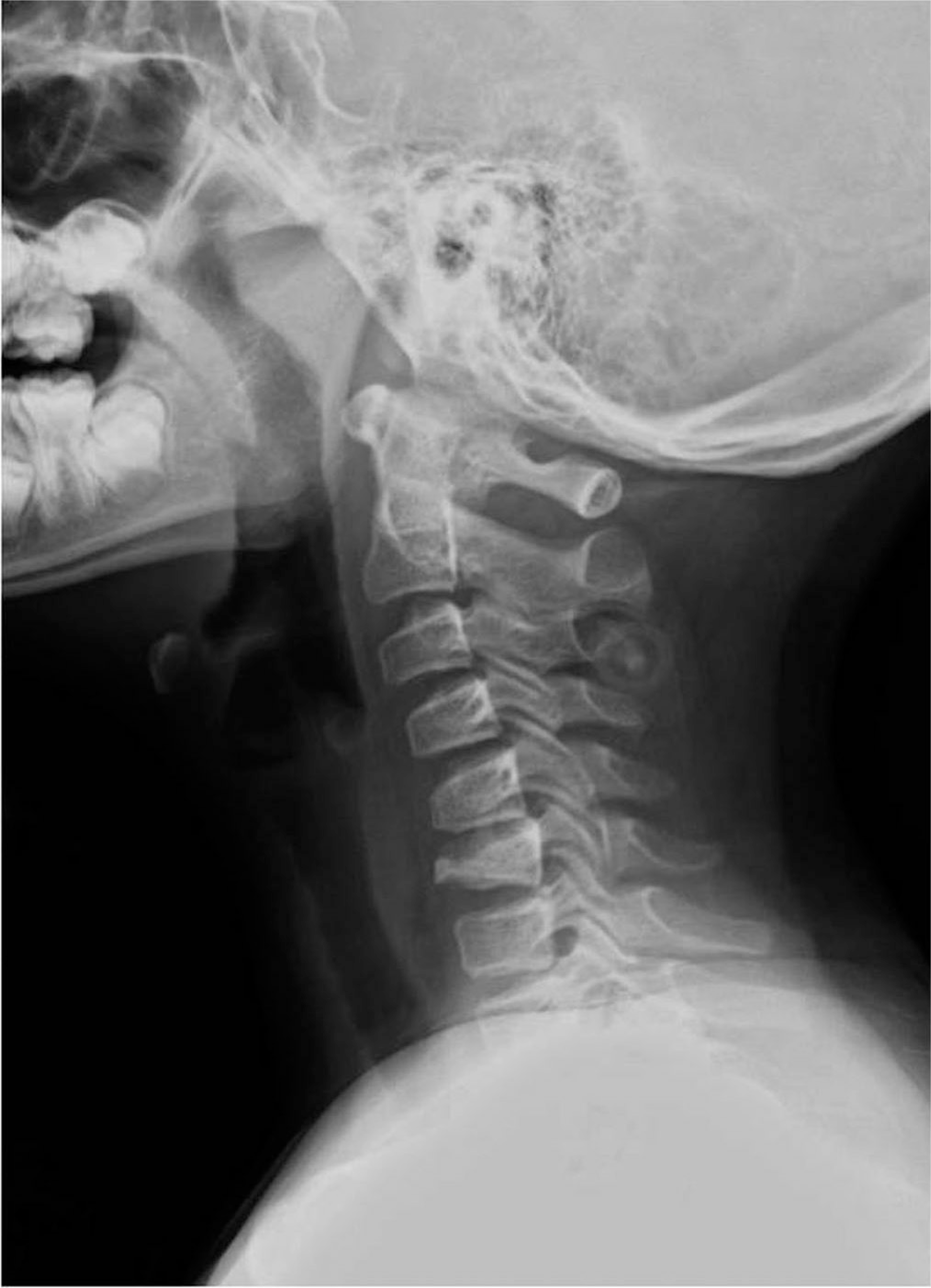
 | Fig. 1.Plain radiograph and CT scan show wedging compression of C6 without retro-pulsion of bony fragment and mild kyphotic angulation of cervical curve. Spinal canal is relatively preserved. |
 | Fig. 2.MRI scan shows marked compression and anterior wedging of C6 spine with minimal spinal canal narrowing. Multifocal high signal intensity on T2WI and STIP image with contrast enhancement are seen in other spines (C3, C5, T7 and T8) suggesting multifocal acute compression fracture. Significant abnormal spinal cord signal is not seen. |
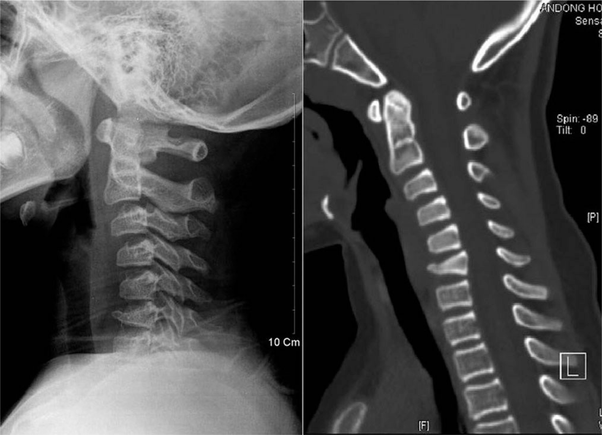
 | Fig. 4.Plain radiograph and CT scan show bilateral symmetrical fractures detaching the pedicles from the body of C2 (Hangman's fracture) and linear fracture lines in the anterior body of the C7 |
 | Fig. 5.MRI scan shows the loss of height of the C3 and C7 body, with hyposignal on T1WI, and hypersignal on T2WI. The compressive myelopathy is not seen in the cervical spinal cord. |

 XML Download