Abstract
Study Design
This is a retrospective study on the clinical availability, diagnosis and treatment of primary psoas muscle abscess.
Objectives
This study investigated the causes and clinical results of patients with primary psoas muscle abscess.
Summary of Literature Review
Primary psoas muscle abscess is not a common disease clinically, but it is a very dangerous disease if the diagnosis and treatment are delayed.
Materials and Methods
Between October 2003 and February 2010, we investigated the symptoms, pathogens, the associated diseases and treatments of 17 patients (11 males and 6 females; mean age: 49.5 years old). We divided patients into the 3 groups According to the treatment options (Group 1: antibiotics alone, Group 2: percutaneous catheter drainage, Group 3: open drainage) and the correlation of the abscess size of each group was analyzed by the Kruskall Wallis method.
Results
The most common complaint was lower back pain (14 patients). Staphylococcus aureus was the most common infectious organism (12 patients). All the patients were treated with broad spectrum antibiotics. Group 1 was composed of 4 patients and the average size of the abscess was 2.3cm (range: 1.2~4.5cm). Group 2 was composed of 7 patients and the average size of the abscess was 7.4cm (range: 3.8~12.2cm). Group 3 was composed of 6 patients and the average size of the abscess was 8.1cm (range: 6.1~14.7cm). There was a significant correlation of the abscess size between each group. (p=0.0007)
Conclusions
The patients diagnosed with primary psoas muscle abscess complained about lower back pain, a febrile sense and gastrointestinal symptoms. Most of the primary psoas muscle abscesses are pyogenic infections. We have to use broad-spectrum antibiotics for the initial treatment. When the occasion demands, additional treatment like percutaneous catheter drainage and open drainage should be considered.
Go to : 
REFERENCES
1.Mynter H. Acute psoitis. Buffalo Med Surg J. 1881. 21:202–10.
2.Ricci MA., Rose FB., Meyer KK. Pyogenic psoas abscess: worldwide variations in etiology. World J Surg. 1986. 10:834–43.

3.Chern CH., Hu SC., Kao WF., Tsai J., Yen D., Lee CH. Psoas abscess: making an early diagnosis in the ED. Am J Emerg Med. 1997. 15:83–8.

4.Mü ckley T., Schü tz T., Kirschner M., Potulski M., Hofmann G., Bü hren V. Psoas abscess: the spine as a primary source of infection. Spine. 2003. 28:E106–13.
5.Walsh TR., Reilly JR., Hanley E., Webster M., Peitzman A., Steed DL. Changing etiology of iliopsoas abscess. Am J Surg. 1992. 163:413–6.

6.Riyad MN., Sallam MA., Nur A. Pyogenic psoas abscess: discussion of its epidemiology, etiology, bacteriology, diagnosis, treatment and prognosis-case report. Kuwait Med J. 2003. 35:44–7.
7.Gruenwald I., Abrahamson J., Cohen O. Psoas abscess: case report and review of the literature. J Urol. 1992. 147:1624–6.

8.Santaella RO., Fishman EK., Lipsett PA. Primary vs secondary iliopsoas abscess. Presentation, microbiology, and treatment. Arch Surg. 1995. 130:1309–13.
9.Bratton RL. Assessment and management of acute low back pain. Am Fam Physician. 1999. 60:2299–308.
11.Baier PK., Arampatzis G., Imdahl A., Hopt UT. The iliopsoas abscess: aetiology, therapy, and outcome. Langenbecks Arch Surg. 2006. 391:411–7.

12.Thongngarm T., McMurray RW. Primary psoa abscess. Ann Rheum Dis. 2001. 60:173–4.
13.Isdale AH., Foley-Nolan DF., Butt WP., Birkenhead D., Wright V. Psoas abscess in rheumatoid arthritis—an inperspicuous diagnosis. Br J Rheumatol. 1994. 33:853–8.

14.van den Berge M., de Marie S., Kuipers T., Jansz AR., Bravenboer B. Psoas abscess: report of a series and review of the literature. Neth J Med. 2005. 63:413–6.
15.Lee KY., Sohn SK., Hwang KS. Comparison of Pyogenic and Tuberculous Spondylitis. J Korean Soc Spine Surg. 1999. 6:443–50.
16.Koo KH., Lee HJ., Chang BS., Yeom JS., Park KW., Lee CK. Differential Diagnosis between Tuberculous Spondylitis and Pyogenic Spondylitis. J Korean Soc Spine Surg. 2009. 16:112–21.

17.Leu SY., Leonard MB., Beart RW Jr., Dozois RR. Psoas abscess: changing patterns of diagnosis and etiology. Dis Colon Rectum. 1986. 29:694–8.
18.Procaccino JA., Lavery IC., Fazio VW., Oakley JR. Psoas abscess: difficulties encountered. Dis Colon Rectum. 1991. 34:784–9.
19.Desandre AR., Cottone FJ., Evers ML. Iliopsoas abscess: etiology, diagnosis, and treatment. Am Surg. 1995. 61:1087–91.
20.Agrawal SN., Dwivedi AJ., Khan M. Primary psoas abscess. Dig Dis Sci. 2002. 47:2103–5.
21.Taiwo B. Psoas abscess: a primer for the internist. South Med J. 2001. 94:2–5.
22.Kim YM., Won CH., Seo JB., Choi ES., Lee HS., Um SM. Pyogenic L4-5 Spondylitis Managed with Percutaneous Drainage Followed by Posterior Lumbar Interbody Fusion: A Case Report. J Korean Soc Spine Surg. 2001. 8:513–9.
Go to : 
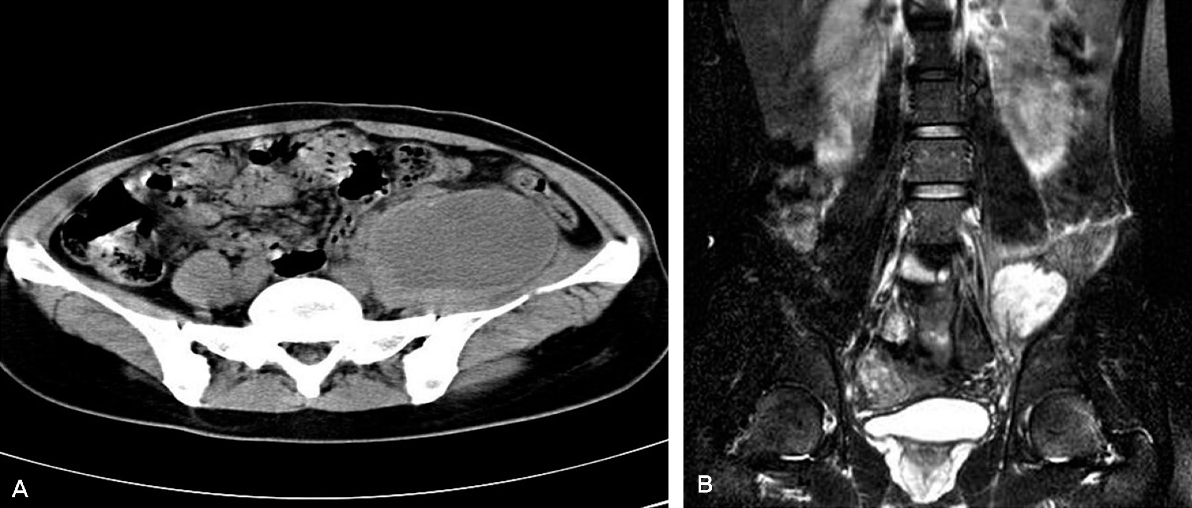
 | Fig.1. (A)Transverse contrast-enhanced CT scan shows a hypodense lesion with enhancing rim in the left psoas muscle, typically an abscess. (B) Coronal T2-weighted MRI scan shows a septated abscess in the left psoas muscle |
 | Fig.2. (A)Coronal contrast-enhanced CT scan shows a large hypodense lesion with enhancing rim in the right psoas muscle, typically an abscess (B) Post 3 days of PCD insertion, Coronal contrast-enhanced CT scan shows a decreased amount of abscess in the right psoas muscle |
Table 1.
Characteristics of patients with a psoas abscess seen in the period october 2003 to february 2010


 PDF
PDF ePub
ePub Citation
 Print
Print


 XML Download