Abstract
Objectives
To analyze our results following simple discectomy of central massive disc herniation focusing on instability for the usefulness of intervertebral fusion.
Summary of Literature Review
Lumbar instability is a complication of central massive disc herniation. However, there is limited evidence on the correlation between lumbar instability and loss of disc material.
Materials and Methods
A total of 25 patients who had undergone discectomy for a single-level lumbar disc herniation were followed up for two years. The clinical group (group A) included 12 patients that had a compromised canal with greater than 50% of the herniated disc, while the central axis of the herniated disc was less than 20% deviated from the center axis of the spinal canal, as seen on MRI. The control group (group B) had 13 patients that had a compromised canal with less than 50% of the herniated disc while their axis was more than 20% deviated from the center axis of the spinal canal. Clinical and radiologic instability, pain and functional disability were compared between the two groups.
Go to : 
REFERENCES
1.Knutsson F. The instability associated with disk degeneration of the lumbar spine. Acta Radiol Scand. 1944. 25:593–609.
2.Dupis PR., Yong-Hing K., Cassidy JD., Kirkaldy Willis WH. Radiologic diagnosis of lumbar spinal instability. Spine. 1985. 10:262–76.
3.Nachlas IW. End-results study of the treatment of herniated nucleus pulposus by excision with fusion and without fusion. J Bone Joint Surg Am. 1952. 34:981–8.
4.Barr JS., Kubik GS., Molloy MK, et al. Evaluation of end results in treatment of ruptured lumbar intervertebral discs with protrusion of nucleus pulposus. Surg Gynecol Obstet. 1967. 125:250–6.
5.Frymoyer JW., Hanley E., Howe J., Kuhlmann D., Matteri R. Disc excision and spine fusion in the management of lumbar disc disease: A minimum ten-year follow-up. Spine. 1978. 3:1–6.
6.LaMont RL., Morawa LG., Pederson HE. Comparison of disc excision with combined disc excision and spinal fusion for lumbar disc ruptures. Clin Orthop. 1976. 121:212–6.
7.Rish BL. A comparative evaluation of posterior lumbar interbody fusion for disc disease. Spine. 1985. 10:855–7.

8.Vaughan PA., Malcolm BW., Maistelli GL. Results of L4–L5 disc excision alone versus disc excision and fusion. Spine. 1988. 13:690–5.

9.Young HH., Love GJ. End results of removal of protruded lumbar intervertebral discs with and without fusion. Am Acad Orthop Surg Inst Course Lecture. 1959. 16:213–6.
10.Takeshima T., Kambara K., Miyata S., Ueda Y., Tamai S. Clinical and radiographic evaluation of disc excision for lumbar disc herniation with and without posterolateral fusion. Spine. 2000. 25:450–6.

11.Satoh I., Yonenobu K., Hosono N., Ohwada T., Fuji T., Yoshikawa H. Indication of posterior lumbar interbody fusion for lumbar disc herniation. J Spinal Disord Tech. 2006. 19:104–8.

12.Tominaga S., Date K., Ouchi K, et al. Comparison between nonfusion and fusion group for lumbar discopathy—Effects of intervertebral fusion. Rinshou Seikei Geka. 1988. 23:236–46.
13.White AH., von Rogov P., Zucherman J., Heiden D. Lumbar laminectomy for herniated disc; a prospective controlled comparison with internal fixationfusion. Spine. 1987. 12:305–7.
14.Eie N. Comparison of the results in patients operated upon for ruptured lumbar discs with and without spinal fusion. Acta Neurochir (Wien). 1978. 41:107–13.

15.Barr JS. Low back and sciatic pain: Result of treatment. J Bone Joint Surg Am. 1951. 33:633–49.
17.Decker HG., Shapiro SW. Herniated lumbar intervertebral disks; results of surgical treatment without the routine use of spinal fusion. AMA Arch Surg. 1957. 75:77–84.
18.Frymoyer JW., Hanley EN Jr., Howe J., Kuhlmann D., Matteri RE. A comparison of radiographic findings in fusion and nonfusion patients ten or more years following lumbar disc surgery. Spine. 1979. 4:435–40.

19.Weber H. Lumbar disc herniation: a controlled, prospective study with ten years of observation. Spine. 1983. 8:131–40.

20.GL Cribb, DC Jaffray, VN Cassar pullicino. Observation on the natural history of massive lumbar disc herniation. J Bone Joint Surg Br. 2007. 89:782–4.
21.Barlocher CB., Krauss JK., Seiler RW. Central lumbar disc herniation. Acta Neurochir (wien). 2000. 142:1369–75.
22.Knop-Jergas BM., Zucherman JF., Hsu KY., DeLong B. Anatomic position of a herniated nucleus pulposus predicts the outcome of lumbar discectomy. J Spinal Disord. 1996. 9:246–50.

23.Walker JL., Schulak D., Murtagh R. Midline disc herniation of the lumbar spine. Southern Med J. 1993. 86:13–7.
24.Ki SC., Choi YS., Kim KS., Kuk WJ. Lumbar Discectomy Using Tubular Retractor and Microendoscopy. J Korean Soc Spine Surg. 2008. 15:265–71.

Go to : 
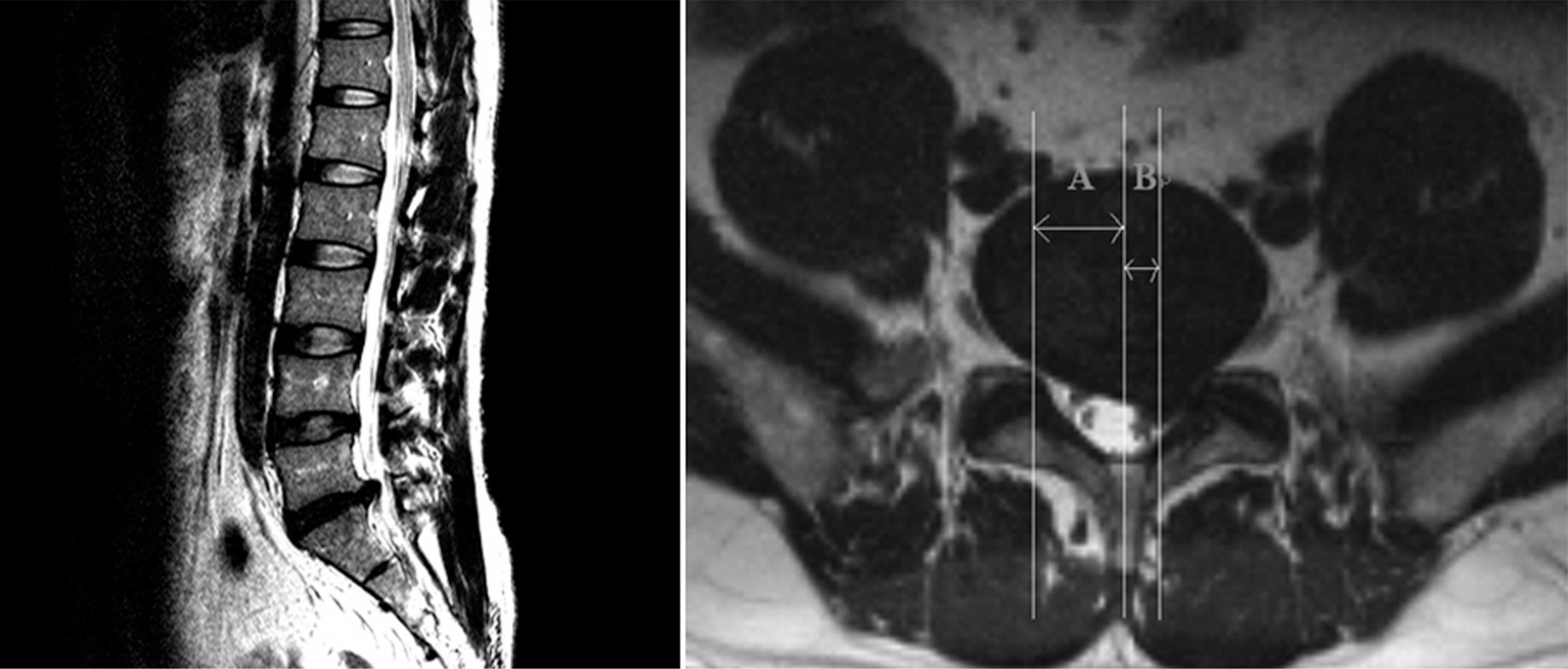
 | Fig. 1.MRI finding of disc herniation and the method of canal compromised area. A Canal compromised (%) = A/A+B × 100 BA (A + B : spinal canal area, A : involved canal area) |
 | Fig. 2.Axial lateralization of disc herniation in MRI Axial lateralization (%) = B/A ⨯ 100 (A : radius of spinal canal, B : length from canal center to axis of herniated disc) |
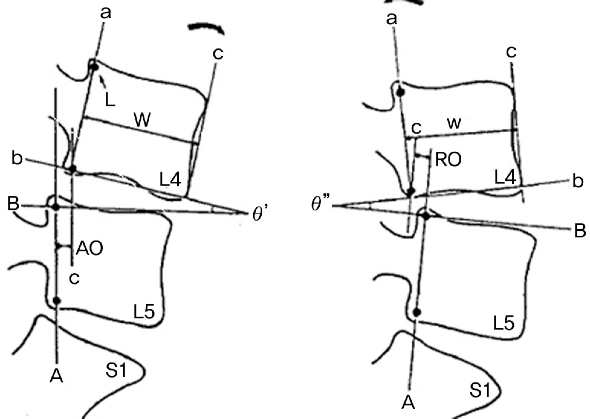
 | Fig. 3.Measurement of angular difference and horizontal displacement on flexion/extension radiogram (Radiographic method of Dupuis and co-workers) Horizontal displacement = AO – RO Angular displacement = θ”-θ’
|
Table 1.
Constitution of the clinical group and control group
| Group A | GroupB | |||||
|---|---|---|---|---|---|---|
| Case (no.) | 12 | 13 | ||||
| Sex (M:F) | 3 : 9 | 11:2 | ||||
| Age (years) | 39.75 | 41.69 | ||||
| Canal compromised (%) | 71.06 | 27.47 | ||||
| Axis lateralization rate (%) | 7.83 | 59.77 | ||||
| Level | L4-L5 | L5-S1 | L4-L5 | L5-S1 | ||
| 11 | 1 | 4 | 9 |
Table 2.
Clinical result of clinical group and control group
| Group A | GroupB | p-value | ||
|---|---|---|---|---|
| Pre-op | ODI | 32.08 | 32.15 | 0.972 |
| VAS | 8.83 | 8.85 | 0.991 | |
| 1 months F/U | ODI | 7.17 | 7.00 | 0.917 |
| VAS | 2.42 | 2.46 | 0.954 | |
| 2 years F/U | ODI | 2.58 | 2.08 | 0.610 |
| VAS | 0.83 | 0.85 | 0.969 |


 PDF
PDF ePub
ePub Citation
 Print
Print


 XML Download