Abstract
Summary of the Literature Review
There are several methods for measuring the compression ratio and kyphotic angle in thoracolumbar fractures, but no definitive measurements and no different values according to the stability have been established.
Objectives
We wanted to compare the compression ratio and kyphosis of thoracolumbar and lumbar fractures according to the radiologic measuring methods and we wanted to analyze their relationship with the stability of fracture.
Materials and Methods
From July 2002 to August 2008, the plain films, CT, MRI and medical records of thoracolumbar and lumbar fracture were reviewed. The compression ratio and kyphotic angle were calculated by several different formulas with using the lateral view of the plain X-ray film, the sagittal reconstruction image of CT and the sagittal image of MRI and the results were compared. Each subject was classified according to both McAfee's classification and the TLISS classification.
Results
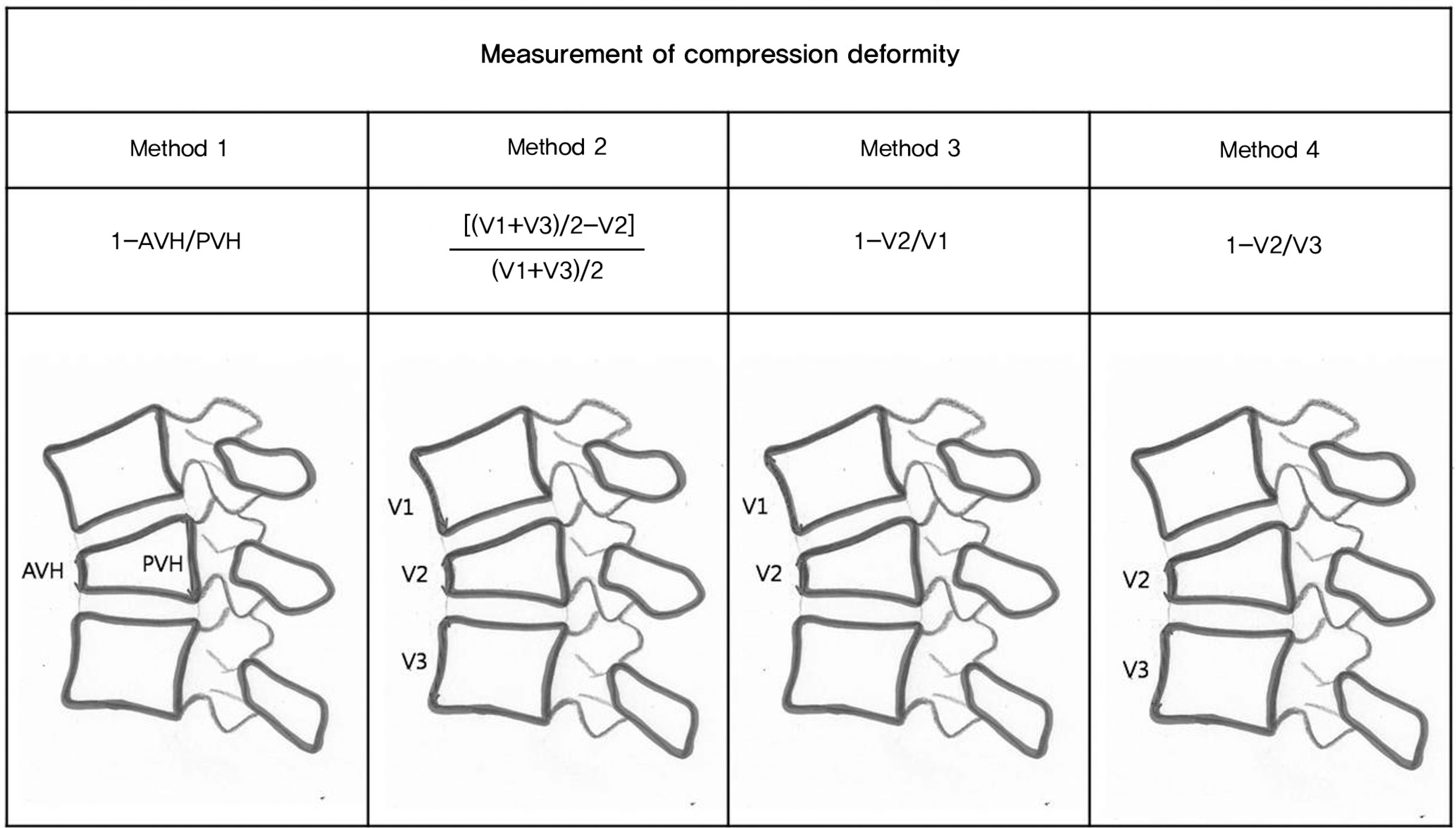
Two hundred forty eight vertebral bodies of 205 thoracolumbar fracture patients were analyzed. The compression ratio according to formula 1, which was calculated as 1-anterior vertebral height/posterior vertebral height, was significantly correlated with Cobb's angle and the local kyphotic angle. There was no significant difference between the Cobb's angle calculated using the lateral X-ray and that using the sagittal view of CT; however, it was significantly less using the sagittal MRI view. The unstable fractures according to McAfee's classification showed a significantly higher compression ratio and kyphotic angle compared to those of the stable fractures.
Conclusions
The compression ratio formula 1 was most significantly correlated with the kyphotic deformity. The unstable fractures showed a mean compression ratio higher than 30%, a mean Cobb's angle of 15° and local kyphotic angle of 18°. The sagittally reconstructed CT was a useful measuring method for the evaluation of kyphotic deformity, and it was more accurate than that of the plain film.
Go to : 
REFERENCES
1.Bohlman HH. Treatment of fractures and dislocations of the thoracic and lumbar spine. J Bone Joint Surg Am. 1985. 67:165–9.

2.DeWald RL. Burst fractures of the thoracic and lumbar spine. Clin Orthop Relat Res. 1984. 189:150–61.

3.Jacobs RR., Casey MP. Surgical management of thoracolumbar spinal injuries: General principles and controversial considerations. Clin Orthop Relat Res. 1984. 189:22–35.

4.Willen J., Anderson J., Toomoka K., Singer K. The natural history of burst fractures at the thoracolumbar junction. J Spinal Disord. 1990. 3:39–46.

5.Mumford J., Weinstein JN., Spratt KF., Goel VK. Thoracolumbar burst fractures: The clinical efficacy and outcome of nonoperative management. Spine. 1993. 18:955–70.
6.Weitzman G. Treatment of stable thoracolumbar spine compression fractures by early ambulation. Clin Orthop Relat Res. 1971. 76:116–22.

8.Krompinger WJ., Fredrickson BE., Mino DE., Yuan HA. Conservative treatment of fractures of the thoracic and lumbar spine. Orthop Clin North Am. 1986. 17:161–70.

9.Gertzbein SD. Scoliosis Research Society. Multicenter spine fracture study. Spine. 1992. 17:528–40.
10.Weidenbaum M., Farcy JP. Surgical management of thoracic and lumbar burst fractures. Bridwell KH, Gordon RL, editors. The textbook of spinal surgery. 2nd ed.New York: Lippincott-Raven;1997. p. 1851.
11.Farcy JP., Weidenbaum M., Glassman SD. Sagittal index in management of thoracolumbar burst fractures. Spine. 1990. 15:958–65.

12.Kuklo TR., Polly DW., Owens BD., Zeidman SM., Chang AS., Klemme WR. Measurement of thoracic and lumbar fracture kyphosis: evaluation of intraobserver, interobserver, and technique variability. Spine. 2001. 26:61–5.
13.Kim SW., Chung YK. Long term follow-up of osteoporotic vertebral fractures according to the morphologic analysis of fracture pattern. J Korean Soc Spine Surg. 2000. 7:611–7.
14.Keynan O., Fisher CG., Vaccaro A, et al. Radiographic measurement parameters in thoracolumbar fractures: a systematic review and consensus statement of the spine trauma study group. Spine. 2006. 31:E156–65.

15.Denis F. The three column spine and its significance in the classification of acute thoracolumbar spinal injuries. Spine. 1983. 8:817–31.

16.Masharawi Y., Salame K., Mirovsky Y, et al. Vertebral body shape variation in the thoracic and lumbar spine: characterization of its asymmetry and wedging. Clin Anat. 2008. 21:46–54.

17.Abdel-Hamid Osman A., Bassiouni H., Koutri R., Nijs J., Geusens P., Dequeker J. Aging of the thoracic spine: distinction between wedging in osteoarthritis and fracture in osteoporosis-a cross-sectional and longitudinal study. Bone. 1994. 15:437–42.
18.Ismail AA., Cooper C., Felsenberg D, et al. Number and type of vertebral deformities: epidemiological characteristics and relation to back pain and height loss. European Vertebral Osteoporosis Study Group. Osteoporos Int. 1999. 9:206–13.
19.McAfee PC., Yuan HA., Fredrickson BE., Lubicky JP. The value of computed tomography in thoracolumbar fractures. An analysis of one hundred consecutive cases and a new classification. J Bone Joint Surg Am. 1983. 65:461–73.

20.Vaccaro AR., Zeiller SC., Hulbert RJ, et al. The thoracolumbar injury severity score: a proposed treatment algorithm. J Spinal Disord Tech. 2005. 18:209–15.
21.Lee HM., Kim DJ., Kim HS., Suk KS., Kim NH., Park SY. Reliability of MRI to detect posterior ligament complex injury in thoracolumbar spinal fractures. J Korean Soc Spine Surg. 2000. 7:70–6.
22.Lau EM., Chan HH., Woo J, et al. Normal ranges for vertebral height ratios and prevalence of vertebral fracture in Hong Kong Chinese: a comparison with American Caucasians. J Bone Miner Res. 1996. 11:1364–8.

23.Tayyab NA., Samartzis D., Altiok H, et al. The reliability and diagnostic value of radiographic criteria in sagittal spine deformities: comparison of the vertebral wedge ratio to the segmental Cobb's angle. Spine. 2007. 32:E451–9.
24.Park BC., Oh CW., Min WK. Anatomical study of lumbar vertebrae and intervertebral space in Korean adults. J Korean Soc Spine Surg. 1999. 6:34–40.
25.Dunn HK. Anterior spine stabilization and decompression for thoracolumbar injuries. Orthop Clin North Am. 1986. 17:113–9.

26.Jacobs RR., Casey MP. Surgical management of thoracolumbar spinal injuries. General principles and controversial considerations. Clin Orthop Relat Res. 1984. 189:22–35.

27.Teng MM., Wei CJ., Wei LC, et al. Kyphosis correction and height restoration effects of percutaneous vertebroplasty. AJNR Am J Neuroradiol. 2003. 24:1893–900.
28.Pradhan BB., Bae HW., Kropf MA., Patel VV., Delamarter RB. Kyphoplasty reduction of osteoporotic vertebral compression fractures: correction of local kyphosis versus overall sagittal alignment. Spine. 2006. 31:435–41.

Go to : 
 | Fig. 1.Measurement of compressive deformity by 4 different methods. ∗AVH: anterior vertebral height, PVH: posterior vertebral height, V1:anterior height of upper vertebra, V2:vertebra of fracture level, V3:anterior height of lower vertebra |
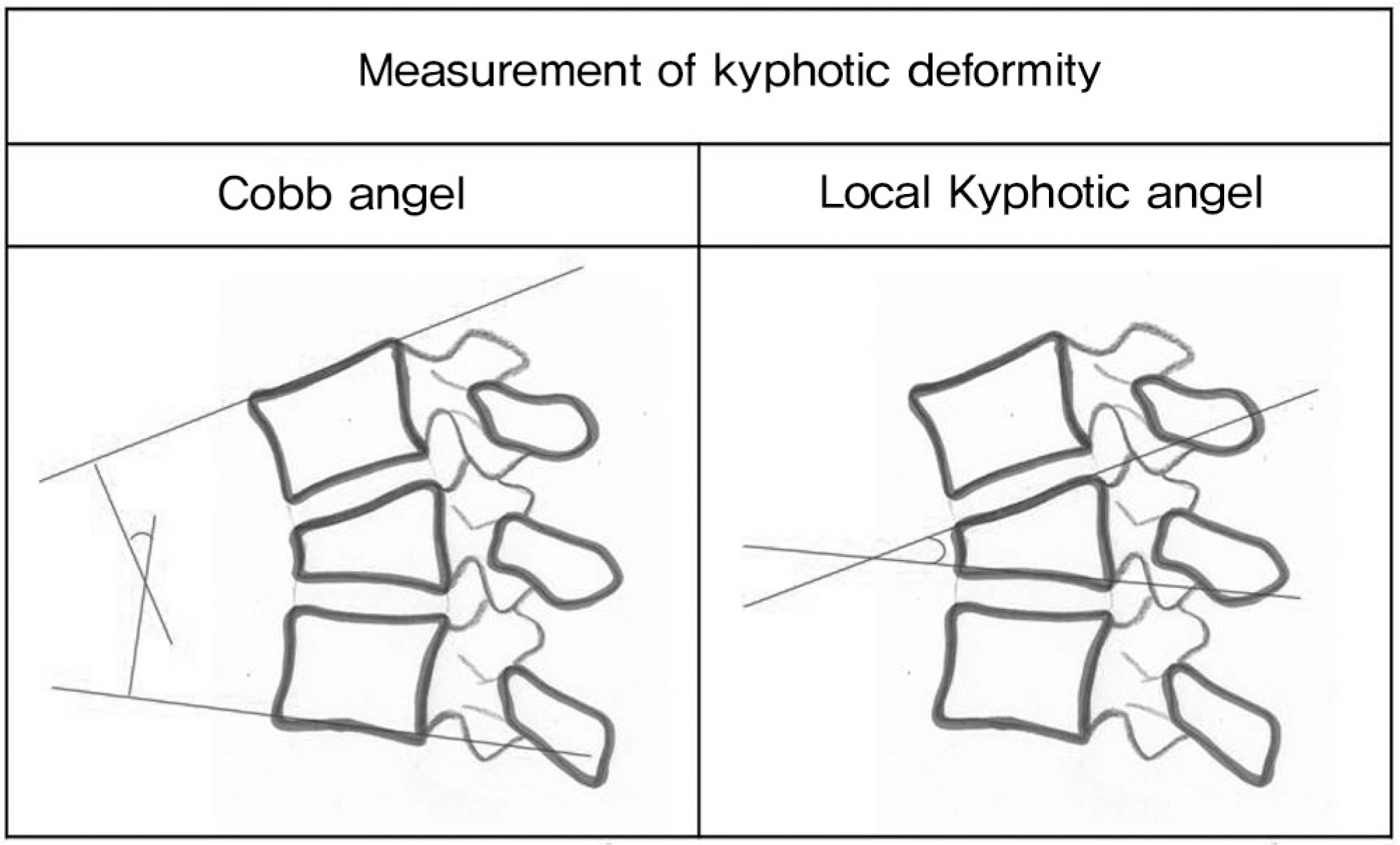
 | Fig. 2.Measurement of kyphotic deformity by 2 different methods(Cobb's angle, local kyphotic angle). |
 | Fig. 3.Examples measuring of local kyphotic angle with lateral view of plain film, CT, T1-weighted MR image. CT demonstrates definite margins of vertebral plate. |
Table 1.
Comparison of measured values depending on stability


 PDF
PDF ePub
ePub Citation
 Print
Print


 XML Download