

 PDF
PDF ePub
ePub Citation
 Print
Print


Abstract
Study Design
A retrospective radiologic and clinical analysis of 48 patients following anterior cervical fusion.
Objectives
To examine the prevalence of symptomatic adjacent segment disease after anterior cervical fusion and determine the risk factors affecting its progression.
Summary of Literature Review
Symptomatic adjacent segment disease appears to occur at a rate of 2% to 3% per year.
Materials and Methods
Forty-eight patients, who were followed up for more than 3 years after anterior cervical arthrodesis, were enrolled in this study. A modification of the Robinson criteria was used for the clinical evaluation and the radiographic grading of Hilibrand was used for the radiology evaluation. Kaplan-Meier survival analysis was used to examine the survival rate. Age, gender, number of fusion segments, preoperative ROM, angle of lordosis, spinal canal diameter and radiologic degeneration of adjacent segments were selected as potential risk factors. The subjects were divided into two groups according to their clinical symptoms.
Results
Symptomatic adjacent segment disease developed in 11 of the 48 patients(22.9%). The disease free survival rates were 88.7%, 82.4% and 58.7% at 5, 10 and 15 years, respectively. A study of the risk factors showed that preoperative degenerative changes and narrow spinal canal in the adjacent segments were significant. However, there were no significant associations with age, gender, number of fusion segments, preoperative ROM and angle of lordosis.
Go to : 
REFERENCES
1.Katsuura A., Hukuda S., Saruhashi Y., Mori K. Kyphotic malalignment afger anterior cervical fusion is one of the factors promoting the degenerative process in adjacent inververtebral levels. Eur Spine J. 2001. 10:320–4.
2.Goffin J., Geusens E., Vantomme N, et al. Long-term follow-up after interbody fusion of the cervical spine. J Spinal Disord. 2004. 17:79–85.

3.Bohlman HH., Emery SE., Goodfellow DB., Jones PK. Robinson anterior cervical discectomy and arthrodesis for cervical radiculopathy. Long-term follow-up of one hundred and twenty-two patients. J Bone Joint Surg Am. 1993. 75:1298–1307.
4.Gore DR., Sepic SB. Anterior cervical fusion for degenerated or protruded discs. A review of one hundred forty-six patients. Spine. 1984. 9:667–71.
5.Smith GW., Robinson RA. The treatment of certain cervical-spine disorders by anterior removal of the intervertebra disc and interbody fusion. J Bone Joint Surg Am. 1958. 40:607–24.
6.Hilibrand AS., Yoo JU., Carlson GD., Bohlman HH. The success of anterior cervical arthrodesis adjacent to a previous fusion. Spine. 1997. 22:1574–9.
7.Hilibrand AS., Carlson GD., Palumbo MA., Jones PK., Bohlman HH. Radiculopathy and myelopathy at segments adjacent to the site of a previous anterior cervical arthrodesis. J Bone Joint Surg Am. 1999. 81:519–28.
8.Eck JC., Humphreys SC., Lim TH, et al. Biomechanical study on the effect of cervical spine fusion on adjacent level intradiscal pressure and segmental motion. Spine. 2002. 27:2431–4.
9.Lopez-Espina CG., Amirouche F., Havalad V. Multilevel cervical fusion and its effect of disc degeneration and osteophyte formation. Spine. 2006. 31:972–8.
10.Herkowitz HN., Kurz LT., Overholt DP. Surgical management of cervical soft disc herniation. A comparison between the anterior and posterior approach. Spine. 1990. 15:1026–30.
11.Yue WM., Brodner W., Highland TR. Long-term results after anterior cervical discectomy and fusion with allograft and plating: a 5-to 11-year radiologic and clinical follow-up study. Spine. 2005. 30:2138–44.
12.Ishihara H., Kanamori M., Kawaguchi Y., Nakamura H., Kimura T. Adjacent segment disease after anterior cervical interbody fusion. Spine J. 2004. 4:624–8.
13.White AA 3rd., Southwick WO., Deponte RJ., Gainor JW., Hardy R. Relief of pain by anterior cervical-spine fusion for spondylosis. A report of sixty-five patients. J Bone Joint Surg Am. 1973. 55:525–34.
14.Park HJ., Kim IG., Kim WK. Changes of Adjacent Segment in Anterior Cervical Fusion. J Kor Soc Spine Surg. 2003. 10:269–76.
15.Wu W., Thuomas KA., Hedlund R., Leszniewski W., Vavruch L. Degenerative changes following anterior cervical discectomy and fusion evaluated by fast spin-echo MR imaging. Acta Radiol. 1996. 37:614–7.
Go to :
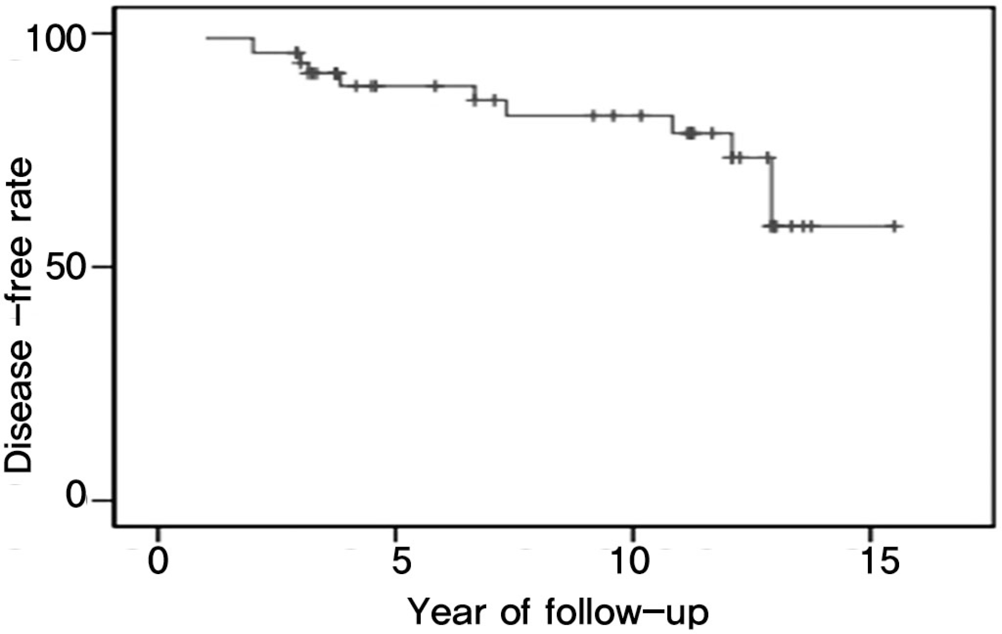
Table 1.
Criteria for the assessment of clinical outcome (modification of Robinson criteria)
Table 2.
Radiographic grading of degenerative change at adjacent levels (Hilibrand)
Table 3.
The difference of the clinical and radiological parameters between symptomatic patients(n=11) and symptom-free patients(n=37)
Table 4.
The difference of the radiological parameters between symptomatic adjacent segments(n=12) and symptom-free adjacent segments(n=84)
 XML Download