

 PDF
PDF ePub
ePub Citation
 Print
Print


Introduction
Antiretroviral therapy (ART) has increased the life expectancy of human immunodeficiency virus (HIV) infected patients. However, the incidence of kidney diseases in HIV-infected patients has increased. Before the ART era, HIV-associated nephropathy (HIVAN) was the most common type of glomerulonephritis in HIV-related kidney diseases, but after the advent of ART, many other types of glomerulonephritis, besides HIVAN, such as mesangial proliferative glomerulonephritis, immunoglobulin A (IgA) nephropathy, and immune-complex glomerulonephritis have been reported (1). In Korea, only 1 case of HIV-related membranous glomerulonephritis has been reported (2) and lupus-like glomerulonephritis has not been reported thus far. Therefore, we report a case of a patient with HIV-related lupus-like glomerulonephritis who showed good response to treatment with ART and steroids.
Case report
A 50-year-old man was brought to the emergency department with a 4-week history of fever, myalgia and weakness. He had unprotected sexual intercourses with multiple partners (HIV status unknown) 3 years ago in Thailand. He also has lost 10 kg over the past 2 months. On arrival, his blood pressure was 120/80 mmHg, heart rate was 90 beats/min, respiratory rate was 20 breaths/min, and body temperature was 39.5℃.
A complete blood count on admission demonstrated a white blood cell (WBC) count of 4,270/mm3, a hemoglobin level of 15 mg/dL, and a platelet count of 65,000/mm3. His liver function test was normal but serum urea nitrogen level was elevated to 41.5 mg/dL, and serum creatinine level to 1.38 mg/dL. Urinalysis showed 2+ protein and red cell count of 11-15/high power field (hpf). Both enzyme-linked immunosorbent assay (ELISA) and western blot revealed the presence of anti-HIV antibodies. The HIV viral load was 960,000 copies/mL and CD4-positive lymphocyte count was 71 cells/mm3. Antibodies to both hepatitis C and hepatitis B surface antigens were absent.
The patient's renal function, which had initially deteriorated, improved with hydration. Acyclovir and penicillin G were given to the patient to treat herpes labialis and neurosypilis, respectively. One week after the initiation of penicillin and acyclovir, follow-up urinalysis revealed proteinuria of 3+ and his serum creatinine level started to increase. Therefore, acyclovir was discontinued and the dose of penicillin was reduced. ART with lamivudine-zidovudine and lopinavir-ritonavir was initiated. Two days after the initiation of ART, defervescence was obtained. There was no evidence of other infection although many tests including blood cultures and abdomino-pelvic CT were performed to find the cause of fever. Creatinine levels increased to 2.87 mg/dL, and blood pressure rose to 140/90 mm Hg. Edema was noted in his pretibial areas. Urinalysis revealed a high red blood cell (RBC) count and a WBC count of 1-2 cells/hpf. The amount of protein measured in a 24-h urine collection was 6.5 g of protein. Therefore, the dose of potentially nephrotoxic drugs were reduced, but the creatinine levels did not decrease to below 2.0 mg/dL. Angiotension-converting enzyme inhibitor (ACEI) was given in order to reduce proteinuria and control his blood pressure.
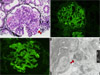
Kidney biopsy was performed to investigate the cause of acute azotemia. Light microscopic analysis of paraffin sections revealed 8 glomeruli with diffuse endocapillary proliferation, 2 epithelial crescents, and 1 segmental sclerosis. Direct immunofluorescence showed a diffuse, coarse granular glomerular capillary wall and mesangial fluorescence for IgA, IgG, IgM, C3, and C1q. Tubuloreticular inclusions were observed in the cytoplasm of endothelial cells (Fig. 1). The findings obtained on pathological examination of these lesions were strongly suggestive of lupus nephritis.
To rule out lupus nephritis, serologic tests for systemic lupus erythematosus (SLE) were performed. The result of a fluorescent antinuclear antibody test was negative, and antibodies to double-stranded DNA were absent. The C3 and C4 concentrations were 107 mg/dL and 42 mg/dL, respectively; they were in the normal range.
There was no serologic evidence of SLE, but renal pathological examination revealed lupus-like features. We diagnosed the patients with HIV-related lupus-like immune complex glomerulonephritis. The dosage of the ART regimen was adjusted to an estimated creatinine clearance rate of 20.9 mL/min. Even though the dose was adjusted, his serum creatinine level increased up to 3.15 mg/dL, and the daily amount of urinary protein excreted did not decrease. Prednisolone at a dosage of 1 mg/kg was added to ART and thereafter, the creatinine level began to decrease, and proteinuria also reduced simultaneously. Prednisolone was maintained at a dosage of 1 mg/kg for 1 month and tapered thereafter for over a year.
One and a half year after the treatment was initiated, his serum creatinine level normalized to 1.2 mg/dL, and the amount of protein measured in a 24-h urine collection decreased to 407 mg/d. The results of analysis performed on the most recent follow-up showed that his CD4-positive T-lymphocyte count was 402 cells/mm3, and the HIV RNA level was below the detectable range.
Discussion
Kidney function has been reported to be abnormal in up to 30% of HIV-infected patients (4). In particular, chronic kidney disease (CKD) and end-stage renal disease (ESRD) are important comorbid conditions prevalent in HIV-infected patients (3). Compared to the other HIV-infected populations, HIV-infected African Americans have a 50-fold higher risk of developing ESRD (3).
HIVAN, a late manifestation of HIV infection, is the most common HIV-associated kidney disease (4). The pathologic finding for HIVAN is focal segmental glomerulonephritis (4).
In the US, lupus-like glomerulonephritis is the second most common type of nephritis in HIV-infected populations (5), but HIVAN accounts for over 60% of HIV-related kidney diseases (4). In contrast, lupus-like nephritis is more common than HIVAN in Europe (5-8). The results of these studies suggest that certain genetic factors may underlie the development of HIV-related renal diseases (7).
Although a considerable number of HIV infections have been reported in Asian countries, only 1 study has reported HIV-related renal disease diagnosed from pathologic findings. In 1995 and 1996 in Thailand, 26 cases of HIV-infected patients with proteinuria (daily protein excreted, >1.5 g) were studied; among these, there were 17 cases of mesangial proliferative glomerulonephritis, 2 cases of IgA nephropathy, 1 case of membranous glomerulonephritis, and 1 case of membranoproliferative glomerulonephritis. Despite the limited data available, the incidence of HIVAN and lupus-like glomerulonephritis is thought to be low in HIV-infected Asians (9).
Lupus-like glomerulonephritis in HIV-infected patients is defined by the presence of "full house" glomerular immunoglobulin and complement deposits (IgG, IgA, IgM, C3, etc.) without serologic and clinical evidence of systemic lupus erythematosus (8,10). It was reported that none of the histopathological findings obtained with electron microscopic analysis are pathognomonic for lupus-like nephritis in HIV infected patients (5). The proportion of mesangial, subendothelial, and subepithelial deposits were found to be similar. However, in 1 study on lupus-like nephritis, tubuloreticular inclusions were observed in all cases (8).
Despite the clinical research conducted in this field, the best treatment modality for HIV-related kidney diseases has not yet been determined (4). However, ART is useful in the treatment of HIV-related renal disease (1,4,10), because HIV infection may cause HIVAN and contribute to the development of other renal diseases (4). In some cases of lupus-like glomerulonephritis, withdrawal of ART has been reported to result in a decrease in renal function (10).
Several retrospective cohort studies have reported significant improvements in renal function and proteinuria in response to steroid treatment (4). However, the optimal duration and intensity of such therapy has not yet been determined. If kidney function deteriorates in patients with HIVAN despite the use of ART, administration of prednisolone therapy at 1 mg per kilogram of body weight per day (maximum dosage, 80 mg/d) for 2 months, followed by tapering of the dosage for the next 2~4 months, should be considered (4).
The patient in this report was diagnosed with acquired immunodeficiency syndrome (AIDS) and AIDS-associated lupus-like immune complex glomerulonephritis, without any serologic evidence of SLE. We didn't find the cause as to why fever continued for about 1 month despite many tests and fever subsided by itself. We presumed that it has been 3 years since he had HIV infection according to his exposure history. He was administered ART and steroids, with which his renal function normalized.
 XML Download