

 PDF
PDF ePub
ePub Citation
 Print
Print


Introduction
Actinomyces species normally colonize the human body. Actinomycosis is a chronic suppurative and granulomatous infectious disease caused by Actinomyces species (1,2). Disruption of normal mucosal barrier is necessary for Actinomyces species to penetrate the subcutaneous tissue and cause an infection (1,3). Although oral mucosa is usually the portal of entry into deeper tissues, actinomycosis localized to the oral mucosa, especially presenting as ulcers of hard palate, is rare (4-7). Besides, to our knowledge, there has been no report on anctinomycosis with esophagogastroduodenoscopy (EGD) as the source of infection.
Herein, we report an unusual case of actinomycosis which presented as ulcers of hard palate with no other predisposing factors except for having undergone EGD.
Case report
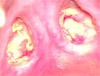
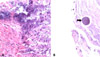
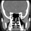
A 49-year-old woman with no significant past medical history was admitted for evaluation of anemia. Except for mildly anemic conjunctiva, all the other physical examinations, including that of the oral cavity, was unremarkable. Initial blood test results were as follows: hemoglobin, 7 g/dL; serum iron, 16 µg/dL; TIBC, 378 µg/dL; ferritin, 3 ng/mL. Peripheral blood smear showed microcytic hypochromic anemia. Although she had no abdominal discomfort, EGD was performed to evaluate the cause of iron deficiency anemia. Gastric ulcers were present on EGD, and she was thus discharged with proton pump inhibitor and ferrous sulfate. On the 6th day after undergoing EGD, she returned to the hospital with pain in the oral cavity. On physical examination, two ulcerative lesions were observed on hard palate (Fig. 1). Biopsy was done under local anesthesia and histologic examination showed acute ulceration and prominent granulation tissue with proliferation of actinomycetes (Fig. 2). Facial computed tomography scan was taken to estimate the extent of the lesions (Fig. 3); mild mucosal irregularity in bilateral hard palate with adjacent air collection was seen but there was no involvement of bony structures. The patient was admitted again and was given intravenous ampicillin 2,500 mg/day for 2 weeks and then discharged with oral ampicillin 2,000 mg/day for nine weeks. During follow-up at the outpatient department, the ulcerative lesion completely resolved and no relapse occurred afterwards.
Discussion
Actinomycosis was first described as a disease by fungus over 100 years ago (1). In the 1960s, Waksman showed Actinomyces to be a gram positive bacterium which is a strict or facultative anaerobe (1,8). Most of the cases of actinomycosis present as chronic illness but it may also present in either acute or subacute forms (3,6,7,9). In 2002, Garelick et al. reported a case on acute postoperative endophthalmitis caused by Actinomyces which developed on the 6th day after uncomplicated phacoemulsification (10). Except for the fact that the patient underwent EGD, we could not find, in our case, any other predisposing factors or procedures that could have caused actinomycosis. It is possible that the endoscope might have caused microdamage to oral mucosa while passing through the oral cavity. However, the duration between the procedure and the occurrence of the symptoms was short. Therefore, it is difficult to say clearly that EGD was the definitive cause of actinomycosis, but with no other evident infection source, the possibility cannot be ruled out. Actinomycosis shows a slight male predominance but there is no racial or geographic predisposition (1,9, 11). Actinomyces species are normal commensals of the oral cavity and cannot penetrate healthy tissue. Thus, mucosal break-down is a prerequisite for infection (1,3,7,12,13). Malnutrition, alcoholism, debilitating states (diabetes, malignancy, immunosuppression, etc.), and surgery which is processed in oral cavity are considered as predisposing factors (1,4,5,7,8). Hematogenous spread is rare and lymphatic spread is believed not to occur due to the large size of the organism (3). Although actinomycosis usually involves the cervicofacial region, localized actinomycosis in the oral mucosa, especially the hard palate, is uncommon (4,7). Most cases of cervical actinomycosis are odontogenic in origin (1). A typical actinomycotic lesion is painless and hard that most commonly presents as a chronic mass (1,4), sometimes mimicking a malignant tumor (1,4,7). The ulcerative lesion is a rare clinical manifestation of actinomycosis (4,6,7). Diagnosis of actinomycosis is difficult because of a general lack of familiarity with the disease and a low success rate in culturing the organism as a result of its fastidious nature (8).
The traditional treatment of actinomycosis is high-dose penicillin for a prolonged period (6 months to 1 year). However, there have been several reports on successful short-term treatment-less than 6 months of antibiotic therapy. In our case, the patient received a total of eleven weeks antibiotic treatment. Patients with actinomycosis may be candidates for individualized courses of therapy, depending on the initial burden of the disease and the clinical and radiological response to therapy (14-16).
In our case, she had no trauma history, intraoral lesion, precipitating factors, nor history of orthognathic surgery. Moreover, the lesions were ulcers in hard palate and thus, actinomycosis was not suspected. It would have been better if the organism had been cultured, but the diagnosis could still be made as actinomycosis by characteristic histologic findings such as sulfur granules which are typical to actinomycosis.
We have reported an unusual case of actinomycosis with no other predisposing factors except for having undergone EGD, which presented as ulcers of hard palate. Actinomycosis can present in a variety of forms and during many different kinds of procedures. Therefore, it is important to have a high index of suspicion in order to make an accurate and timely diagnose. Early investigation and treatment can allow the patient to make a quick and full recovery without complications.
 XML Download