

 PDF
PDF ePub
ePub Citation
 Print
Print


Introduction
Dengue fever is the most important arboviral infection in the world, with an estimate of 100 million cases per year and 2.5 billion people at risk (1). Since dengue fever is not endemic in Korea, it usually does not occur naturally without any extrinsic cause. However, due to the increase in people traveling abroad, the cases of imported dengue fever are increasing (2-5).
The common symptoms in patients with dengue fever are malaise, fever, musculoskeletal pain, headache, nausea, vomiting, and abdominal pain. However, about one-third of patients develop one or more complications that include bleeding, seizures, acute renal failure, and dengue shock syndrome (6, 7). Recently, reports on other uncommon clinical complications such as acute myocarditis (8), acute hepatic failure (9), and dengue encephalitis (10) have increased. There also have been reports on cases resembling surgical emergencies like acute pancreatitis (11), acute acalculous cholecystitis (12), and gastrointestinal bleeding (13). In addition, there have been reports on dengue enteritis mimicking appendicitis (14,15) but, no reports on such cases have been documented in Korea. We experienced a case of enteritis that mimicked acute appendicitis in dengue fever. We believe that this is the first case report of such complication in Korea.
Case report
A 36-year-old male presented to the emergency department with 3 days' history of low grade fever, headache, and abdominal pain after making a trip to Boracay island in the Philippines 10 days ago, where he stayed for 4 days. He denied having any sexual contact during the trip. He had no past medical history and was not taking any medications. Except for fever, his vital signs were stable. His conjunctiva was slightly injected but, no petechia or purpura could be observed on his skin. He complained of severe pain in the right iliac fossa region and watery diarrhea. Complete blood cell count showed thrombocytopenia (platelet count of 143×106/mm3) with leucopenia (white blood cell count 2.37×10⁶/mm3). Hemoglobin and hematocrit were normal. Dengue serology was not conducted on the first day of admission and initially, acute appendicitis was suspected and intravenous quinolone was started. However, computed tomogram (CT) of abdomen and pelvis revealed a normal appendix. Tests for malaria and typhoid fever were negative. Results of blood cultures and urine culture for bacterial infection, as part of the initial sepsis work-up, were also negative.
On the next day, the patient began to complain of a headache which was generalized, dull aching, and non-throbbing in nature. It was transiently relieved by non-steroidal anti-inflammatory drugs. Although he seemed to be somewhat slightly confused, there were no evident signs of meningeal irritation. CT scan of the brain was normal. Cerebrospinal fluid (CSF) examination for cell count, gram staining, india ink staining, protein, and CSF glucose/blood glucose ratio were all within normal limits. Therefore, the patient was given a conservative treatment with intravenous fluids and analgesics.
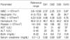
On the third day of admission, the patient developed petechial rash on both legs. Subsequent daily hemogram monitoring (Table 1) showed sustained leucopenia (white blood cell count of 2.25×10⁶/mm3) and thrombocytopenia (platelet count of 108×106/mm3). Hematocrit was normal throughout the hospital stay. Dengue fever was suspected and the diagnosis was confirmed by IgM capture enzyme-linked immunosorbent assay against Dengue virus (MAC-ELISA) (Panbio, Australia) which was analyzed with the sample collected on the fifth hospital day. Serum tests for Hantaan virus and serum agglutination for leptospirosis was all negative. The patient showed clinical improvement with symptomatic treatment and was discharged on the sixth hospital day. During the follow-up period of four-weeks, he was completely recovered.
Discussion
Dengue fever is an acute febrile disease found in the tropics, and is caused by four closely related virus serotypes of the genus Flavivirus, family Flaviviridae. This disease manifests as a sudden onset of severe headache, muscle and joint pain, fever, and rash. Gastritis can also occur with or without abdominal pain, nausea, vomiting, and diarrhea. Rarely, some cases present with an aggressive abdominal pain which can be misdiagnosed as acute abdomen. Although the pathogenesis of abdominal pain in dengue fever is not clearly understood, lymphoid follicular hyperplasia (14, 15), which causes lymphadenopathy in dengue fever, seems to play an important role and it has been observed on the histopathologic examination from the specimen obtained after appendectomy. The aforementioned findings could be a possible explanation for subserosal fluid collection and thickened gall bladder associated with dengue fever (15, 16). Other plausible mechanism is that plasma leakage through damaged capillary endothelium could provoke symptoms mimicking appendicitis (1).
Geographically speaking, dengue virus is the most widely spread arthropod-born viruses. It is especially prevalent in the Americas, the Pacific islands, and on continental Asia. Previously, there have been reports on the outcome of domestic surveillance data regarding dengue positive rate (5) and clinical features of imported dengue fever (2, 3), which included rhabomyolysis (4).
In our case, the symptoms and signs were mostly similar to what have been previously described for dengue fever (7). However, gastrointestinal symptoms, particularly nausea and vomiting, were unusual. Leucopenia and thrombocytopenia were also present. Although liver involvement is commonly seen (17), the patient's transaminases were normal throughout the illness. In most of the cases, dengue fever can be diagnosed by suggestive clinical features but in our case, it was masked by acute abdominal pain and tenderness. The diagnosis of dengue enteritis was based solely on clinical findings and not on histological or serologic results. In a case like this, we are faced with a diagnostic dilemma. Therefore, it is important to take dengue fever into consideration when making a differential diagnosis on acute abdomen. It may help to prevent unnecessary surgical intervention in dengue fever. Dengue has an incubation period of 5-8 days followed by fever, violent headache, and chills; rash develops 3-4 days later (18). The patient showed similar disease progression. We used IgM captured ELISA to confirm dengue virus infection. The IgM ELISA has a high sensitivity and specificity for dengue viral infection (19). The limitation of this case report is that the serum was not collected as recommended by WHO guideline which states that serologic test should be performed within 5 days of symptom onset and serologic follow up should be done after 1 week. This case is the first report in Korea on dengue fever, the clinical features of which were suggestive of acute appendicitis.
 XML Download