

 PDF
PDF ePub
ePub Citation
 Print
Print


INTRODUCTION
In the early 2000s, in vitro blood tests measuring production of interferon (IFN)-γ by T-cells exposed to antigens highly specific to the Mycobacterium tuberculosis antigens ESAT-6 and CFP-10 were introduced to detect latent tuberculosis infection (LTBI). These included a whole-blood IFN-γ enzyme-linked immunosorbent assay (ELISA) and an enzyme-linked immunospot assay (ELISPOT), collectively referred to as IFN-γ release assays (IGRAs) (1). Unlike the tuberculin skin test (TST), which had been the only diagnostic tool for LTBI since the early 1930s, IGRAs are not affected by prior Bacille Calmette-Guerin (BCG) vaccination nor by nontuberculous mycobacteria (NTM) infection (with the exception of Mycobacterium kansasii, Mycobacterium szulgai, and Mycobacterium marinum), thus yielding greater specificity than TST (2). Promising results in the diagnosis of LTBI and active tuberculosis (TB) infection by both tests have been reported (3-9). On the basis of these data, the U.S. Food and Drug Administration approved the use of a commercialized whole-blood interferon-γ ELISA assay, QuantiFERON-TB Gold (QFT-G; Cellestis, Carnegie, Australia), for the diagnosis of active TB and LTBI (10). A commercialized ELISPOT assay, T-SPOT.TB assay (Oxford Immunotec, Oxford, UK), was also approved for diagnostic use in Europe (11, 12).
One concern in routine clinical practice is to distinguish patients with active TB from LTBI. Several studies have reported that IFN-γ production by lymphocytes challenged by M. tuberculosis-specific antigens was proportional to M. tuberculosis activity (13-17). Goletti et al. compared the results of different IGRAs in patients with clinically suspected TB according to outcome: culture-proven TB versus alternative diagnosis established or resolution of clinical symptoms without drugs active against TB (15). In that study, IFN-γ levels measured using whole blood enzyme-linked immunosorbent assay were significantly higher in patients with active TB than TST-positive or negative controls. However, the quantitative results of either T-SPOT.TB or QuantiFERON-TB Gold were not reported.
It was, therefore, hypothesized that by using the QuantiFERON-TB Gold In-Tube (QFT-IT) test, which is a more advanced test than QFT-G (18), it would be possible to establish the threshold IFN-γ levels, permitting the distinction of active TB from LTBI. Thus, a study was designed to compare IFN-γ levels of QFT-IT between active TB and LTBI patients admitted to Severance Hospital (Seoul, South Korea) between July 2006 and August 2007. The incidence of active TB in South Korea is intermediate (96/105 cases per year), and BCG vaccination at birth has been mandatory (19).
MATERIALS AND METHODS
1. Study design
Records of 314 patients older than 15 years who had QFT-IT performed because of suspected LTBI or active TB between July 2006 and August 2007 at Severance Hospital, Yonsei University College of Medicine (a 2000-bed tertiary care teaching hospital), were reviewed retrospectively. The study was approved by the local research ethics committee. The following data were collected by reviewing the records: results of QFT-IT, results of acid-fast stain and Mycobacterium culture, pathology results, chest X-ray findings, other medical conditions, and treatments given. We compared IFN-γ levels of QFT-IT between individuals with active TB and LTBI before anti-TB treatment.
2. QFT-IT
Peripheral venous blood was collected into three evacuated QFT-IT tubes, one containing TB antigens (ESAT-6, CFP-10, and TB 7.7), a positive control tube containing phytohemagglutinin, and a negative control tube, and processed for 4 hours. ELISA for IFN-γ was performed according to the manufacturers instructions. Samples were incubated for 20 hours. A positive result was defined as TB antigen minus negative control IFN-γ equal to or greater than 0.35 IU/mL and increment of negative control IFN-γ level to 25% or more. An indeterminate result was defined as either a negative control IFN-γ level of >8.0 IU/mL or a positive control IFN-γ response of <0.5 IU/mL with a TB antigen minus a negative control IFN-γ level of either <0.35 IU/mL or <25% of the negative control value. Because the IFN-γ ELISA cannot accurately quantify levels greater than 10 IU/mL, such levels were treated as 10 IU/mL during analysis.
3. Definitions and diagnoses
1) Active TB
Final diagnoses were made on the basis of all the clinical, radiological, microbiological, and pathological information collected. Active TB was defined as culture positive for TB (culture confirmed TB), diagnosed histologically, or obvious symptoms and signs appropriate for diagnosis, which responded clinically and radiologically to a full course of first-line anti-TB drugs (clinical TB).
2) Immunocompromised subjects
Immunocompromised subjects were defined as those with hematological malignancy, aplastic anemia, chronic renal failure, primary immunodeficiency diseases, HIV infection or those receiving immunosuppressive drugs (anticancer chemotherapy, post-transplantation, receiving >15 mg/day prednisolone for >1 month).
3) LTBI subjects
LTBI subjects were defined as patients with positive result of QFT-IT and without evidence of active tuberculosis.
RESULTS
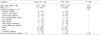
Of the 314 persons screened for this study, 158 persons were eligible. The number of active TB patients and LTBI patients was 82 and 76, respectively. The result of QFT-IT was positive in 66 of 82 (80.5%) patients, negative in 15 of 82 (18.3%) patients, and indeterminate in 1 of 82 (1.2%) patients with active TB. Of the 76 LTBI patients, those with high risk for LTBI were 29 patients (38.1%). Of the 82 active TB patients, one patient was excluded because of indeterminate QFT-IT result. Baseline characteristics for the remaining 157 patients are shown in Table 1. Of the 81 remaining active TB patients, 38 patients (38/81, 46.9%) had culture-confirmed pulmonary TB, 2 patients (2/81, 2.5%) had culture-confirmed extrapulmonary TB, 31 patients (31/81, 38.3%) had Clinical pulmonary TB, and 10 patients (10/81, 12.3%) had Clinical extrapulmonary TB.
1. Active TB vs. LTBI
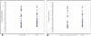
Subjects with active TB (n=81) and LTBI (n=76) were included. Mean±SD IFN-γ levels were 4.96±3.98 IU/mL (range -0.08-10) for subjects with active TB and 4.11±3.57 IU/mL (range -0.35-10) for subjects with LTBI. The quantitative results of QFT-IT on IFN-γ levels between all the subjects with active TB and those with LTBI were not statistically significant (P=0.16).
2. Culture confirmed active TB vs. LTBI
In subjects with culture confirmed active TB (n=40), mean±SD IFN-γ level was 4.54±4.05 IU/mL (range 0.08-10). The difference on the quantitative results of the QFT-IT between subjects with culture confirmed active TB and those of LTBI was also statistically insignificant (P=0.554) (Fig. 1B).
DISCUSSION
The aim of the present study was to determine whether quantitative analysis using QFT-IT could differentiate between active TB and LTBI. The quantitative results of the QFT-IT of subjects with active TB (culture confirmed TB and clinical TB) were not higher than those with LTBI. Our results showed that there was no relationship between the activity of M. tuberculosis infection and the production of IFN-γ by lymphocytes, as assessed by QFT-IT. The result of the present study wasnt consistent with that of several studies that have reported that IFN-γ production by lymphocytes challenged with M. tuberculosis-specific antigens was proportional to M. tuberculosis activity (13-17). There was another report about the use of IGRAs in distinguishing active TB from LTBI (17). In that study, mean (range) sfu number of subjects with active TB was 107.4±56.0 (1-207), that of subjects with LTBI with a TST induration diameter of >5 mm was 53.6±60.3 (7-239), and that of subjects with LTBI with a TST induration diameter of ≤5 mm was 18.9±27.1 (7-143). Mean sfu numbers of active TB were statistically different from that of LTBI with a TST induration diameter of >5 mm and those of LTBI with a TST induration diameter of ≤5 mm. The authors assessed the cut-off value of sfu, measured by T.SPOT.TB, to distinguish active TB from LTBI. In that study, the T.SPOT.TB has a sensitivity of 82.8% and specificity of 74.2% for distinguishing active TB from LTBI at a threshold of 49 sfu. Discrepancy between results of that study and those of the present study might be explained by several factors. First, in that study, LTBI was defined as the positive result of T.SPOT.TB without clinical or radiological evidence of TB but we defined LTBI as the positive QFT-IT without evidence of active TB. Second, the authors compared the values of sfu, measured by T.SPOT.TB between culture-positive TB and LTBI but we compared the IFN-γ levels, measured by QFT-IT. Because the IFN-γ ELISA cannot accurately quantify values of more than 10 IU/ml, such inaccuracy might be another factor. Other reports in Korea have explored the use of IGRAs in the diagnosis of active TB. Lee et al. studied 87 subjects with active TB (culture proven in 63%) and 131 young students with low risk of LTBI. Sensitivity of 86.2% (95% CI, 77.1-92.7) and specificity of 87.0% (95% CI, 80.0-92.2) at a threshold of 0.13 IU/mL for QFT-G in the diagnosis of active TB were reported (13). Kang et al. studied 144 subjects with suspected pulmonary TB. They reported 49% specificity (95% CI, 37-61) and 89% sensitivity (95% CI, 79-96) at a threshold of 0.35 IU/mL for QFT-G in the diagnosis of active pulmonary TB (20). The association of these results is debatable because the definition of LTBI is arbitrary and the exact proportions of LTBI in two reports are unknown.
To our knowledge, the present study is the first study to determine whether quantitative analysis using QFT-IT can discriminate between active TB and LTBI in an intermediate-burden country. However, the present study had several limitations. First, the definition of LTBI was arbitrary. In reality, we define LTBI as patients with positive result of QFT-IT without evidence of active tuberculosis. Because an arbitrary definition was used to define LTBI in the present study (positive QFT-IT result without evidence of active TB), LTBI patients defined in the present study might not have included LTBI patients with negative results with QFT-IT but might have included healthy individuals with positive results of QFT-IT instead. Because there is no gold standard for the diagnosis of LTBI, it was difficult to resolve this limitation. Second, the present study is retrospective; we evaluated the subjects based on clinical records. For some subjects, there was no BCG vaccination history. So the effect of BCG vaccination on QFT-IT results remains unresolved. Because it is known that QFT-G is minimally affected by BCG vaccination, we presumed that the QFT-IT results in the present study also might have been minimally affected (2).
In summary, we showed that the production of IFN-γ measured by QFT-IT showed no correlation between its level and the activity of M. tuberculosis infection. These results suggest that measuring IFN-γ using QFT-IT might not be useful for distinguishing active TB from LTBI. A large, prospective study will be necessary to confirm that patients with high IFN-γ levels are not at higher risk of having active TB.
 XML Download