Abstract
Background
Pericardiocentesis is simple procedure in itself but has been associated with serious complications. In this study we assess the mortality and morbidity of 2-dimentional contrast echocardiographically directed pericardiocentesis.
Method
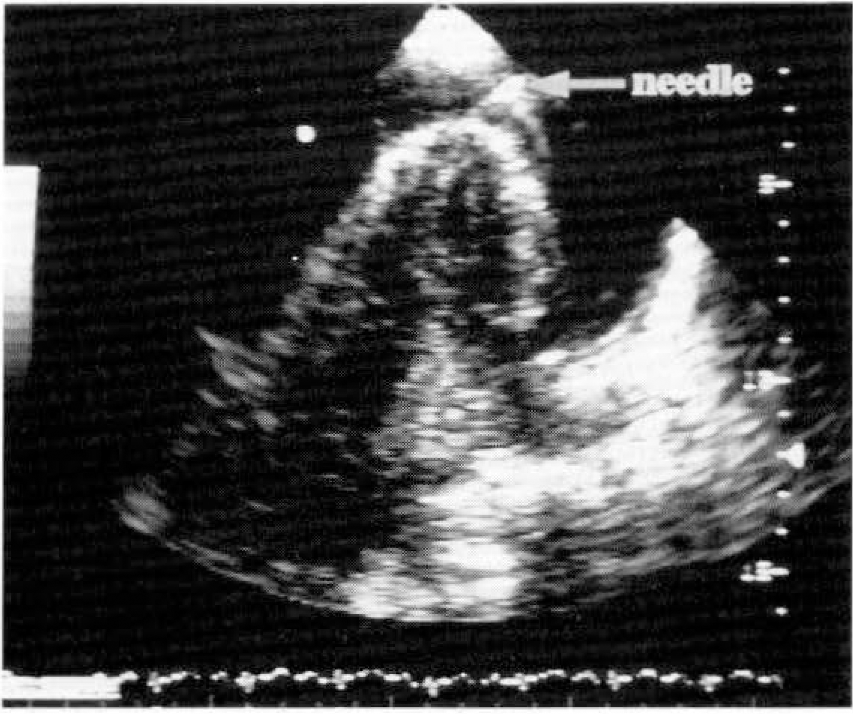
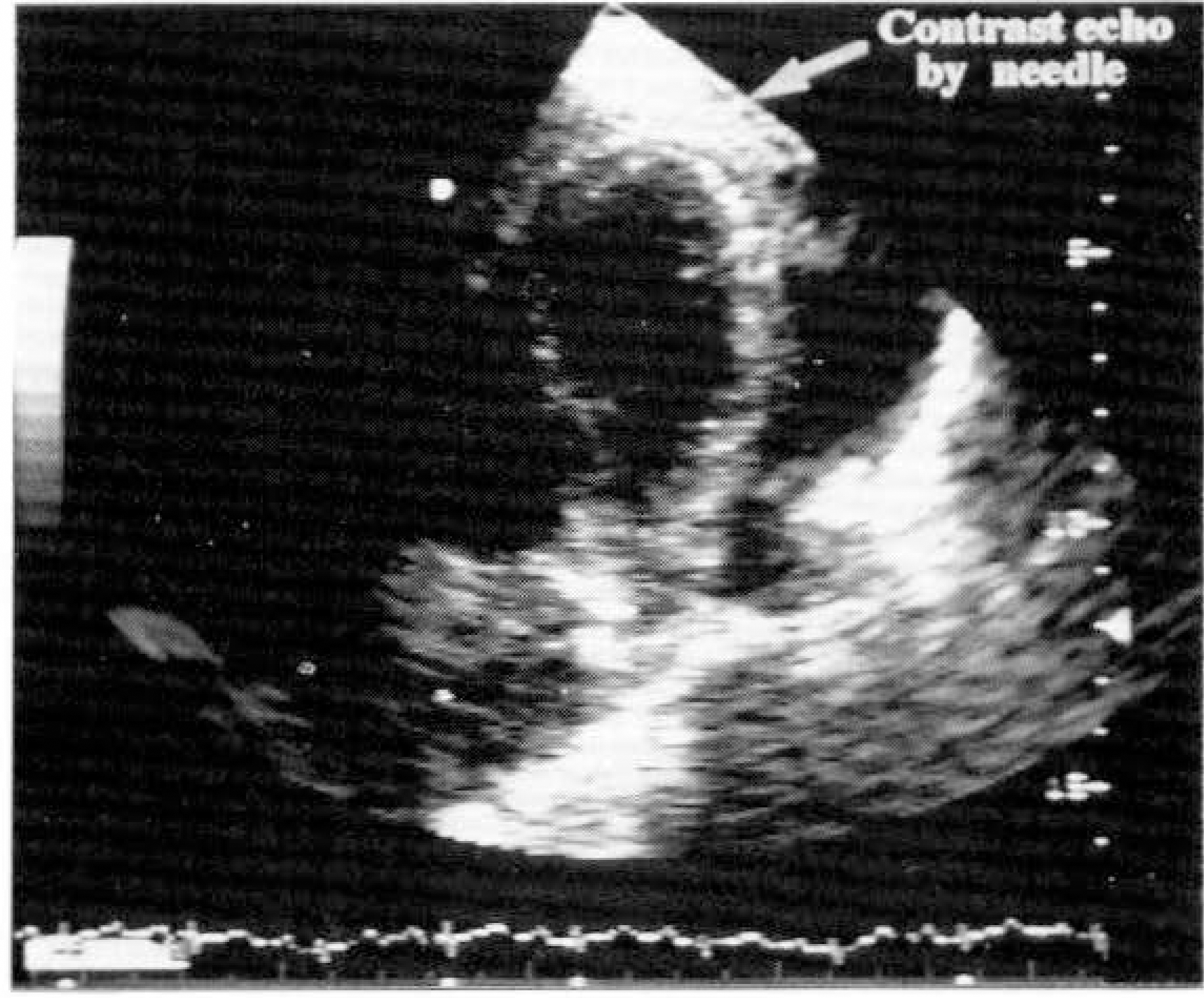
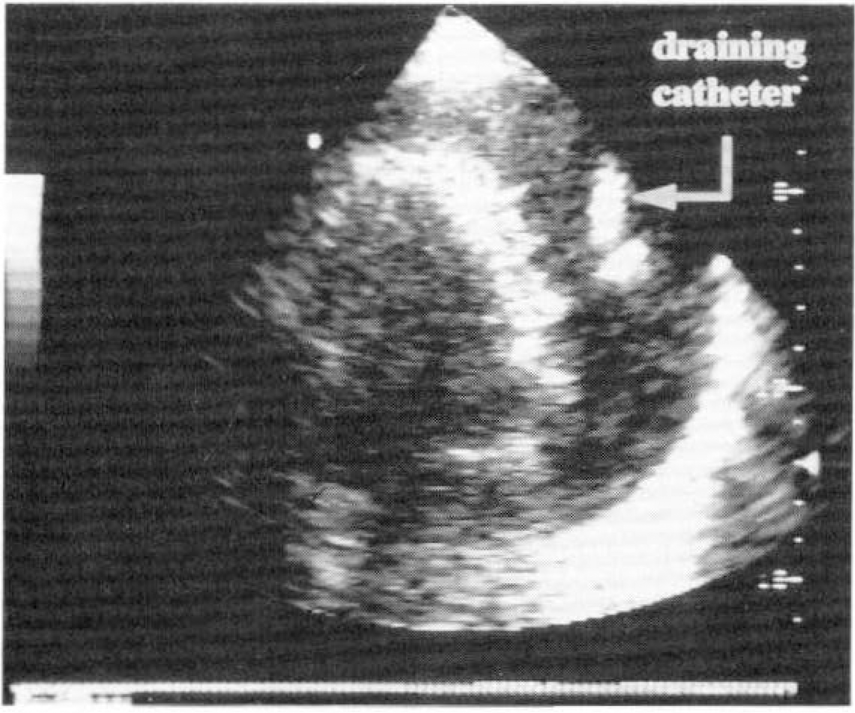
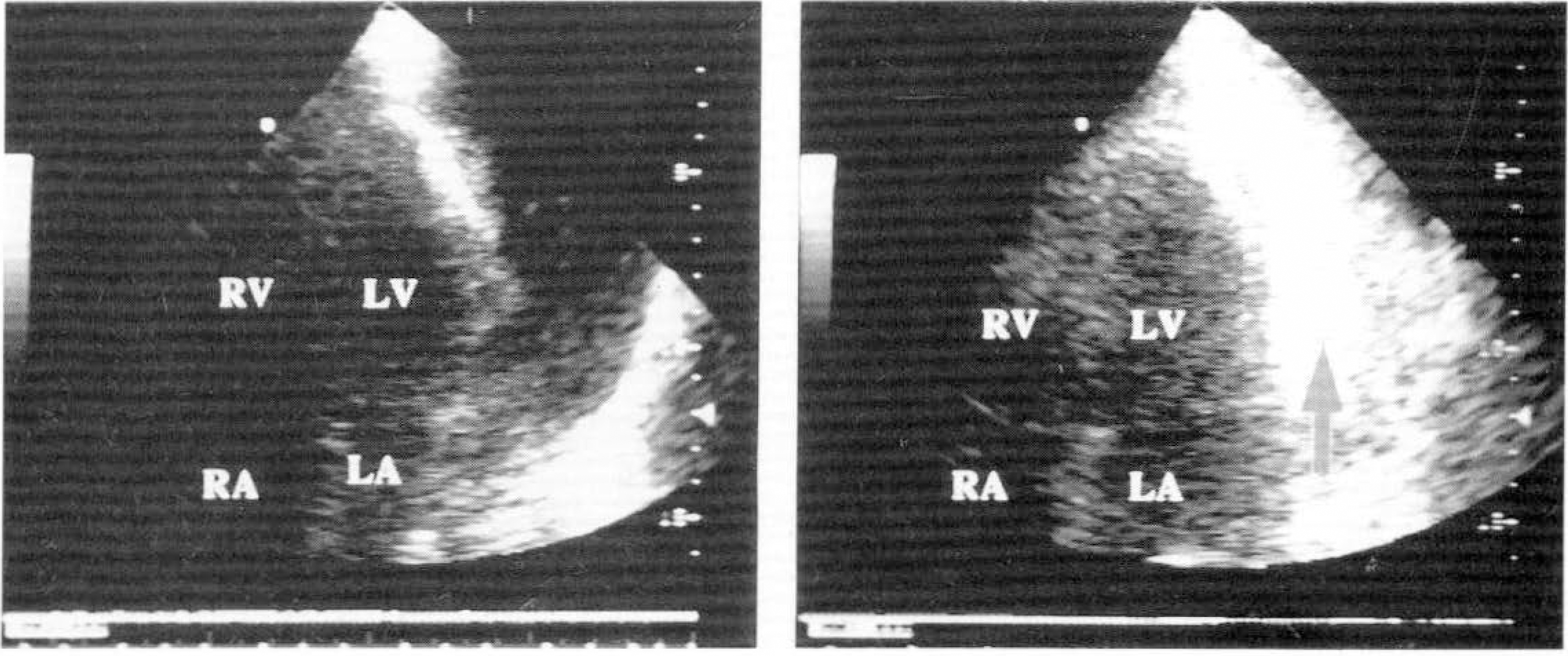
Fourteen patients(8 males and 6 females, mean age: 55 years) with pericardial effusion were included in this study. The 2-dimensional echocardiographic transducer was placed at the apex and a 4-chamber view was obtained. When return of fluid was obtained at least 10cc were aspirated and discarded, then 5cc of agitated saline solution were injected through the explorating needle and cloud of echoes indicated the position of the needle. When its position in the pericardial sac was confirmed, the needle was replaced by a catheter, then 5cc of agitated saline solution were injected through the catheter and cloud of echoes confirm the location of the catheter.
Results
In all 14 patients a satisfactory apical 4-chamber view was obtained. The echocardiogram showed explorating needle in all patients but did'nt show exact location. The echocardiographic contrast effect produced by hand-agitated saline was seen in the pericardiac sac in all patients. The contrast study confirmed the position of the needle and catheter in the pericardial cavity in all patients. The life-threatening complications were not developed.
References
1). Schlant RC, Alexander RW. Hurst's The Heart. 8th Ed. p. 1675–1676. New York: McGRAW-HILL. INC;1994.
3). Morin Je, Hollomby D, Gonda A, Long R, Dobell RC. Manegement of Uremic Pericarditis: A report of 11 patients with Cardiac Tamponade and a Review of the Literature. Annal Thorac Surg. 22:588–592. 1976.
4). Guberman BA, Fowler NO, Engel PJ, Gueron M, Allem JM. Cardiac Tamponade in Medical Patients. Circulation. 64:633–640. 1981.

5). Wong B, Murphy J, Hassenein K, Dunn M. The Risk of Pericardiocentesis. Am J Cardiol. 44:1110–1114. 1979.

6). Clarke DP, Cosgrove DO. Real-time Ultrasound Scanning in the Planning and Guidence of periocardiocentesis. Clinical Radiology. 38:119–122. 1987.
7). Chandraratna PAN, Reid CL, Mimalasuriya A, Kawanishi D, Rahimtoola SH. Application of 2-Dimentional Contrast Studies During Pericardiocentesis. Am J Cardiol. 52:1120–1122. 1983.
8). Preis LK, Taylor GJ, Martin RP. Traumatic Pericardiocentesis: Two-Dimensional Echocardiographic Visualization of an Unfortunate Event. Arch intern Med. 142:2327–2329. 1982.
9). Chandraratna PAN, First j, Langevin E, O'Dell R. Echocardiographic contrast studies during Pericardiocentesis. Ann Intern Med. 87:199–200. 1977.

10). Kiopatrick ZM, Chapman CB. On Pericardiocentesis. Am J Cardiol. 16:722–727. 1965.
11). Bishop LH Jr, Estes EH Jr, McIntosh HD. The Electrocardiogram as a Safeguard in Pericardiocentesis. JAMA. 162:264–265. 1956.

12). Feigenbaum H, Waldhausen JA, Hyde LP. Ultrasound Diagnosis of Pericardial Effusion. JAMA. 191:711–714. 1965.

13). Feigenbaum H, Zaky A, Waldhausen JA. Use of Ultrasound in the Diagnosis of Pericardial Effusion. Ann of Intern Med. 65:443–452. 1966.

14). Tajik AJ. Echocardiography in Peficardial Effusion. Am J Med. 63:29–40. 1977.
15). Goldberg BB, Pollack HM. Ultrasonically Guided Pericardiocentesis. Am J Cardiol. 31:490–493. 1973.

16). Martin RP, Rakowski H, French J, Popp RL. Localization of Pericardial Effusion with Wide Angle Phased Array Echocardiography. Am J Cardiol. 42:904–912. 1978.

17). Callahan JA, Seward JB, Nishimura RA, Miller Jr FA, Reeder GS, Shub C, Callahan MJ, Schattenberg TT, Tsjik AJ. Two-Dimensional Echocardiographically Guided pericardiocentesis: Experience in 117 Consecutive Patients. Am J Cardiol. 55:476–479. 1985.

18). Callahan JA, Seward JB, Holmes Jr DR, Smith HC, Reeder GS, Miller Jr FA. Enhanced safety of Two-Dimensional Echocardiography-Directed Pericardiocentesis: A technique of Choice. J Am Coll Cardiol. 1(2):738. 1983.
19). Taavirsainen M, Bondestam S, Mankinen P, Pitkaranta P, Tierala E. Ultrsound guidence for pericardiecentesis. Acta Radiologica. 32:9–11. 1991.
20). Chandraratna PAN. Echocardiography and Doppler Ultrasound in the Evaluation of Pericardial Disease. Circulation. 84(suppl I):I–303-I-310. 1991.
21). Schuster AH, Nanda NC. Pericardiocentesis induced intrapericardial thrombus: Detection by two-dimentional echocardiography. Am Heart Journ. 104:308–311. 1982.
22). Susini G, Pepi M, Sisillo E, Bortone F, Salvi L, Barbier P, Fiorentini C. Percutatneous pericardiocentesis Versus Subxiphoid Pericardiotomy in Cardiac Tamponade Due to Postoperative pericardial Effusion. Journal of Cardiothoracic and Vascular Anesthesia. 7:173–183. 1993.
23). Duvernoy O, Borowiec J, Helmius G, Erikson U. Complication of percutaneous pericardiocentesis under fluoroscopic guidence. Acta Radiologica. 33:309–313. 1992.


 PDF
PDF ePub
ePub Citation
 Print
Print


 XML Download