Abstract
Background
Analysis of mitral flow velocity pattern provides useful variables in the assessment of left ventricular diastolic dysfuntion, but are affected by loading conditions or presence of atrial fibrillation. Thus we assessed intraventricular diastolic flow velocity profile in order to assessment of left ventricular diastolic dysfuntion.
Methods
The study population consisted of 20 subjects with normal left ventricular function(including 7 patients with atrial fibrillation only), 15 patients with hypertensive heart disease, and 14 patients with dilated cardiomyopathy. The flow velocity pattern at the mitral tip was recorded simultaneously with regional pulsed Doppler diastolic velocity patterns at 1,2, or 3 cm from the mitral tip toward the apex.
Results
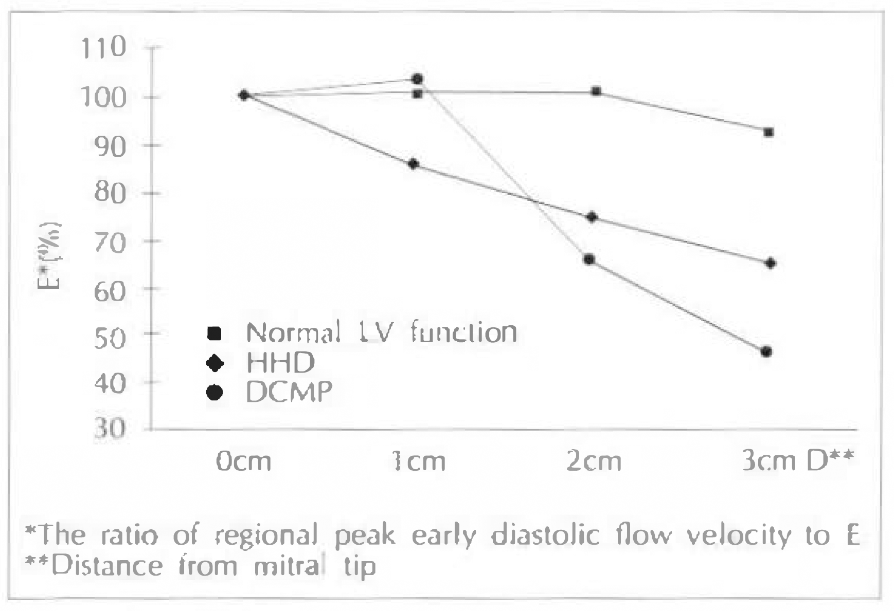
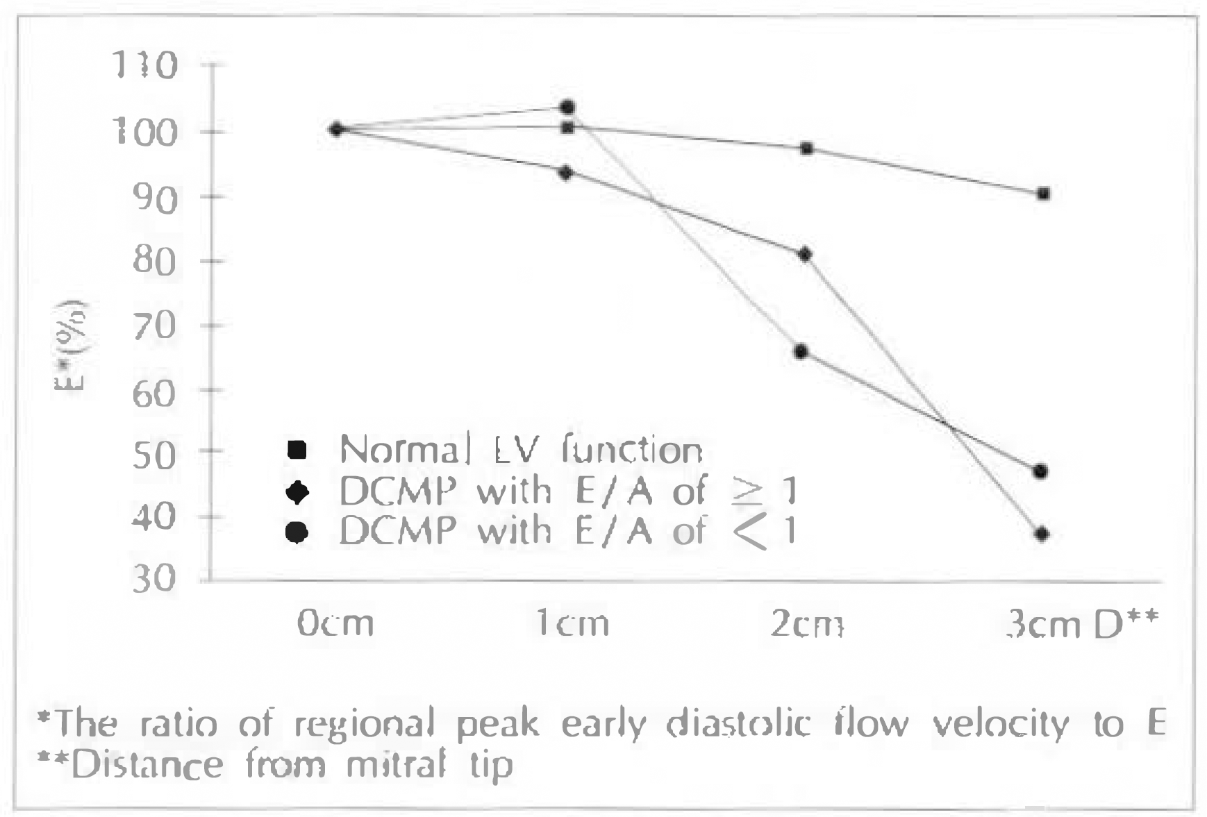
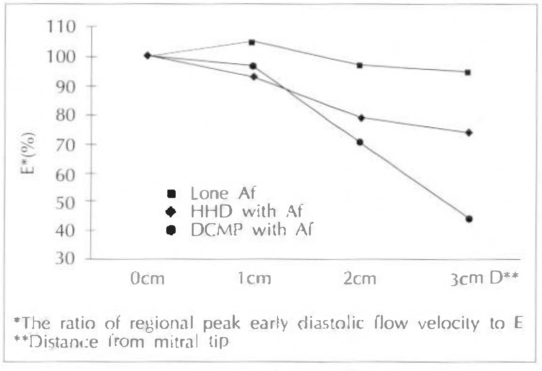
In the normal subjects, early diastolic flow velocity at the mitral tip was maintained at the positions 1 to 3cm away from the tip into the left ventricular cavity. In patients with dilated cardiomyopathy or hypertensive heart disease, peak early diastolic flow velocity decreased from the mitral tip toward the apex more progressively than in the subjects with normal left ventricular function. The same findings were obtained in selected patients group with atrial fibrillation or a normalized mitral flow velocity pattern.
Go to : 
References
1). Hanrath P, Marthey DG, Siegert R, Bleifeld W. Left ventricular relaxation and filling pattern in different form of left ventricular hypertrophy: An echocardiographic study. Am J Cardiol. 45:15. 1980.
2). Inouye I, Massie B, Loge D, Topic N, Silverstein D, Simpson P, Tubau J. Abnormal left ventricular filling: An early finding in mild to moderate systemic hypertension. Am J Cardiol. 53:120. 1984.

3). Snider AT, Gidding SS, Rocchini AP, Rosenthal A, DickII M, Crowley DC, Peters J. Doppler evaluation of left ventricular diastolic filling in children with systemic hypertension. Am J Cardiol. 56:921. 1985.

4). Liebson PR, Savage DD. Echocardiography in hypertension. Echocardiography. 3:181. 1986.
5). Dreslinski GR, Frohlich ED, Dunn FG, Messerli FH, Suarez DH, Reisin E. Echocardiographic diastolic ventricular abnormality in hypertensive heart disease: Atrial emtying index. Am J Cardiol. 47:1087. 1981.
6). Rokey R, Dvo LC, Zoghbi WA, Limacher MC, Qvinones MA. Determination of parameters of left ventricular diastolic filling with pulsed Doppler echocardiography: Comparision with cineangiography. Circulation. 71:543. 1985.
7). Thomas JD, Weyman AE. Echocardiographic Doppler evaluation of left ventricular diastolic function. Circulation. 84:977. 1991.
8). Topol EJ, Traill TA, Fortuin NJ. Hypertensive hypertrophic cardiomyopathy of the elderly. N Engl J Med. 312:277–283. 1985.

9). Takenaka K, Dabestani A, Gardin JM, Russell D, Clark S, Allfie A, Henry WL. Left ventricular filling in hypertrophic cardiomyopathy: A pulsed Doppler echocardiographic study. J Am Coll Cardiol. 7:1263–1271. 1986.

10). Stoddard MF, Pearson AC, Kern MJ, Ratcliff J, Mrosek DG, Lavovitz AJ. Left ventricular diastolic function: Comparison of pulsed Doppler echocardiographic and hemodynamic indexes in subjects with and without coronary disease. J Am Coll Cardiol. 13:327–336. 1989.
11). Thomas JD. Assessment of diastolic heart failure by echocardiography. Heart Failure. 7:195–204. 1991.
12). Sagie A, Benjamin EJ, Galderisi M, Larson MG, Evans JC, Fuller DL, Lehman B, Levy D. Echocardiographic assessment of left ventricular structure and diastolic filling in elderly subjects with borderline isolated systolic hypertension(the Framingham Heart Study). Am J Cardiol. 72:662–665. 1993.
13). Bonow RO, Bacharach SL, Green MV, Kent KM, Rosing DR, Lipson LC, Leon MB, Epistein SE. Impaired left ventricular diastolic filling in patients with coronary arterial disease: Assessment with radionuclide angiography. Circulation. 64:2. 1981.
14). Pouleur H, Rousseau MF, Van Eyll C, Gurne O, Hanet C, Charlier AA. Impaired regional diastolic distensibility in coronary artery disease: Relations with dynamic left ventricular compliance. Am Heart J. 112:721. 1986.

15). Miller TR, Grossman SJ, Schectman KB, Biello DR, Ludbrook PA, Ehsani AA. Left ventricular diastolic filling and its association with age. Am J Cardiol. 58:531–535. 1986.

16). Zoghbi WA, Roley R, Limacher MC, Quinones MA. Assessment of left ventricular diastolic filling by two-dimensional echocardiography. Am Heart J. 113:1108–1113. 1987.

17). Bareiss P, Facello A, Constantinesco A, Demangeat JL, Brunot B, Arbogast R, Roul G. Alterations in left ventricular diastolic function in chronic ischemic heart failure. Assessment by radionuclide angiography. Circulation. 81:11171–11177. 1990.
18). Clements IP, Sinak LJ, Gibbons RJ, Brown ML, O'Connor MK. Determination of diastolic function by radionuclide ventriculography. Mayo Clin Proc. 65:1007–1019. 1990.

19). Spirito P, Maron BJ. Doppler echocardiography for assessing left ventricular diastolic function. Ann Intern Med. 109:122–126. 1988.

20). Nishimura RA, Abel MD, Hatle LK, Tajik AJ. Assessment of diastolic function of the heart: Backgroud and current applications of Doppler echocardiography. Part II. Clinical studies. Mayo Clin Proc. 64:181–204. 1989.
21). Stauffer JC, Gaasch WH. Recognition and treatment of left ventricular diastolic dysfuction. Prog Cardiovasc Dis. 32:319–332. 1990.
22). DeMaria AN, Wisenbaugh TW, Smith MD, Harrison MR, Berk MR. Doppler echocardiographic evaluation of diastolic dysfunction. Circulation. 84:1288–1295. 1991.
23). Spirito P, Maron BJ. Influence of aging on Doppler echocardiographic indices of left ventricular diastolic function. Br Heart J. 59:672–679. 1988.

24). Masuyama T, Kodama K, Nakatani S, Kitabatake A. Effects of atrioventricular interval on left ventricular diastolic filling assessed with pulsed Doppler echocardiography. Cardiovasc Res. 23:1034–1042. 1989.

25). Myreng Y, Nitter-Hauge S. Age-dependency of left ventricular filling dynamics and relaxation as assessed by pulsed Doppler echocardiography. Clin Physiol. 9:99–106. 1989.

26). Kitzman DW, Sheikh KH, Beere PA, Philips JL, Higgmbotham MB. Age-related alterations of Doppler left ventricular filling indexes in normal subjects are independent of left ventricular mass, heart rate, contractility and loading conditions. J Am Coll Cardiol. 18:1243–1250. 1991.

27). Voutilainen S, Kupari M, Hippelainen M, Karppinen K, Ventila M, Heikkila J. Factors influencing Doppler indexes left ventricular filling in healthy persons. Am J Cardiol. 68:653–659. 1991.
28). Benjamin EJ, Levy D, Anderson KM, Wolf PA, Plehn JF, Evans JC, Comai K, Fuller DL, Sutton MS. Determinants of Doppler indexes of left ventricular diastolic filling in normal subjects(the Framingham Heart Study). Am J Cardiol. 70:508–515. 1992.
29). Galderisi M, Benjamin EJ, Evans JC, D'Agostino RB, Fuller DL, Lehman B, Levy D. Impact of heart rate and PR interval on Doppler indexes of left ventricular diastolic filling in early cohort(the Framingham Heart Study). Am J Cardiol. 72:1183–1187. 1993.
30). Stoddard MF, Tesuda K, Thomas M, Dillon S, Kupersmith J. The influence of obesity on left ventricular filling and systolic function. Am Heart J. 124:694–699. 1992.

31). Beppu S, Izumi S, Miyatake K, Nagata S, Park YD, Sakakibara H, Nimura Y. Abnormal blood pathways in left ventricular cavity in acute myocardial infarction: experimental observations with special reference to regional wall motion abnormality and hemostasis. Circulation. 78:157–164. 1988.

32). Brun P, Tribouilloy C, Duval AM, Iserin L, Meguira A, Pelle G, Dubois-Rande JL. Left ventricular flow propagation during early diastolic filling is related to wall relaxation: A color M-mode Doppler analysis. J Am Coll Cardiol. 20:420–432. 1992.
Go to : 
 | Fig. 1.Comparison of ratio of regional peak early diastolic flow velocity to E among subjects with normal LV function, patients with hypertensive heart disease (HHD), and Patients with dilated cardiomyopathy (DCMP).
Values are mean.
|
 | Fig. 2.Comparison of ratio of regional peak early diastolic flow velocity to E among subjects with normal LV function, patients with dilated cardiomyopathy (DCMP) with E/A ratio of > 1, and with E/A ratio of <1.
Values are mean.
|
 | Fig. 3.Comparison of ratio of regional peak early diastolic flow velocity to E among patients with Af only, patients with hypertensive heart disease(HHD), and Af, and patients with dilated cardiomyopathy (DCMP) and with Af.
Values are mean.
|
Table 1.
Clinical characteristics and echocardiographic measurements
| Normal LV function | HHD | DCMP | |
|---|---|---|---|
| Number | 20(13 ± 7†) | 15(9 ± 6†) | 14(9 ± 5†) |
| Age(yr) | 43 ± 15 | 58 ± 18 | 51 ± 9 |
| HR(beats/min) | 71 ± 13 | 77 ± 14 | 76 ± 14 |
| LVEDd(mm) | 50 ± 7 | 49 ± 6 | 66 ± 4∗1) |
| LVEDs(mm) | 34 ± 4 | 33 ± 5 | 55 ± 7∗,1) |
| EF(%) | 61 ± 8 | 63 ± 6 | 28 ± 5∗,1) |
Table 2.
Doppler parameters of LV diastolic function
| Normal LV function | HHD | DCMP | |
|---|---|---|---|
| n=13 | n=9 | n=9 | |
| IVRT(ms) | 79 ± 10 | 92 ± 9∗ | 105 ± 35∗ |
| E(cm/sec) | 68 ± 20 | 59 ± 5 | 66 ± 13 |
| A(cm / sec) | 52 ± 13 | 67 ± 15 | 65 ± 21 |
| E/A | 1.2 ± 0.6 | 0.9 ± 0.4 | 1.0 ± 0.9 |
| DT(ms) | 180 ± 21 | 238 ± 17 | 175 ± 51 |
Table 3.
Indexes of mitral flow pattern and echocardiogram in normal LV function and patients with DCMP
| Normal LV function with sinus rhythm n=13 | DCMP with sinus rhythm | ||
|---|---|---|---|
| E/A<1 | E/A>1 | ||
| n=13 | n=6 | n=6 | |
| IVRT(ms) | 79 ± 10 | 127 ± 34∗ | 82 ± 41† |
| E(cm/sec) | 68 ± 20 | 45 ± 8∗ | 72 ± 31† |
| A(cm/sec) | 52 ± 13 | 55 ± 12 | 27 ± 13∗,† |
| E/A | 1.2 ± 0.6 | 0.8 ± 0.3 | 2.7 ± 1.2† |
| LVEDd(mm) | 50 ± 8 | 64 ± 9∗ | 68 ± 8∗ |
| LVEDs(mm) | 33 ± 6 | 55 ± 6∗ | 54 ± 6∗ |
| EF(%) | 63 ± 7 | 28 ± 7∗ | 27 ± 9∗ |


 PDF
PDF ePub
ePub Citation
 Print
Print


 XML Download