

 ePub
ePub Citation
 Print
Print


| J Korean Soc Echocardiogr. 1996 Dec;4(2):138-144. Korean. Published online December 31, 1996. https://doi.org/10.4250/jkse.1996.4.2.138 | |
| Copyright © 1996 Korean Society of Echocardiography | |
| Seung-Woo Park, Jae-Choon Ryu, Hyeon-Cheol Kwon, Shin-Bae Joo, June Soo Kim, Duk-Kyung Kim, Sang-Hoon Lee, Kyung Pyo Hong, Jeong Euy Park and Won Ro Lee | |
| Division of Cardiology, Cardiovascular Institute, Samsung Medical Center, Seoul, Korea. | |
|
Abstract
| |
|
Background
Left ventricular diastolic function has been evaluated mainly by Doppler measurement of mitral inflow. It depends on the hypothesis that mitral inflow is determined by left ventricular relaxation. However, mitral inflow can be affected not only by left ventricular funciton, but also by left atrial function. The purpose of this study is to know whether the measurement of left ventricular wall velocity could evaluate the left ventricular diastolic function or not.
Method
In 39 people of normal left ventricular systolic function, we measured Doppler variables of mitral inflow at apical 4 chamber view and posterior wall velocity of left ventricle at parasternal short axis view. Then, we divided them into 3 groups by Doppler criteria of mitral inflow such as isovolumic relaxation time(IVRT), deceleration time(DT), and the ratio between early mitral inflow velocity(E) and late mitral inflow velocity(A), i.e. E/A ratio. We defined the normal group(group A) as the all 3 Doppler criteria of mitral inflow are within normal limit, abnormal group(group C) as the all 3 variables of Doppler criteria of mitral inflow are prolonged, and borderline group(group B) as 1 or 2 variable are prolonged. We compared myocardial wall velocity pattern among 3 groups.
Result
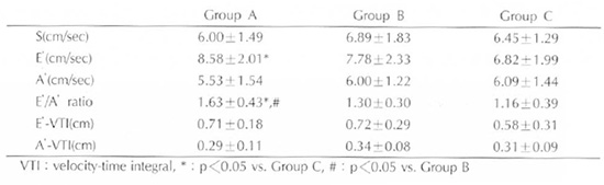
1) The myocardial wall velocity is composed of 3 parts, systolic velocity(S), early diastolic velocity(E′) and late diastolic velocity(A′). 2)
Conclusion
Early diastolic wall velocity and E'/A' ratio decreased in abnormal relaxation group compared to those in normal group. However, E'/A' ratio is more than 1 even in abnormal relaxation group whereas E/A ratio < 1 in mitral inflow is a characteristic finding of abnormal relaxation. |
|
Keywords: Myocardial wall velocity; Relaxation abnormality |
|