Abstract
Objectives
Primary cardiac tumors are rare, being found in approimately 1 in 10,000 to 33 in 1,000 routine autopsies in patients of all ages. The purpose of this study is to evaluate the status of primary cardiac tumor in Korea, their clinical and pathological characteristics. We analysed our 13 cases of primary cardiac tumors confirmed by operative findings, and all cases which were published in several literatures.
Method
Thirteen cases of primary cardiac tumors confirmed by pathologic findings from 1982 in Keimyung university hospital, and 116 cases of published data from 1962 were reviewed their pathologic and clinical findings.
Results
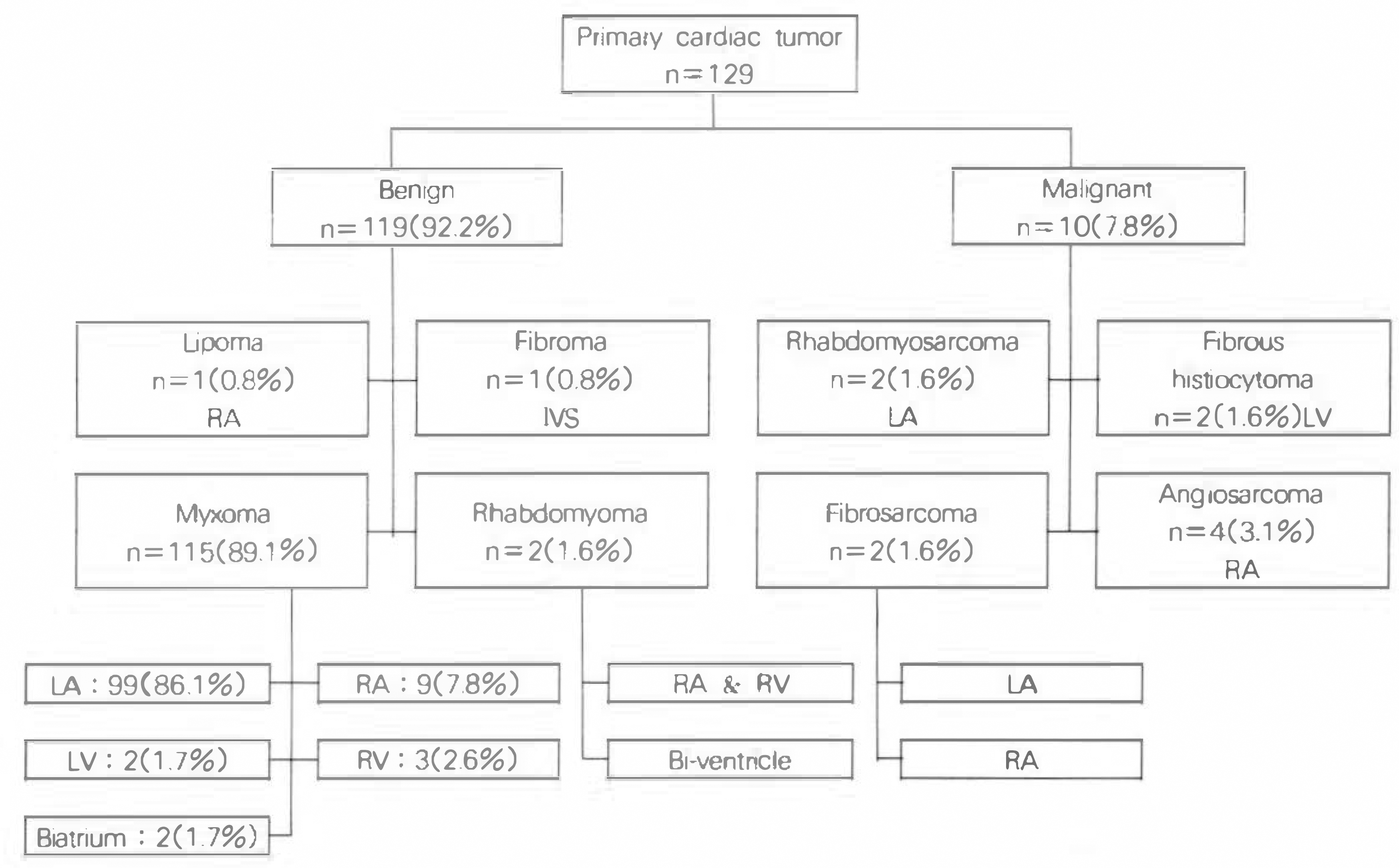
One hundred and twenty nine cases were included in this study. The age was ranged from 15 days to 75 years old. 45 cases(35%) were male and 84(65%) were female. 119 cases(92.2%) were revealed benign tumor: 115 myxoma, 2 rhabdomyoma, 1 lipoma and 1 fibroma. 10 cases(7.8%) were malignant tumors: 4 angiosarcoma, 2 fibrous histiocytoma, 2 rhabdomyosarcoma, 2 fibrosarcoma. The most common site of benign tumor was left atrium, and of malignant tumor was right atrium.
References
1). Smith C. Tumors of the heart. Arch, pathol. Lab. Medicine. 110:1. 1986.
2). Richadson JV, Brandt B, Doty DB, Eorenhaff JL. Surgical treatment of atrial myxomas: Early & late result of operations and review of literatures. Ann Thorac Surg. 28:364. 1979.
3). Straus R, Merliss R. Primary tumor of the heart. Arch Pathol Lab Med. 39:74. 1945.
4). McAllister HA. Primary tumors of the heart and pericardium. Curr Probl Corrdil. 4:1. 1979.
5). Wold LE, Lie JT. Cardiac myxomas. Am J Pathol. 101:208. 1980.
7). Goldstein S, Mahoney E. Right ventricular fibrosarcoma causing pulmonic stenosis. Am J Cardiol. 21:328. 1968.

8). 차준갑 · 이명진 · 서상현 · 홍숭록 • 홍필훈. 좌심방 점액종 치협례 대한흉부외과학회지. 2:73. 1969.
9). 박영배 ’ 이명욱 · 김성연 · 권인순 • 서정돈 • 이영 우· 이성호· 안광필· 김종환·한만청·김용일; 좌섬방 점액종의 1례 보고 순환기. 7:47. 1977.
10). 김근호 • 지행욕 • 정윤채 • 이종배 • 오철수 · 김영 태 • 김기홍 • 김춘원: 좌심방 정액종 의개심술 절제 치 험 례. 대 한흉부외과학회지. 10:164. 1977.
11). 이성행 · 이길노 • 이광숙 · 윤채호 · 김규태: 우심 방에 발생한 원발성 조직섬유종 대한흉부외과학회 지. 10:173. 1977.
12). 김상현 • 노준량 • 김종환 · 서경필 · 이영균. 화싱 방내 점액종 치험 2례. 대한흉부외과학회지. 11:58. 1978.
13). 윤여준 • 조범구 • 홍숭록 • 이웅구 • 김태승 • 최인 준. 좌심방에 발생한 섬유성 점액종 치험 1례. 대 한흉부외 과학회지. 11:135. 1978.
14). 유수웅 • 이학중 • 김대 한 • 김병열 • 김주이 • 강 정 호·이정호·유영선·박문향·박효숙;좌심방 점 액종의 치 험 1례. 대한흉부외과학회지. 11:348. 1978.
15). 안 혁. 심방 정액종4례 보고 대한흉부외과학회지. 12:23. 1979.
16). 채종욱 • 이종태 ’ 한숭세 • 김규태 • 이성행. 좌심 방 정액종의 치험 1례. 대한흉부외과학회지. 13:250. 1980.
17). 김형묵 • 김주현 • 노중기 • 김광택. 좌심방 점액종 l례 보고. 대한흉부외과학회지. 13:256. 1980.
18). 정재복 • 김성순 • 이웅구 • 차홍도. 좌심방 점액종 3례 대 한내과학회 잡지. 23:531. 1980.
19). 심원홈 • 정낭식 · 조숭연 • 이웅구 • 황영남 • 최규 식 • 흥필훈. 우심방 접액종 l례. 순환기. 12:179. 1980.
20). 장 명 • 이철주 · 김광호 • 홍숭록. 재발된 좌심방 점액종의 치험 1례. 대한흉부외과학회지. 14:260. 1981.
21). 오병회 • 박 정식 ‘ 최윤식 • 서정동 • 이영우 • 노준 량: 2연성 섬에코도로 진단된 화심실 점액종 l례 보고. 대 한내과학회 잡지. 24:613. 1981.
22). 이영욱 • 최윤식, 서정동 • 이영우 • 이영균 • 김종 환 • 노준량 • 한만청. 원발성 심장점액종의 입상적 고찰, 대 한내 과학회 잡지. 24:46. α. 1981.
23). 김동철 • 송 정상 ‘ 배종화 • 김영식 • 노준량. 우심 실 점액종 l례 보고. 대한내과학회잡지. 24:626. 1981.
24). 염 욱 • 이영란. 심장 점액중 대한흉부외파학회지. 15:98. 1982.
25). 안병회 • 이호완 • 이동준. 양심방에 발생한 점액종 치험 1례 대한흉부외과학회지. 15:107. 1982.
26). 윤종경 • 강영회 • 양영선 • 이홍식 • 김종성. 심 Echo도로 확진된 우심방 점액종 1예 보고 언제의학. 3:393. 1982.
27). 이명욱 · 박영배 · 최윤식 • 서정돈 • 이영우 • 노준 량 • 박재량. 석회화된 점액종 2례 보고 순환기. 13:245. 1983.
28). 조규도 • 김세화. 좌심방 점액종 2례 보고. 대한흉 부외 과학회 지. 15:402. 1982.
29). 박재길 • 송인성 • 이홍균. 거대 우심실 정액종 1례 보고 대한흉부외과학회지. 16:470. 1983.
30). 정경영 • 조범구 • 홍숭록 • 흥필훈. 심장 점액종 치 험 16례 보고. 대한흉부외과학회지. 16:485. 1983.
31). 임중규 • 변정섭 • 김 석 주 • 임준영 • 임숭찬 • 이동 준: Bilateral atrial myxoma l예 보고. 순환기. 13:257. 1983.
32). 김병주 • 왕영펼 • 곽문섭 • 김세화 • 이홍균. 심장 종양 6례 보고. 대 한흉부외 과학회 지. 18:667. 1985.
33). 김영대 • 서봉관 • 권오훈 • 이혁엽 • 이명욱 • 서 정 돈 • 이영우 • 노준량· 지제근. 수술후 재발한 좌싱 방 정액종 l혜 순환기. 15:507. 1985.
34). 김치정 • 도문홍 • 권오훈 · 오병회 • 이명묵 • 박영 배 • 최윤식 • 서정돈 • 이영우. 심장 점액종의 임상 적 관찰 순환기. 15:671. 1985.
35). 한병선 ’ 정덕용 • 한균인 • 임숭평 • 홍장수 • 이영. 화심방 접액총 2례 보고. 대한흉부외과학회지. 19:429. 1986.
36). 권영주 • 서세웅 • 김성구 · 좌심방에서 발생한 원발 성 섬유성육종 순환기. 17:389. 1987.
37). 오세웅 • 김병석 • 한영숙 • 이선회. 좌심방 점액종 2례 보고 대한흉부외과학회지. 20:809. 1987.
38). 노태훈 • 김원곤 • 조규석 · 박 주 철 · 유세영. 감염 된 좌심방 정액종 치험례 대한흉부외과학회지. 20:570. 1987.
39). 이윤우 · 박영우 · 최석구 • 유원상 • 구본일 · 박국 양 • 이홍섭 • 김창호 · 윤귀옥 • 고일향. 화심방 점 액종 치험 1례 보고. 인제의학. 8:433. 1987.
40). 강연식 • 정경영 • 조범구 • 홍숭록 • 소동문: 원발 성 심장종양의 수술적 치료-22례 보고 대한흉부 외과학회지. 22:11. δ. 1989.
41). 김용수 ‘ 김혁 • 이준영 • 이재원 • 강정호 • 지행 옥 • 김근호. 섬장 점액종의 외과적 고찰-임상 경 험 및 장기성적. 대한흉부외과학회지. 21:518. 1988.
42). 박찰호 • 이양행 • 강인득 • 우종수 • 조광현. 화심 방 점액종 2례 보고 대한흉부외과학회지. 21:131. 1988.
43). 이선호 • 조항보 • 이규환 • 이항 • 이근수 • 박병 태 • 박문향 • 이중달. 심실중격에서 기원한 심장의 원발성 섬유총 1예. 대한내과학회잡지. 31:101. 1988.
44). 라찬영 • 최세 영 • 박창권 · 이광숙 • 유영선. 심 장 점액종의 외과적 치료, 대한흉부외과학회지. 22:781. 1989.
45). 막종원 • 박상섭 • 류지윤 • 박철호 • 우종수 • 조광 현 • 이경순. 식장내 악성 섬유성 조직구종. 대한흉 부외과학회지. 22:29. 끼. 1989.
46). 김상섭 · 주1봉덕, 박순창. 섬에코도로 확인된 좌 심방 점액종을 동반한 Marjan 증후군 1례 순환기. 20:4342. 1990.
47). 이선회 • 분석환 • 조규도 • 조건현 • 왕영필 • 팍문 섭 · 김세화 • 이홍균. 성장 점액종의 외과척 치료 대한흉부외과학회지. 23:1158. 1990.
48). 나국주 • 허선 • 김상형 • 이동준. 성장 정액종의 임 상적 고찰. 대한흉부외과학회지. 23:1168. 1990.
49). 심재영 • 최영석, 임진수 • 최영호 • 장정수. 심 방 점액종. 대한흉부외과학회지. 23:501. 1990.
50). 이선회 • 문석환 • 조규도 • 조건현 • 왕영필 • 곽운 섭, 김세화 • 이흥균. 심장점액종의 외과척 치료. 대한흉부외과학회지. 23:1158. 1990.
51). 조상록 • 김용진 • 노준량 • 서경필. 심장내 횡문근 종의 수술 치료-2혜 보고. 대한흉부외과학회지. 24:1138. 1991.
52). 차경 태 • 홍민수 • 최 병 철 • 이 섭 • 유환국 • 허 용·안욱수·김병렬·이정호·유회성;원발성 심 장종양에 대한 외과적 치험 대한흉부외과학회지. 24:701. 1991.
53). 김택진 • 김광택 • 김형욱 • 원남회 • 안태훈 • 노영 무. 좌심방내에 발생한 악성 섬유성 조직구종 치험 1 례. 대 한흉부외과학회 지. 24:35. 낀. 1991.
54). 박영훈 • 남상민 • 이상호 • 최재웅 • 안태훈 • 신익 균. 폐 색전중을 동반한 다발성 우심방 접액종 l예. 순환 기. 24:1034. 1992.
55). 김욱성 • 안 혁. 심장의 왼발성 횡푼근육종, 대한 흉부외과학회지. 26:715. 1993.
56). 정일영 • 전회재 • 최필조 • 합시영 • 성시찬 • 우종 수. 심장내 원발성 지방중 1례 보고. 대한흉부외과 학회 지. 27:310. 1994.
57). Carney JA. Difference between nonfamilial and familial myxoma. Am J of Surg Pathol. 9:53. 1985.
58). McAlliter HA, Fenolio JJ. Tumors of the cardiovascular system. In atlas of Tumor Pathology. Washington DC: Armed Forces Institude of Pathology Fasc;15 2nd series. 1978.
59). Richard RW. Tumors of the heart, review of the subject and report of one hundred and fifty cases. Arch Pathol. 51:98. 1951.
60). Gerbode F, Kerth WJ, Hill JD. Surgical management of tumors of the heart. Surgery. 61:94. 1967.
61). Sutton MG, Mercier LA, Giuliani ER, Lie JT. Atrial myxomas, a review of the litrature. Ann Thorac Surg. 28:354. 1979.
62). Oh JK, Seward JB, Tajik AT. The Echo Manual. 1st Ed. p. 187. Little: Brown and Company;1994.
63). Greenwood WF. Profile of atrial myxoma. Am J Cardiol. 21:367. 1968.
64). MacGregor CA, Cullen RA. The syndrome of fever, anemia, and high sedimentation rate with atrial myxoma. Brit MJ. 2:1991. 1959.
65). Currey HFL, Mathews JA, Robinson J. Right atrial myxoma mimicking a rheumatic disorder. Brit MJ. 1:547. 1967.

66). Silverman J, Olwin JS, Graehinger JS. Cardiac myxomas with systemic embolization. Circulation. 26:99. 1962.

67). Silverman NA. Primary cardiac tumors. Ann Surg. 191:127. 1979.
68). Heath D, Mackimon J. Pulmonary hypertension due to myxoma of the right atrium. Am Heart J. 68:227. 1964.

69). Keenan PJM, Morton P, Kane H. Right atrial myxoma and pulmonary embolism. Br Heart J. 8:510. 1982.
70). Yang HI, Wasielewski JF, Lee W, Lee E, paik Y. Angiosarcoma of the heart: Ultrastructural study. Cancer. 47:72. 1981.

71). Janigan DT, Husain A, Robinson NA. Cardiac angiosarcomas. A review and a case reprot. Cancer. 57:852. 1986.
72). Hannah H, Eisemann G, Hiszvzynskyj R, Winsky M, Cohen L. Invasive atrial myxoma documentation of malignant potential of cardiac myxoma. Am Heart J. 104:881. 1982.
73). Hattler BJ Jr, Fuchs JCA, Cosson R, sabiston DC Jr. Atrial myxoma, and evaluation of clinical and laboratory manifestations. Ann Thorac Surg. 10:65. 1970.
74). Petsas AA, Gottieb S, Kingsley B, Segal BL, Lyerburg RJ. Echocardiographic diagnosis of left atrial myxoma. Usefulness of suprasternal approach. Br Heart J. 38:627. 1978.

75). Duncan WJ, Rowe RD, Freedom RM, Izukawa T, Olley PM. Space-occupying lesions of the myocardium: Role of two-dimensional echocardiography in detection of cardiac tumors in children. Am Heart J. 104:780. 1982.

76). Abrams HL, Adams DF, Grant HA. The radiology of tumors of the heart, Radiol Clin North Am. 9:299. 1971.
77). Pendyck G, Pierce EC, Borron MG, Lukbom SB. Embolization of left atrial myxoma after transseptal cardiac catheterization. Am J Cardiol. 30:569. 1972.
78). Godwin JD, Axel L, Adams JR, Schiller NB, Simpson PC Jr, Gertz EW. Computed tomography: A new method for diagnosing tumor of the heart. Circulation. 63:448. 1981.

79). Go RT, O'Donnel JK, Underwood DA, Feiglin DH, Salcedo EE, Pantoja M, MacIntyre WJ, Meaney TF. Comparison of gated cardiac MRI and 2D echocardiography of intracardiac neoplasms. A. J. Radiol. 145:21. 1985.
80). Lin TK, STech JM, Eckert WG, Lin JJ, Farrha SJ, Hagan CT. Pericardial angiosarcoma stimulating pericardial effusion by echocardiography. Chest. 73:881. 1978.
81). Shin MS, Kirklin JK, Cain JB, Ho KJ. Primary angiosarcoma of the heart: CT characteristics. Am J Roentogenol. 148:267. 1987.

82). Semb BK. Surgical considerations of treatment of cardiac myxoma. J Thorac Cardiovasc. Surg. 87:251. 1984.
83). Becker RC, Loeffler JS, Leopold KL, Underwood DA. Primary tumors of the heart: A review with emphasis on diagnosis and potential treatment modalities. Semin Surg Oncol. 1:161. 1985.

84). Dein JR, First WH, Stinson EB, et al. Primary cardiac neoplasm: Early and late result of surgical treatment in 42 patients. J Thorac Cardiovasc Surg. 93:502. 1987.
85). Kabbani S, Cooley DA. Atrial myxoma surgical considerations. J Thorac Cardiovasc Surg. 65:731. 1973.
86). Larrieu AJ, Jameson WRE, Tyers GFO, et al. Primary cardiac tumors; experience with 25 cases. J Thorac Cardiovas Surg. 83:339. 1982.
87). Hermann MA, Shankerman RA, Edmunds WD, Shub C, Shaff HV. Primary cardiac angiosarcoma: A clinicopathologic study of six cases. J Thorac Cardiovasc Surg. 103:655. 1992.
Fig. 1.
Apical four-chamber view of two-dimensional echocardiography showing hyperechogenic mass(arrow) in right atrium(Angiosarcoma).

Fig. 2.
Horizontal view of transesophageal echocardiography showing hyperechogenic mass(arrow) in right atrium(Angiosarcoma).

Fig. 3.
Chest CT scan of angiosarcoma revealed a low density mass lesion in right atrium with irregular margin and left pleural effusion.

Fig. 4.
Microscopic finding showing polygonal tumor cells are made up large nuclei & scanty cytoplasm with frequent mitoses(×400. H & E stain).

Table 1.
Clinical manifestation of 13 patients
Table 2.
Physical examination & Laboratory findings of 13 patients
Table 3.
Diagnosis & outcome of 13 patients
Table 4.
Site of tumor origin of 13 patients
Table 5.
Review of primary cardiac tumor of korea


 PDF
PDF ePub
ePub Citation
 Print
Print


 XML Download