

 PDF
PDF ePub
ePub Citation
 Print
Print


Abstract
Background
As atrial fibrillation occurs in the patients with hypertensive heart disease, the functional abnormalities of heart become more severe and the risk of systemic and cerebral embolization increases. In this study we checked left atrial dimension and other parameters with echocardiography in hypertensive patients with atrial fibrillation.
Methods
In an attempt to define the difference in left atrial dimension and the relation between left atrial size and various echocardiographic parameters, M-mode echocardiography was used to study 63 hypertensive patients, 33 patients with atrial fibrillation, and 30 patients with sinus rhythm.
Results
The mean left atrial dimension was 51.7 ± 8.7mm in hypertensive patients with atrial fibrillation versus 39.3 ± 8.4mm in hypertensive patients with sinus rhythm(P < 0.001). In hypertensive patients with atrial fibrillation left atrial dimension was over 50mm in 82%(27 of 33). Left atrial size had positive correlations with end-systolic and -diastolic internal dimension of left ventricle(LV), LV mass and E-point septal separation, and negative correlations with fractional shortening and ejection fraction of LV.
References
1). 박헌식 · 정의룡 · 류제근 이봉혈 검신우 – 채성 철 – 선재은 – 박의현 숭모판 협착증에서 성방세 동의 동반과 좌성방의 크기. 한국성초음파학회지. 1:195–200. 1993.
2). Devereux RB, Alonzo DR, Lutas EM, Gottlieb GJ, Campo E, Sachs I, Reichek N. Echocardiographic assessment of left ventricular hypertrophy: comparison to necropsy findings. Am J Cardiol. 57:450–8. 1986.

3). Petersen P. Thromboembolic complications of atrial fibrillation and their prevention: a review. Am J Cardiology. 65:24c–28c. 1990.
4). Forfar JC, Miller HC, Toft AD. Occult thyrotoxicosis: A correctable cause of “idiopathic” atrial fibrillation. Am J Cardiol. 44:9–12. 1979.
5). Kopecky SL, Gersh BJ, McGoon MD, Whisnant JP, Holmes DR, Ilstrup DM, Frye RL. The natural history of lone atrial fibrillation. A population-based study over three decades. N Eng J Med. 317:669. 1987.
6). Kannel KB, Abbott RD, Savage DD, McNamara PM. Epidemiologic features of chronic atrial fibrillation. The Framingham Study. N Eng J Med. 306:1018–22. 1982.
7). Inouye J, Massie B, Loge D, et al. Abnormal left ventricular filling: an early finding in mild to moderate systemic hypertension. Am J Cardiol. 53:120–6. 1984.
8). Fouad FM, Slominski JM, Tarazi RC. Left ventricular diastolic function in hypertension: relation to left ventricular mass and systolic function. J Am Coll Cardiol. 3:1500–6. 1984.
9). Henry WL, Morganroth J, Pearlman AS. Relation between echocardiogra-phically determined left atrial size and atrial fibrillation. Circulation. 53:273–9. 1976.
10). Selzer A. Atrial fibrillation revisited. N Engl J Med. 306:1044–5. 1982.
11). Probst P, Goldschlager N, Selzer A. Left atrial size and atrial fibrillation in mitral stenosis: factors influencing their relationship. Circulation. 48:1282–7. 1973.
12). Wolf PA, Dawber TR, Thomas HE Jr, Kannel WB. Epidemiologic assessment of chronic atrial fibrillation and risk of stroke: The Framingham Study. Neurology. 28:973–7. 1978.
13). Hinton RC, Kistler JP, Fallon JT, Friedlich AL, Fisher CM. Influence of atrial fibrillation on incidence of systemic embolism. Am J Cardiol. 40:509–13. 1977.
14). Onundarson FT, Thorgeirsson G, Jonmundsson E, Sigfusson N, Hardarson T. Chronic atrial fibrillation-epidemiologic features and 14 year follow-up: a case control study. Eur Heart J. 8:521–7. 1987.
15). Sahn DJ, DeMaria A, Kisslo J, Weyman A. Recomendations regarding quantitation in M-mode echocardiography: Results of a survey of echocardiographic measurements. Circulation. 58:1072–83. 1978.
16). Devereux RB, Reichek N. Echocardiographic determination of left ventricular mass in man: anatomic validation of the method. Circulation. 55:613–8. 1977.
17). Davies MJ, Pomerance A. The morphological basis of atrial fibrillation in man. Br Heart J. 34:520–4. 1972.
18). Petersen P, Kastrup J, Brinch K, Godfredsen J, Boysen G. Relation between left atrial dimension and duration of left atrial fibrillation. Am J Cardiol. 60:382–4. 1987.
19). Brand FN, Abbott RD, Kannel WB, Wolf PA. Characteristics and prognosis of lone atrial fibrillation. 30-year follow-up in the Framingham Study. JAMA. 254:3449. 1985.
20). Sanfillippo AJ, Abascal VM, Sheehan M, Oertel LB, Harrigan P, Hughes RA, Weyman AE. Atrial enlargement as a consequence of atrial fibrillation. A prospective echocardiographic study. Circulation. 82:792. 1990.
21). Cabin HS, Clubb S, Hall C, Permutter RA, Feinstein AR. Risk for systemic embolization of atrial fibrillation without mitral stenosis. Am J Cardiol. 65:1112–1116. 1990.
22). Stroke prevention in Atrial Fibrillation Investigators. Predictors of thromboembolism in atrial fibrillation: I. Clinical features of patient at risk. Ann Intern Med. 116:1–5. 1992.
23). Benjamin EJ, D'Agostino RB, Belanger AJ, Wolf PA, Levy D. Left atrial size and the risk of stroke and death: The Framingham Heart Study. Circulation. 92:835–841. 1995.
24). Predictors of thromboembolism in atrial fibrillation. II. Echocardiographic features of patients at risk. The Stroke Prevention in Atrial Fibrillation Investigators. Ann Intern Med. 116:6–12. 1992.
25). Levy D, Labib SB, Anderson KM, Christiansen JC, Kannel WB, Castelli WP. Determinations of sensitivity and specificity of electrocardiographic criteria for left ventricular hypertrophy. Circulation. 81:815–20. 1990.
26). Bennett DH, Evans DW. Correlation of left ventricular mass determined by echocardiography with vectorcardiographic and electrocardiographic voltage measurement. Br Heart J. 36:981–7. 1974.
27). Savage DD, Drayer JIM, Henry WL. Echocardiographic assessment of cardiac anatomy and function in hypertensive patients. Circulation. 59:623–32. 1979.
28). Levy D, Anderson KM, Savage D, Kannel WB, Christiansen JC, Castelli WP. Echocardiographically detected left ventricular hypertrophy: prevalance and risk factors. The Framingham Heart Study. Ann Intern Med. 108:7–13. 1988.
29). Levy D, Garrison RJ, Savage DD, Kannel WB, Castelli WP. Prognostic implications of echocardiographically determined left ventricular mass in the Framingham Heart Study. New Engl J Med. 322:1561–1566. 1990.
30). Miller JT, O'Rourke RA, Crawford MH. Left atrial enlargement: an early sign of hypertensive heart disease. Am Heart J. 116:1048–1051. 1988.
31). Woythaler JN, Singer SL, Kwan OL. Accuracy of echocardiography versus electrocardiography in detecting left ventricular hypertrophy: comparison with post-mortem mass measurements. J Am Coll Cardiol. 2:305–11. 1983.
32). Simek CL, Feldman MD, Haber HL, Wu CC, Jayaweera AR, Kaul S. Relationship between left ventricular wall thickness and left atrial size: comparison with other measures of diastolic function. J Am Soc Echocardiogr. 8:37–47. 1995.
33). Matsuzaki M, Tamitani M, Toma Y. Mechanism of augmented left atrial pump function in myocardial infarction and essential hypertension evaluated by left atrial pressure-dimension relation. Am J Cardiol. 67:1121–6. 1991.
34). Matsuda Y, Toma Y, Moritani K. Assessment of left atrial function in pateints with hypertensive heart disease. Hypertension. 8:779–85. 1986.
35). Abildscor JA, Millar K, Burgess MJ. Atrial fibrillation. Am J Cardiol. 28:263–7. 1971.
36). Azevedo J, Arroja I, Amado P, Carmona AKJ, Marques J, Araoujo V. Left atrial dimension decrease after atrial fibrillation cardioversion(abst). Circulation. 86(suppl 1):1–663. 1992.
Fig. 1.
Frequencies of atrial fibrillation(AF) and sinus rhythm(SR) in hypertensive patients(HT) according to left atrial dimension.
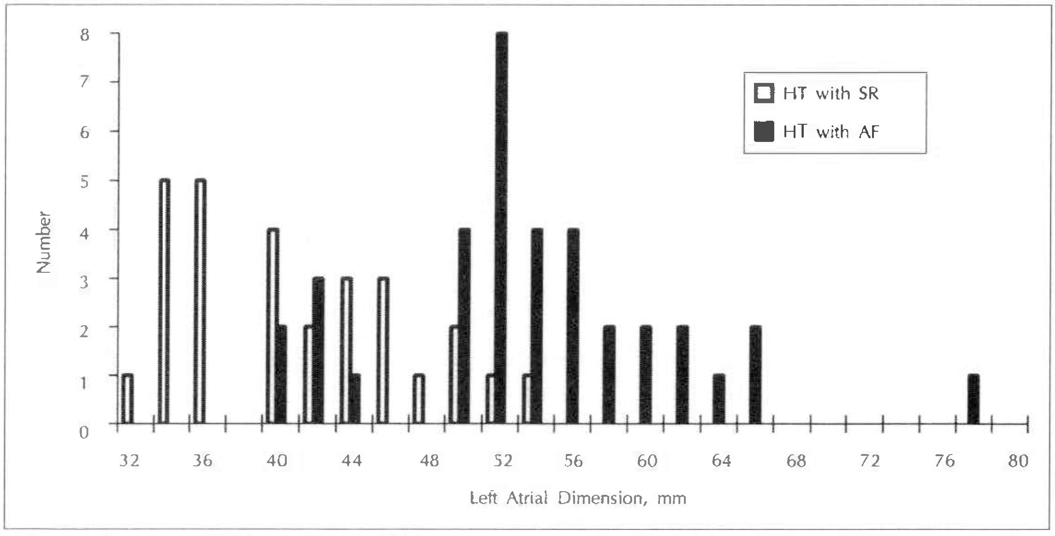
Fig. 2.
Correlation between left ventricular end systolic dimension(LVIDs) and left atrial dimension(LAD).
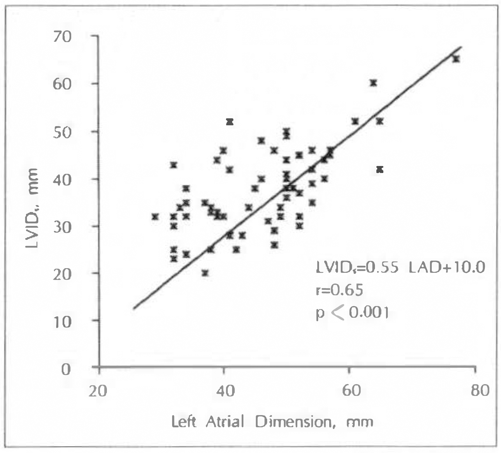
Fig. 3.
Correlation between left ventricular end diastolic dimension(LVIDd) and left atrial dimension(LAD).
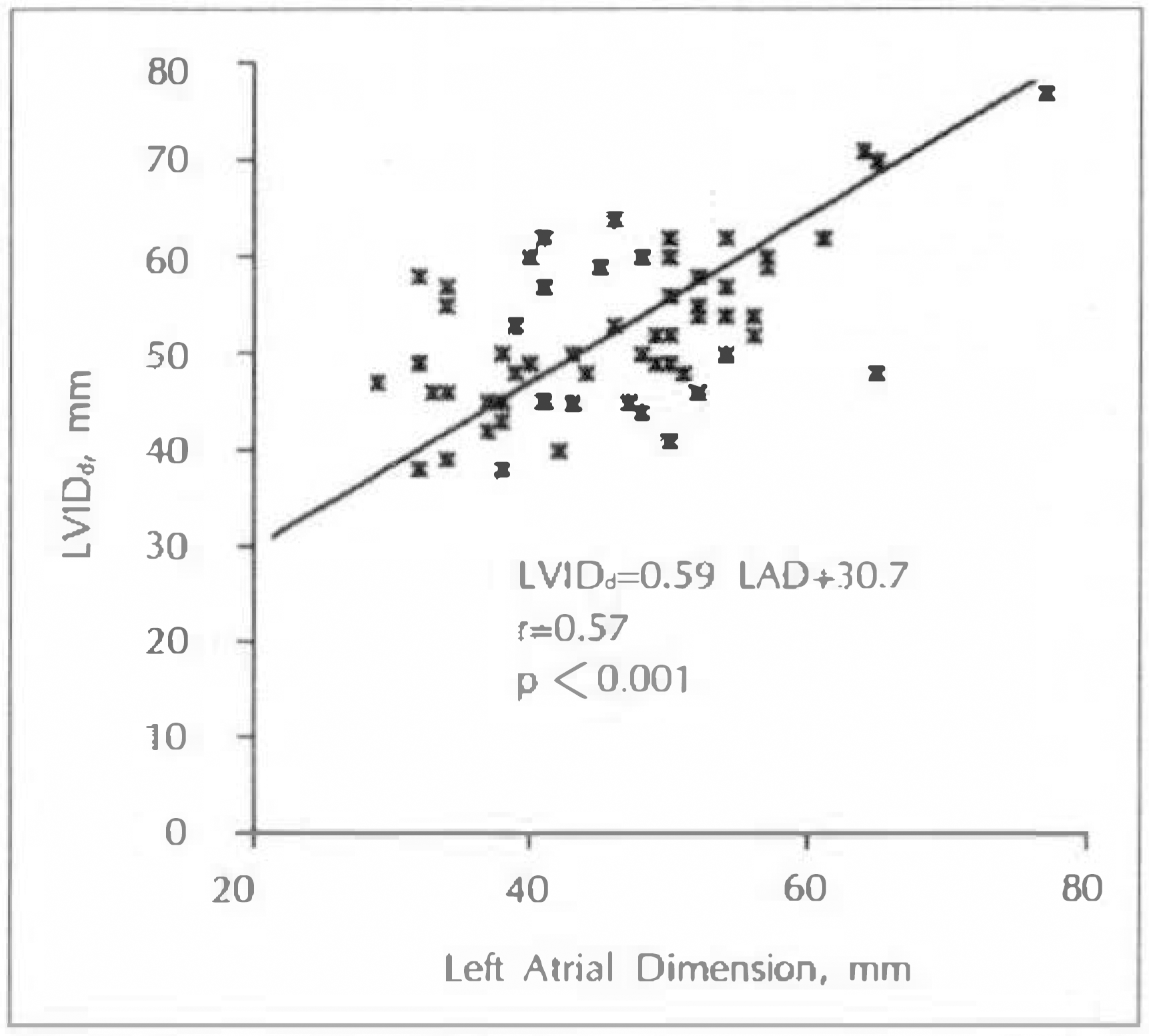
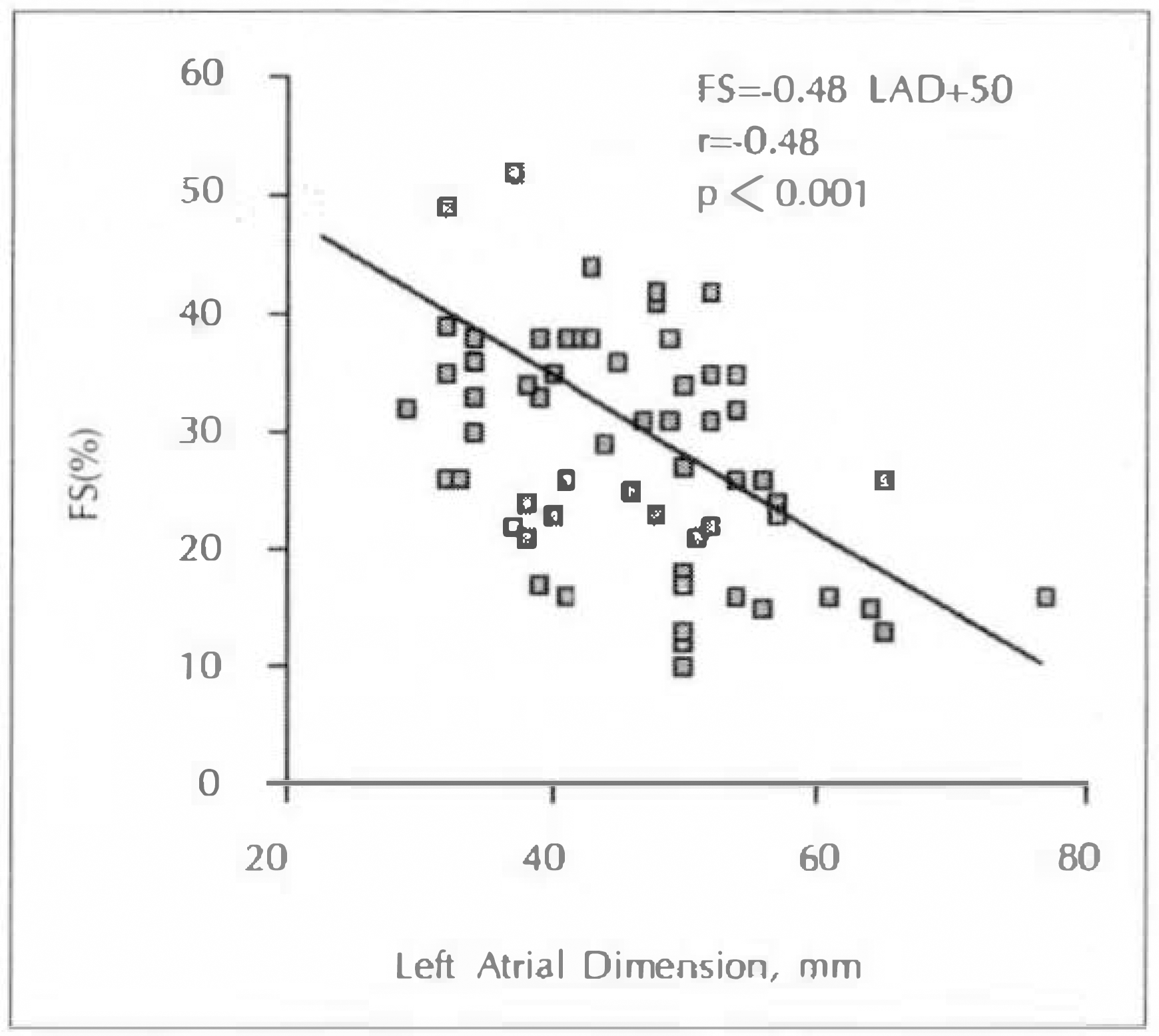
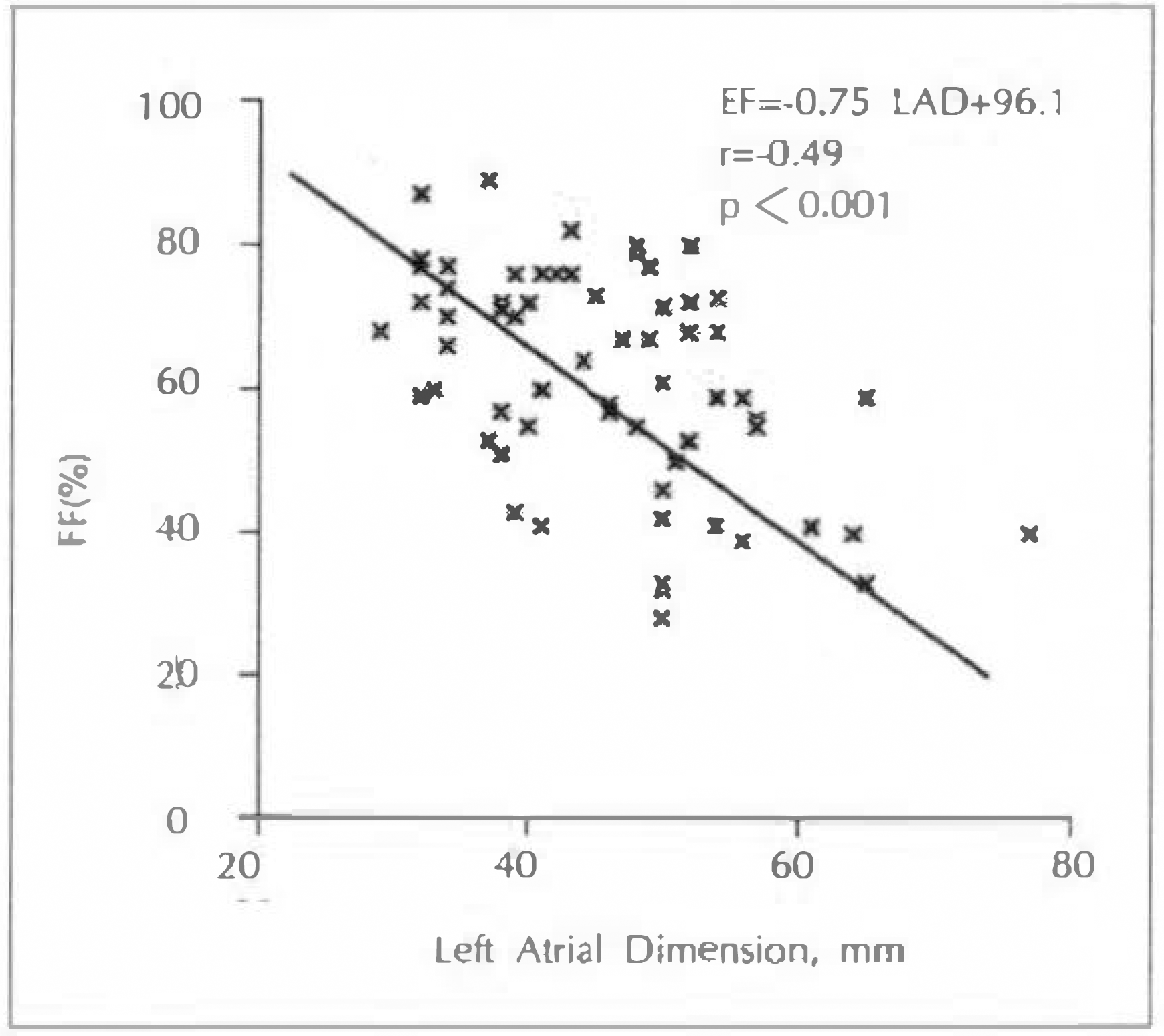
Table 1.
Clinical characteristics in the hypertensive patients
Table 2.
Blood pressure and heart rate in the hypertensive patients
| Characteristics | HT with AF (n=33) | HT with SR (n=30) | P value |
|---|---|---|---|
| SBP, mmHg | 153 ± 17 | 165 ± 17 | <0.01 |
| DBP, mmHg | 96 ± 12 | 104 ± 13 | <0.05 |
| MBP, mmHg | 115 ± 12 | 124 ± 13 | <0.01 |
| HR, beats/min | 94 ± 23 | 70 ± 18 | < 0.001 |
Table 3.
Chief complaints on initial visit in the hypertensive patients
Table 4.
Combined diseases in the hypertensive patients
| HT with AF (n=33) | HT with SR (n=30) | |
|---|---|---|
| Angina | 3(15.2%) | 4(13.3%) |
| Old myocardial infarction | 2(6.1%) | 2(6.7%) |
| Diabetes mellitus | 5(15.2%) | 1(3.3%) |
| Cerebrovascular accident | 6(18.2%) | 4(13.3%) |
Table 5.
Heights of the electrocardiographic voltages in the hypertensive patients
| HT with AF | HT with SR | P value | |
|---|---|---|---|
| SV1–2, mm | 18 ± 9 | 15 ± 5 | NS |
| RV5–6, mm | 24 ± 8 | 21 ± 7 | NS |
| SVY2 + RV5–6, mm | 43 ± 15 | 36 ± 9 | <0.05 |
Table 6.
ST-T Wave changes in the hypertensive patients
| HT with AF | HT with SR | |
|---|---|---|
| (n=33) | (n=30) | |
| ST depression in V5–6 | 7(21.2%) | 2(6.7%) |
| T-inversion in V5–6 | 9(27.3%) | 4(13.3%) |
Table 7.
Echocardiographic Measurements in the Hypertensive Patients
RV indicates right ventricular dimension; LVIDs, left ventricular systolic dimension; LVIDd, left ventricular diastolic dimension; LAD, left atrial dimension: STd, septal thickness; LVPW, left ventricular posterior wall thickness; EPSS, E-point septal separation; FS, fractional shortening; EF, ejection fraction; LVM, left ventricular mass; LVMI, left ventricular mass index. All values are expressed as mean ± SD.
Table 8.
Regression equations relating to various echocardiographic parameters to left atrial dimensions
 XML Download