Journal List > J Korean Soc Echocardiogr > v.3(2) > 1075196

Ha, Chung, Lim, Kwan, Kim, Oh, Lee, Jang, Lee, Shim, Cho, and Kim: Doppler Echocardiographic Evaluation of Diastolic Function in Different Patterns of Ventricular Hypertrophy and Topography in Essential Hypertension

Abstract

Background

Left ventricular hypertrophy and diastolic dysfunction have been well established in patients with essential hypertension. The left ventricular adaptation to hypertension represents a variety of spectrums with complex changes in topography. Left ventricular topography and diastolic and systolic function were evaluated in 48 patients with essential hypertension(group 1) and 33 age-matched normal controls(group 2) by transmitral inflow and pulmonary venous flow patterns assessed by Doppler echocardiography. The following Doppler parameters were measured: early(E) and late(A) peak velocities and velocity integrals, E/A ratio, deceleration time of E wave; systolic(S) and diastolic(D) peak velocities and velocity integrals of pulmonary venous flow, S/D ratio, and atrial reversal peak velocity.

Results

1) There were no siginificant differences of age, sex, heart rate, and body surface area between the two groups.
2) Systolic and diastolic blood pressures and left atrial dimension and left ventricular mass index and relative wall thickness were significandy increased in group 1. Deceleration time and isovolumic relaxation time were significandy increased in group 1(231.9 ± 47.6msecs vs 202.0 ± 40.7msecs, 106.7 ± 21.4msecs vs 83.2 ± 14.3msecs, p < 0.05, respectively), whereas E/A ratio was decreased compared with group 2(1.08 ± 0.35 vs 1.43 ± 0.44, p < 0.05). Time-velocity integral of atrial reversal pulmonary venous flow was significantly increased in group 1(1.5 ± 0.5 vs 1.3 ± 0.4, p < 0.05).
3) In group 1, 47.9% had concentric hypertrophy, 41.7% had eccentric hypertrophy and 10.4% had normal left ventricular mass index and left ventricular dimension.
4) In concentric hypertrophy, thickess of ventricular septum and posterior wall were significantly increased, whereas left ventricular end-diastolic dimension was significantly decreased compared with those of patients with eccentric hypertrophy.
5) There were no significant differences of peak E velocity, deceleration time of E wave, E/A ratio, time-velocity integral of E and A wave, isovolumic relaxation time between patients with concentric hypertrophy and eccentric hypertrophy.
6) There were no significant differences of pulmonary venous flow pattern between concentric hypertrophy and eccentric hypertrophy.
7) Systolic blood pressure was identified as an independent factor which associated with left ventricular mass index.

Conclusion

There were no significant differences in diastolic functional indices assessed by Doppler profiles of transmitral inflow and pulmonary venous flow between the patients with concentric hypertrophy and eccentric hypertrophy. The supine systolic blood pressure was identified as an independent factor associated with left ventricular mass index.

References

1). Casale PN, Devereux RB, Milner M, Zullo G, Harshfield GA, Pickering TG, Laragh JH. Value of echocardiographic measurement of left ventricular mass in predicting cardiovascular morbid events in hypertensive men. Ann Intern Med. 105:173–178. 1986.
crossref
2). Koren MJ, Devereux RB, Casale PN, Savage DD, Laragh JH. Relation of left ventricular mass and geometry to morbidity and mortality in uncomplicated essential hypertension. Ann Intern Med. 114:345–352. 1991.
crossref
3). Levy D, Garrison RJ, Savage DD, Kannel WB, Castelli WP. Prognostic implications of echocardiographically determined left ventricular mass in the Framingham heart study. N Engl J Med. 322:1561–1566. 1990.
crossref
4). Shepherd RFJ, Zachariah PK, Shub C. Hypertension and left ventricular diastolic function. Mayo Clin Proc. 64:1521–1532. 1989.
crossref
5). Grossman W, Jones D, McLaurin LP. Wall stress and patterns of hypertrophy in the human left ventricle. J Clin Invest. 56:56–64. 1975.
crossref
6). Strauer BE. Structural and functional adaptation of the chronically overloaded heart in arterial hypertension. Am Heart J. 114:948–957. 1987.
crossref
7). 조 정 휘 – 김 권상 김 영 식 · 송정상 u�종화. 고혈 암 환자에서 Doppler싱 초음따도플 이 용한 화 싱 실 확장기 기 능 에 관 한 연구. 순 환 기. 17:621–626. 1987.
8). 정 영 호 신 순철 · 양숭진 · 박상진 · 검 숭관 · 조정 관 · 박종춘 · 강정채. 좌심 설 비 대 가 중등도 고힐압 환자에서 좌 심 실 이 완기없 는기 능경증에 관및 한 연구 순환기 1. 7:627–636. 1987.
9). 이 영 – 홍경표 ‘ 고 혈 앙 에 서 의 이 완 기 심 장기 능 순환기. 18:621–634. 1988.
10). 정 진 원 · 박양규 박욱규. 본태성 고 혈 압 환자에 서 도플러 섬초음파플 이 용한 좌섭실 충혈 지표들 에 관한 연구. 순환기. 20:335–341. 1990.
11). 이 동철 한동신 · 유원상 • 정 상인과 고혈압 환 자 에 서 연 령 과 좌 싱 싱 비 후가 좌 심 설 이 완 기 기 능에 미치는 영 향에 관한 연구. 순환기. 21:92–99. 1991.
12). 강덕회 신 길 자. 고혈압 환자에서 도플러 싱 초응­ 파를 이 용한 좌우싱 실 이 완기 능에 관한 연구. 대 한 내과학회잡지. 45:291–298. 1993.
13). 임석아 정혜경 · 박시훈, 신 길 자 · 이 우 형. 고혈 압 환자에서 심 초음파를 이 용한 좌싱 설 비 대 유형 에 관한 연구. 순환기. 25:423–433. 1995.
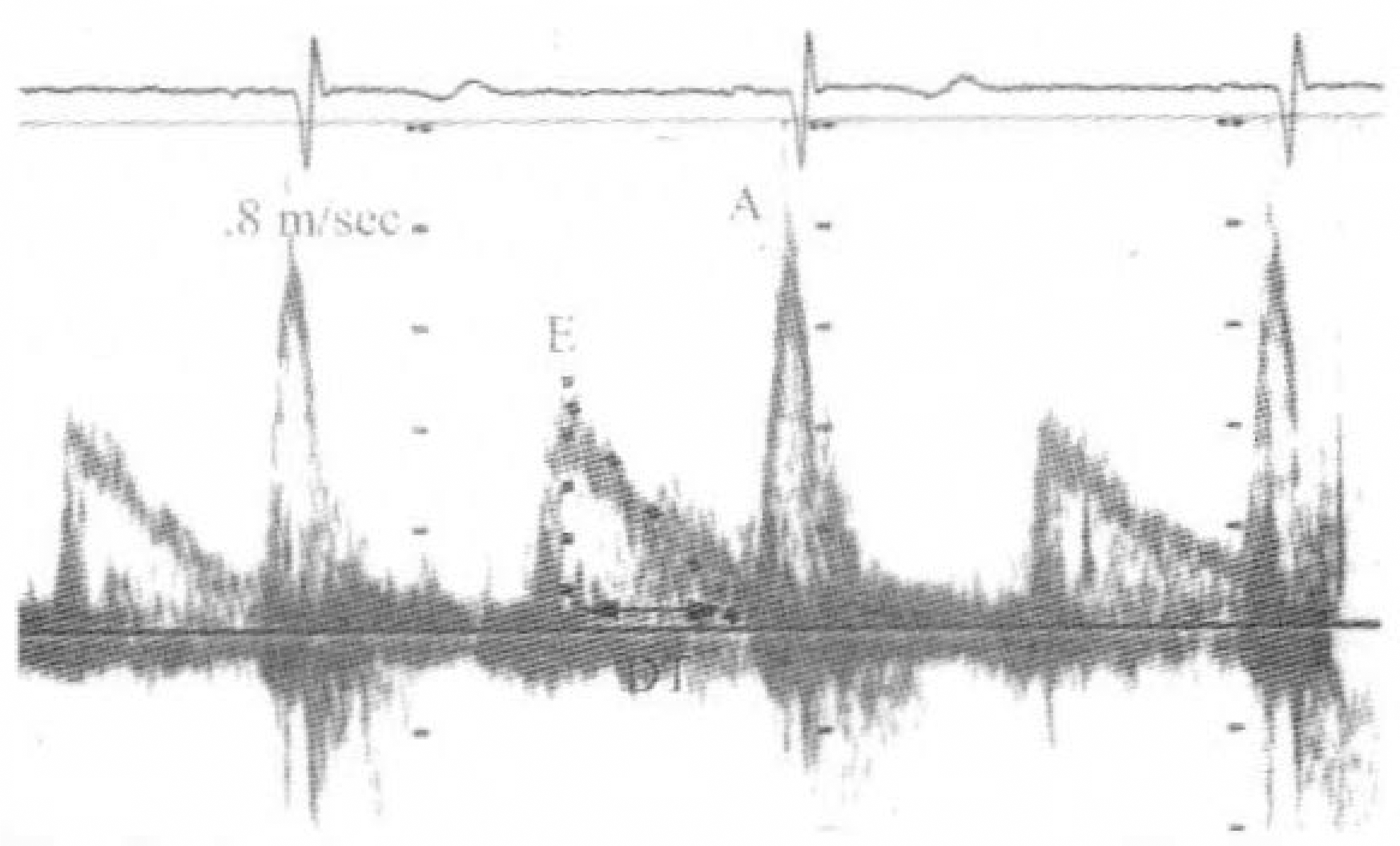
14). Nishimura RA, Abel MD, Hatle LK, Tajik AJ. Relation of pulmonary vein to mitral flow velocities by transesophageal Doppler echocardiography: effect of different loading conditions. Circulation. 81:1488–1497. 1990.
crossref
15). Kuecherer HF, Muhiudeen IA, Kusumoto FM, et al. Estimation of mean left atrial pressure from transesophageal pulsed Doppler echocardiography of pulmonary venous flow. Circulation. 82:1127–1139. 1990.
crossref
16). Kuecherer HF, Kusumoto F, Muhiudeen I A, Cahalan MK, Schiller NB. Pulmonary venous flow patterns by transesophageal pulsed Doppler echocardiography: relation to parameters of left ventricular systolic and diastolic function. Am Heart J. 122:1683–1693. 1991.
crossref
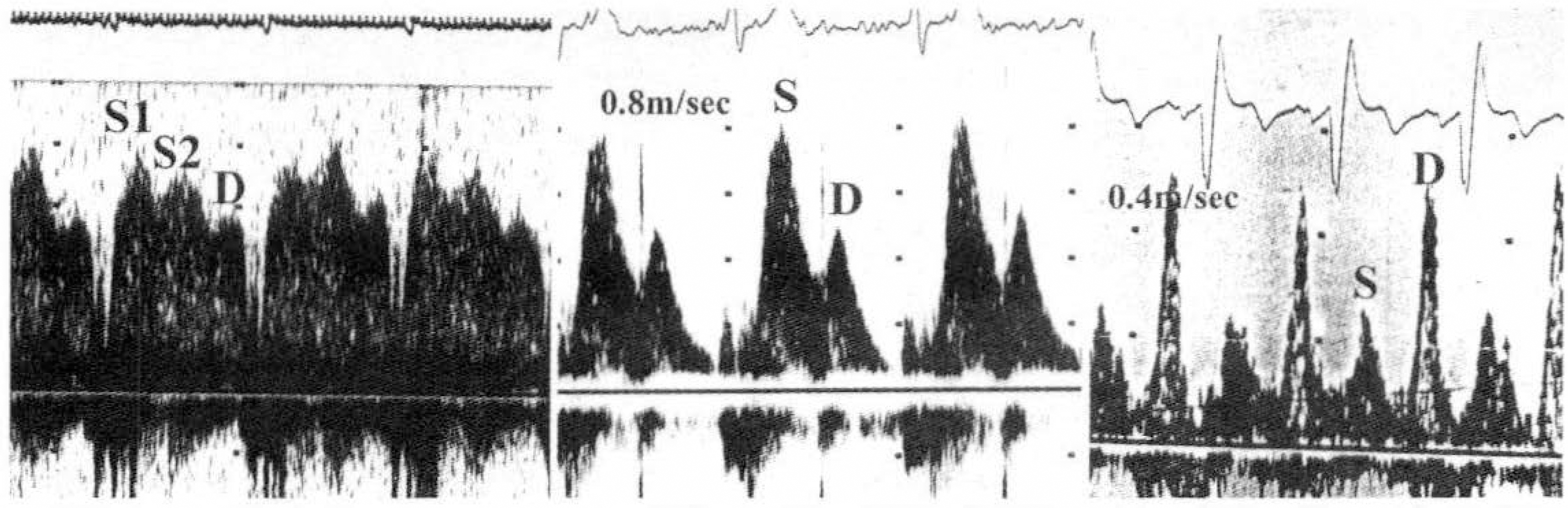
17). Klein AL, Tajik AJ. Doppler assessment of pulmonary venous flow in healthy subjects and in patients with heart disease. J Am Soc Echocardiogr. 4:379–392. 1991.
crossref
18). Keren G, Sherez J, Megidish R, Levitt B, Laniado S. Pulmonary venous flow pattern, its relationship to cardiac dynamics: a pulsed Doppler echocardiographic study. Circulation. 1105–1112. 1985.
19). Rossvoll R, Hatle LK. Pulmonary venous flow velocities recorded by transthoracic Doppler ultrasound: relation to left ventricular diastolic pressure. J Am Coll Cardiol. 21:1687–1696. 1993.
20). Devereux RB, Reichek N. Echocardiographic determination of left ventricular mass in man. Circulation. 55:613–618. 1977.
21). Devereux RB, Alonso DR, Lutas EM, et al. Echocardiographic assessment of left ventricular hypertrophy: comparison to necropsy findings. Am J Cardiol. 57:450–458. 1986.
crossref
22). Devereux RB, Pickering TG, Harshfield GA, et al. Left ventricular hypertrophy inpatients with hypertension: importance of blood pressure response to regularly recurring stress. Circulation. 68:470–476. 1983.
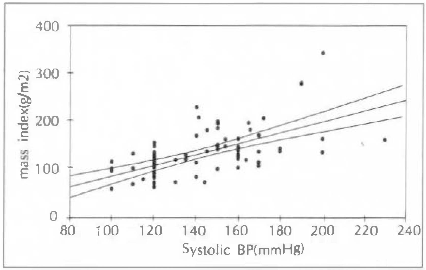
23). Drayer JI, Weber MA, DeYoung JL. BP as a determinant of cardiac left ventricular muscle mass. Arch Intern Med. 143:90–92. 1983.
crossref
24). Ren J-F, Hakki A-H, Kotler MN, Iskandrian AS. Exercise systolic blood pressure: a powerful determinant of increased left ventricular mass in patients with hypertension. J Am Coll Cardiol. 5:1224–1231. 1985.
crossref
25). Kobrin I, Oigman W, Kumar A, et al. Diurnal variation of blood pressure in elderly patients with essential hypertension. J Am Geriatr Soc. 32:896–899. 1984.
crossref
26). Frohlich ED, Tarazi RC. Is arterial pressure the sole factor responsible for hypertensive cardiac hypertrophy? Am J Cardiol. 44:959–963. 1979.
crossref
27). Frohlich ED. Hemodynamics and other determinants in development of left ventricular hypertrophy. Fed Proc. 42:2709–2715. 1983.
28). Walford GD, Spurgeon HA, Lakatta EG. Diminished cardiac hypertrophy and muscle performance in older compared with younger adult rats with chronic atrioventricular block. Cir Res. 63:502–511. 1988.
crossref
29). Isoyama S, Wei JY, Izumo S, Fort P, Schoen FJ, Grossman W. Effect of age on the develoment cardiac hypertrophy produced by aortic constriction in the rat. Circ Res. 61:337–345. 1987.
30). Cornoni-Huntley J, LaCroix AZ, Havlik RJ. Race and sex differentials in the impact of hypertension in the United States: the National Health and Nutrition Examination Survey I Epidemiologic Follow-up Study. Arch Intern Med. 149:780–788. 1989.
crossref
31). Holt JP, Rhode EA, Kines H. Ventricular volumes and body weight in mammals. Am J Physiol. 215:704–715. 1968.
crossref
32). Weber KT, Janick JS, Pick R, et al. Collagen in the hypertrophied, pressure overloaded myocardium. Circulation. 75:(. (Suppl I):):. 140–147. 1987.
33). Swynghedauw B, Delcayre C, Cheav SL, Callens-El Amrani F. Biological basis of diastolic dysfunction of the hypertensive heart. Eur Heart J. 13:(. (Suppl D):):. 2–8. 1992.
34). Limas CJ, Cohn JN. Defective calcium transport by cardiac sarcoplasmic reticulum in spontaneous hypertensive rats. Cir Res. 40:(. (Suppl I):):. 1162–1169. 1977.
35). Bache RJ. Effects of hypertrophy on the coronary circulation. Prog Cardiovasc Dis. 31:403–440. 1988.
crossref
36). Philips RA, Goldman ME, Ardeljan M, Arora R, Eison HB, Buyan Y, Krakoff LR. Determinants of abnormal left ventricular filling in early hypertension. J Am Coll Cardiol. 14:979–985. 1989.
37). Kapuku GK, Seto S, Mori H, Mori M, Utsunomia T, Suzuki S, Oku Y, Yano K, Hashiba K. Impaired left ventricular filling in borderline hypertensive patients without cardiac structural changes. Am Heart J. 125:1710–1716. 1993.
crossref
38). Nagano R, Masuyama T, Lee JM, Yamamoto K, Naito J, Mano T, Kondo H, Hori M, Kamada T. Transthoracic Doppler assessment of pattern of left ventricular dysfunction in hypertensive heart disease: combined analysis of mitral and pulmonary venous flow velocity patterns. J Am Soc Echocardiogr. 7:493–505. 1994.
crossref
39). Ganau A, Devereux RB, Roman MJ, DeSimone G, Pickering TG, Saba PS, Vargiu P, Simongini I, Laragh JH. Patterns of left ventricular hypertrophy and geometric remodeling in essential hypertension. J Am Coll Cardiol. 19:1550–1558. 1992.
crossref
40). Ren JF, Pancholy SB, Iskandrian AS, Lighty Jr GW, Mallavarapu C, Segal BL. Doppler echocardiographic evaluation of the spectrum of left ventricular diastolic dysfunction in essential hypertension. Am Heart J. 127:906–913. 1994.
crossref
41). Chenzbraun A, Pinto FJ, Popylisen S, Schnittger I, Popp RL. Filling patterns in left ventricular hypertrophy: a combined acoustic quantification and Doppler study. J Am Coll Cardiol. 23:1179–1185. 1994.
crossref

Fig. 1.
Geometric profiles in hypertensive patients divided by left ventricular mass index and relative wall thickness.
jkse-3-168f1.tif
Fig. 2.
Correlation between left ventricular mass index and systolic blood pressure.
jkse-3-168f2.tif
Fig. 3.
Doppler transmitral flow velocity recording showing Doppler measurements used in this study. E: left ventricular early diastolic peak velocity, A: atrial peak velocity, DT: deceleration time of E wave.
jkse-3-168f3.tif
Fig. 4.
Representative tracings of pulmonary venous flow patterns. A) biphasic forward systolic flow, B) systolic predominant pattern, C) diastolic predominant pattern.
jkse-3-168f4.tif
Table 1.
Clinical and M-mode echocardiographic data of 48 hypertensive patients and 33 normal controls
  Group 1(N=48) Group 2(N=33) p value
Age 53.8 ± 8.9 46.5 ± 12.0 0.098
Sex(Male/Female) 10/38 8/25 0.72
BSA(m2) 1.62 ± 0.29 1.62 ± 0.17 0.991
Systolic BP(mmHg) 160.4 ± 20.4 116.8 ± 7.2 0.000
Diastolic BP(mmHg) 100.5 ± 10.7 76.5 ± 6.3 0.000
Heart rate(beat/min) 64.9 ± 9.6 69.6 ± 10.3 0.074
Septal thickness(mm) 11.2 ± 2.5 8.0 ± 1.6 0.000
Posterior wall thickness 10.8 ± 2.3 8.3 ± 1.5 0.000
Relative wall thickness 2.2 ± 0.4 1.7 ± 0.3 0.000
LVEDD(mm) 49.8 ± 4.0 48.4 ± 3.3 0.097
LA dimension(mm) 35.4 ± 5.4 33.3 ± 6.6 0.179
FS(%) 38.3 ± 7.5 36.1 ± 5.2 0.122
LVEF(%) 71.0 ± 8.9 67.9 ± 6.2 0.075
LV mass(g) 264.1 ± 90.0 166.2 ± 40.5 0.000
LV mass index(g/m2) 158.6 ± 51.2 102.5 ± 24.0 0.000

BSA: body surface area, BP: blood pressure, LVEDD: left ventricular end diastolic dimension, LA: left atrium, FS: fractional shortening, LVEF: left ventricular ejection fraction

Table 2.
Doppler echocardiographic data of 48 hypertensive patients and 33 normal controls
  Group 1(N=48) Group 2(N=33) p value
E(m/sec) 0.74 ± 0.18 0.72 ± 0.17 0.553
A(m/sec) 0.73 ± 0.20 0.53 ± 0.14 0.000
E/A ratio 1.08 ± 0.35 1.43 ± 0.44 0.000
TVIE(cm) 11.9 ± 2.9 11.6 ± 2.9 0.651
TVIA(cm) 10.1 ± 13.2 6.4 ± 2.1 0.128
DT(msec) 231.9 ± 47.6 202.0 ± 40.7 0.006
IVRT(msec) 106.7 ± 21.4 83.2 ± 14.3 0.000
S(m/sec) 0.59 ± 0.13 0.53 ± 0.16 0.053
D(m/sec) 0.40 ± 0.11 0.43 ± 0.13 0.315
Ar(m/sec) 0.23 ± 0.04 0.23 ± 0.07 0.692
TVIS(cm) 15.5 ± 4.3 13.8 ± 3.9 0.068
TVID(cm) 9.0 ± 3.1 8.8 ± 2.9 0.704
TVIAr(cm) 1.5 ± 0.5 1.3 ± 0.4 0.006

E: diastolic early peak velocity, A: diastolic atrial peak velocity, TVIE: time velocity integral of E wave, TVIA: time velocity integral of A wave, DT: deceleration time of E wave, IVRT: isovolumic relaxation time, S: peak systolic forward velocity of pulmonary venous flow, D: peak diastolic forward flow velocity of pulmonary venous flow, Ar: peak velocity of atrial reversal pulmonary venous flow, TVIS, TVID, and TVIAr: time velocity integral of S, D, and Ar wave.

Table 3.
Clinical and M-mode echocardiographic data of concentric and eccentric hypertrophy in 48 hypertensive patients
  Concentric hypertrophy(N=23) Eccentric hypertrophy(N=20) p value
Age 53.2 ± 7.2 54.4 ± 11.3 0.667
Sex(Male/Female) 6/17 4/16 0.861
BSA(m2) 1.69 ± 0.17 1.55 ± 0.39 0.11
Systolic BP(mmHg) 163.7 ± 20.0 159.6 ± 23.5 0.526
Diastolic BP(mmHg) 99.8 ± 8.9 103.6 ± 12.5 0.000
Heart rate(beat/min) 66.2 ± 10.3 63.9 ± 10.1 0.074
Septal thickness(mm) 12.5 ± 2.3 10.7 ± 1.8 0.007
Posterior wall thickness(mm) 12.2 ± 2.4 9.8 ± 1.2 0.000
Relative wall thickness 2.5 ± 0.4 1.9 ± 0.2 0.000
LVEDD(mm) 49.1 ± 4.2 51.5 ± 3.3 0.044
LA dimension(mm) 36.5 ± 5.0 34.5 ± 6.2 0.239
FS(%) 36.8 ± 7.1 40.9 ± 7.3 0.076
LVEF(%) 69.3 ± 9.1 74.1 ± 8.1 0.081
LV mass(g) 302.3 ± 100.4 248.3 ± 54.8 0.032
LV mass index(g/m2) 179.3 ± 58.2 150.4 ± 27.6 0.043

BSA: body surface area, BP: blood pressure, LVEDD: left ventricular end diastolic dimension, LA: left atrium, FS: fractional shortening, LVEF: left ventricular ejection fraction

Table 4.
Doppler echocardiographic data of concentric and eccentric hypertrophy in 48 hypertensive patients
  Concentric hypertrophy(N=23) Eccentric hypertrophy(N=20) p value
E(m/sec) 0.74 ± 0.17 0.75 ± 0.20 0.917
A(m/sec) 0.74 ± 0.22 0.72 ± 0.19 0.698
E/A ratio 1.05 ± 0.32 1.11 ± 0.40 0.622
TVIE(cm) 11.7 ± 3.1 12.0 ± 3.0 0.787
TVIA(cm) 12.8 ± 19.1 7.9 ± 2.0 0.264
DT(msec) 241.8 ± 46.3 223.5 ± 50.0 0.225
IVRT(msec) 108.3 ± 17.2 109.4 ± 21.1 0.850
S(m/sec) 0.59 ± 0.14 0.58 ± 0.11 0.796
D(m/sec) 0.39 ± 0.10 0.43 ± 0.13 0.308
Ar(m/sec) 0.23 ± 0.04 0.24 ± 0.06 0.839
TVIS(cm) 14.9 ± 4.4 15.6 ± 4.4 0.614
TVID(cm) 8.4 ± 3.2 10.0 ± 3.2 0.105
TVIAr(cm) 1.6 ± 0.5 1.4 ± 0.3 0.231

E: diastolic early peak velocity, A: diastolic atrial peak velocity, TVIE: time velocity integral of E wave, TVIA: time velocity integral of A wave, DT: deceleration time of E wave, IVRT: isovolumic relaxation time, S: peak systolic forward velocity of pulmonary venous flow, D: peak diastolic forward flow velocity of pulmonary venous flow, Ar: peak velocity of atrial reversal pulmonary venous flow, TVIS, TVID, and TVIAr: time velocity integral of S, D, and Ar wave.

TOOLS
Similar articles