

 PDF
PDF ePub
ePub Citation
 Print
Print


Abstract
Background
The radionuclide scintigraphy with Thallium201 or Tc99m sestamibi(MIBI) is the most commonly used method in assessing myocardial perfusion in the clinic. Recently myocardial contrast echocardiography(MCE) has been reported as a new method to evaluate the status of myocardial perfusion. But the objectivity of the MCE was not verified completely. Thus this study was undertaken to evalute the usefulness of MCE by comparing the myocardial opacification grades of MCE and scintigraphic perfusion grades using Tc99m MIBI.
Method
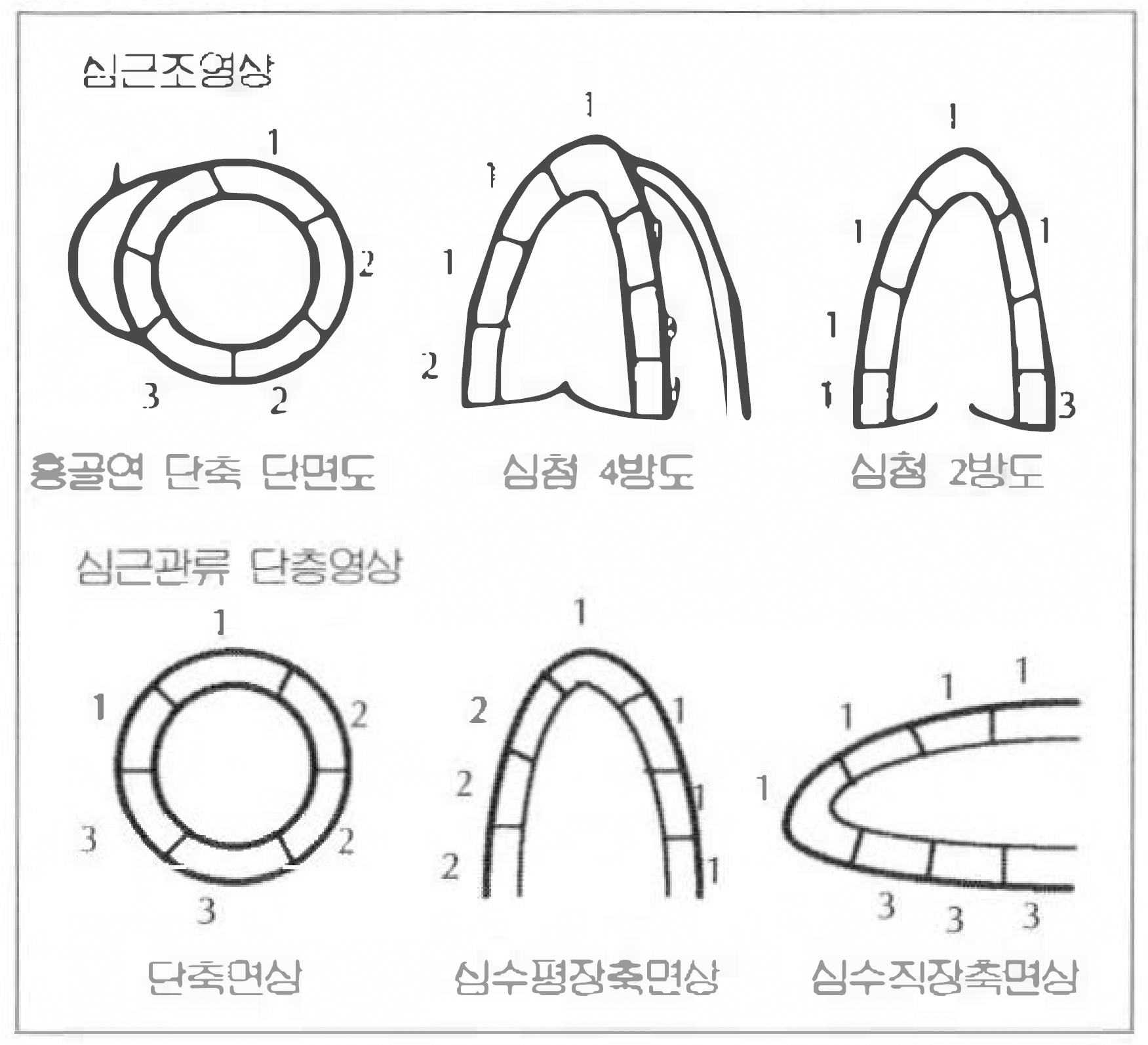
MCE and MIBI scintigraphy was carried out 2 weeks after acute myocardial infarction with a mean time interval of 2 days in 15 patients(M: F=13: 2, mean age: 59 yrs). Scintigraphic myocardial tomographic image at short axis, vertical and horizontal long axis were obtained after 15 mCi of Tc”m MIBI injection in resting state. MCE was done in the catheterization laboratory on the completion of coronary angiography by injecting sonicated Hexabrix(4cc to left and 3cc to right coronary artery) in parasternal short, apical 4 and 2 chamber view. To anaylaze the MCE images and SPECT images, the left ventricle in 3 views were divided into 20 segments and the myocardial opacification and MIBI perfusion grades were scored in each segment. The myocardial segments were classified into 3 coronary artery territoris and opacification indexes and MIBI perfusion indexes were calculated in each coronary artery territories. The comparison of myocardial opacification indexes and MIBI perfusion indexes was done in the whole patients and by location of infarction site.
Result
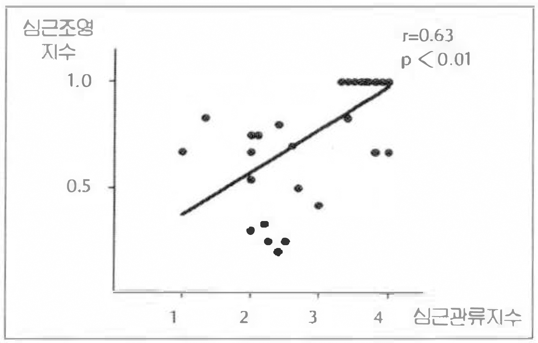
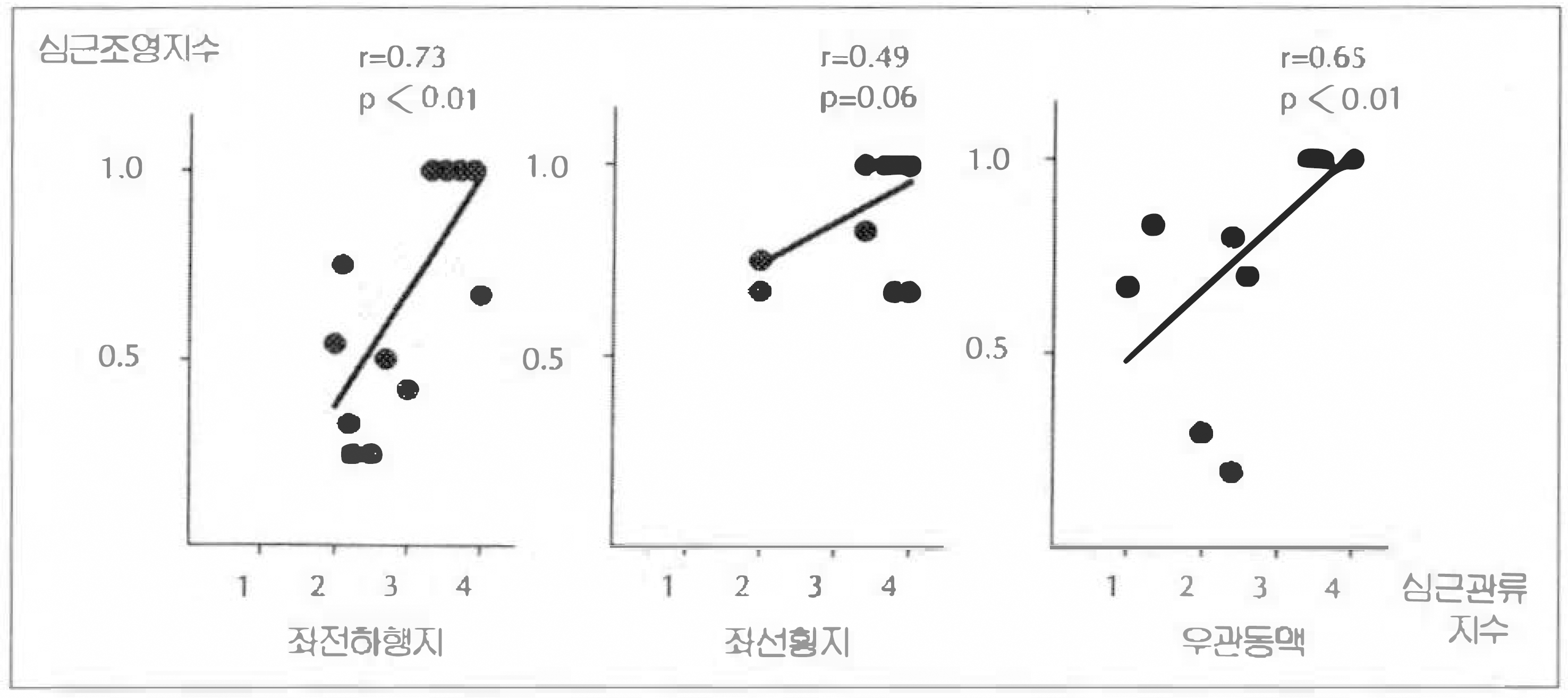
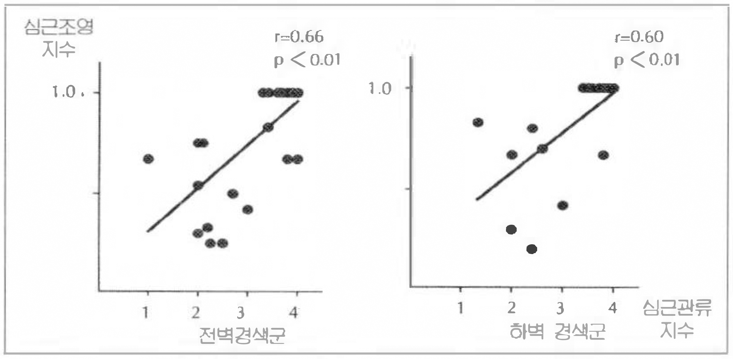
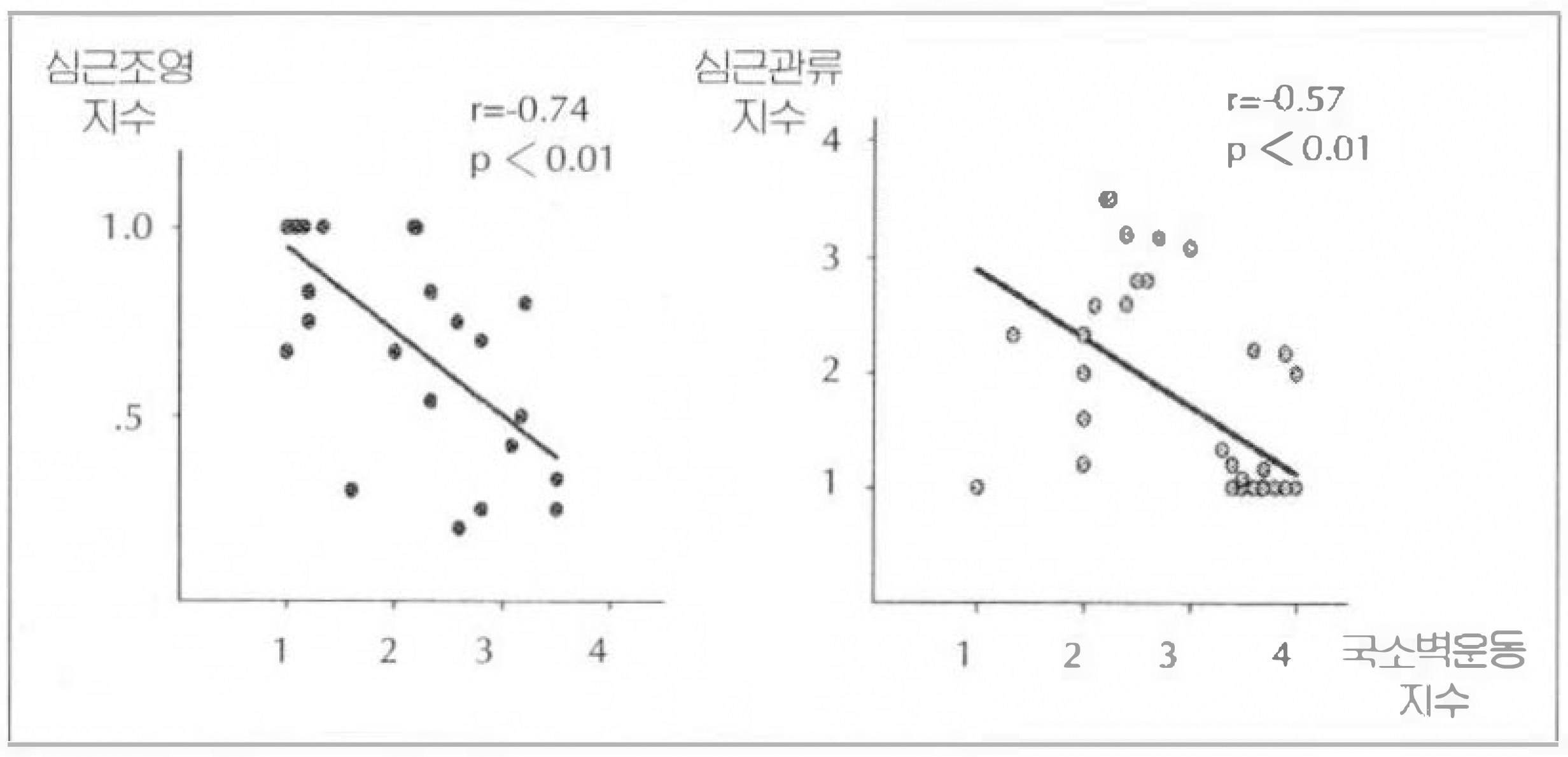
The total number of analyzed myocardial segments was 289. The correlation of myocardial opacification indexes and MIBI perfusion indexes was r=0.63(p < 0.01). The correlation between MCE index and MIBI index was higher in left anterior descending artery territory(r=0.73, p < 0.01) than left circumplex artery territory(r=0.49, p=0.06) and right coronary artery territory(r=0.65, p < 0.01). The correlations between MCE index and MIBI index was not influenced by infarction site(r=0.66, p < 0.01 in anterior wall infarction, r=0.60, p < 0.01 in inferior wall infarction). MCE had a better correlation with the degree of regional wall motion abnormality than MIBI myocardial scintigraphy.
References
1). Kloner RA, Ganote CE, Jennings RB. The “No-Reflow” phenomenon after temporary coronary occlusion in the dog. J Clin Invest. 54:1496. 1974.

2). Kloner RA, Ellis SG, Lamge R, Braunwald E. Studies of experimental coronary artery reperfusion: Effects on infarct size, myocardial function, biochemistry, ultrastructure and microvascular damage. Circulation. 68(Suppl I):I–8. 1983.
3). Gruppo Italiano per lo Studio Della Streptochinasci Nell Infarcto Miocardico(GISSI). Effectiveness of intravenous thrombolytic treatment in acute myocardial infarction. Lancet. 1:397. 1986.
4). Touchstone DA, Beller GA, Nygaard TW, Tedesco C, Kaul S. Effects of successful intravenous reperfusion therapy on regional myocardial function and geometry in humans: A tomographic assessment using two-dimensional echocardiography. J Am Coll Cardiol. 13:1506. 1989.
5). Belenkie I, Knudtson ML, Roth DL, Hansen JL, Traboulsi M, Hall CA, Manyari D, Filipchuck NG, Schnurr LP, Rosenal TW, Smith ER. Relation between flow grade after thrombolytic therapy and the effect of angioplasty on left ventricular function: A prospective randomized trial. Am Heart J. 121:407. 1991.
6). Ragost M, Camarano G, Kaul S, Powers ER, Sarembock IJ, Gimple LW. Microvascular integrityindicates myocellular viability in patients with recent myocardial infarction. Circulation. 89:2562. 1994.
7). Dilsizian V, Bnow RO. Current diagnostic techniques of assessing myocardial viability in patients with hibernating and stunned myocardium. Circulation. 87:1. 1993.
8). Tamaki N, Kawamoto M, Tadamura E, Magata Y, Tonekura Y, Mohara R, Sasayama S, Nichimura K, Ban T. Prediction of reversible ischemia after revascularization: Perfusion and metabolic studies with positron emission tomography. Circulation. 91:1697. 1995.
9). Brunken RC, Mody FV, Hawkins RA, Nienaber C, Phelps M, Schelbert HR. Positron emission tomography delects metabolic viability in myocardium with persistent 24-hour single-photon emission computed tomography 201TI defects. Circulation. 86:1357. 1992.
10). Dilsizian V, Arrighi JA, Diodati JG, Quyyumi AA, Alavi K, Bacharah SL, Marin-Neto JA, Katsiyiannis PT, Bonow RO. Myocardial viability in patients with chronic coroanry artery disease. Circulation. 89:578. 1994.
11). Moore CA, Smucker ML, Kaul SF. Myocardial contrast echocardiography in humans: 1. safety-a comparison with routine coronary arteriography. J Am Coll Cardiol. 8:1066. 1986.
12). Grill HP, Brinker JA, Taube JC, Walford GD, Midel MG, Flaherty JT, Weiss JL. Contrast echocardiographic mapping of collateralized myocardium in humans before and after coronary angioplasty. J Am Coll Cardiol. 16:1594. 1990.
13). Lim YJ, Nanto S, Masuyama T, Kodama K, Kohama A, Kitabatake A, Kamada T. Coronary collaterals assessed with myocardial contrast echocardiography in healed myocardial infarction. Am J Cardiol. 66:556. 1990.
14). Sabia PJ, Powers ER, Jayaweera AR, Rogosta M, Kaul S. Functional significance of collateral blood flow in patients with recent acute myocardial infarction: A study using myocardial contrast echocardiogrpahy. Circulation. 85:2080. 1992.
15). Villanueva FS, Glasheen WP, Sklenar J, Kaul S. Characterization of spatial patterns of flow within the reperfused myocardium by myocardial contrast echocardiography. Corculation. 88:2596. 1993.
16). Zwehl W, Areeda J, Schwartz G, Feinstein S, Ong K, Meerbaum S. Physical factors influencing quantitation of two-dimensional contrast echo amplitudes. J Am Coll Cardiol. 4:157. 1984.
17). Roval D, DeMaria AN, L'abbate A. Myocardial contrast echo effect: The dilemma of oroanry blood flow and voulme. J Am Coll Cardiol. 26:12. 1995.
18). Austin RE, Aldea GS, Coggins DL, Flynn AE, Hoffman JIE. Profound spatial heterogeneity of coronary reserve. Cir Res. 67:319. 1990.
19). Heyndricky GR, Amano J, Patrick TA, Manders WT, Rogers GG, Rosendorff C, Vatner SF. Effects of coronary artery reperfusion on regional myocardial blood flow and function in conscious baboons. Circulation. 71:1029. 1985.
20). Kloner RA, Rude RE, Carlson N, Maroko PR, De-Boer LWV, Braunwald E. Ultrastructural evidence of microvascular damage and myocardial cell injury after coroanry artery occlusion: which comes first? Circulation. 62:945. 1980.
21). Reisner SA, Ong LS, Lichtenberg GS, Shapiro JR, Amico AF, Allen MN, Meltzer RS. Quantitative assessment of the immediate results of coronary angioplasty by myocardial contrast echpcardiography. J Am Coll Cardol. 13:852. 1989.
22). Jr JR. Myocardial perfusion-contraction matching: Implications for coronary heart disease and hibernation. Circulation. 83:1076. 1991.
23). Ito H, Tomooka T, Sakai N, Yu H, Higashino Y, Fujii K, Masuyama T, Kitabatake A, Minamino T. Lack of myocardial perfusion immediately after successful thrombolysis. Circulation. 85:1699. 1992.
24). Porter TR, D'Sa A, Pesko L, Turner C, Nath A, Verovec GW, Nixon JV. Usefulness of myocardial contrast echocardiography in detecting the immediate changes in anterograde blood flow reserve after coronary angioplasty. Am J Cardiol. 71:893. 1993.
25). Schelbert HR. Different roads to the assessment of myocardial viability: Lessons from PT for SEPCT. Circulation. 1894:1995.
26). Elhendy A, Geleijnse ML, Roelandt JRTC, Domburg RT, Cornel JH, TenCate FJ, Postma-Tjoa J, Reijs AEM, El-Said GM, Fioretto PM. Evaluation by quantitative 99m-technetium MIBI SEPCT and echocardiography of myocardial perfusion and wall mition abnormalities in patients with dobutamine-induced ST-segment elevation. Am J Cardiol. 76:441. 1995.
 XML Download