

 PDF
PDF ePub
ePub Citation
 Print
Print


Abstract
Background
The diagnosis and estimation of severity of infundibular pulmonic stenosis (PS) is difficult by two-dimensional or M-mode echocardiography. In continuous wave(CW) Doppler signal valvular PS is seen as a parabolic velocity profile but infundibular PS is seen as a velocity signal with its peak in late, systole. The purpose of this study is that Doppler echocardiography can discriminate valvular PS from infundibular PS and assess the severity of infundibular obstruction.
Method
Six patients with valvular and infundibular PS by Doppler echocardiography and cardiac catheterization were studied. All had been undergone surgery. We compared their Doppler echocardiographic and cardiac catheterization finding with surgical finding.
Result
1) CW Doppler signal of infundibular PS had lesser peak velocity(2.84m/sec vs 4.33m/scc, p<0.05) and later systolic peak in velocity(AT/ET ratio: 0.73 vs 0.51, p<0.05) than that of valvular PS.
2) Pressure gradient across the infundibular obstruction measured by Doppler echocardiography correlated well with that by cardiac catheterization(r=0.89, p<0.05).
3) Operation finding showed infundibular PS in 5 case but not one and coexisting other anomaly such as ASD in 4 case, TOF in 1 case, PAPVR in 1 case.
References
1). Weyman AE. Principles and Practice of Echocardiography. 2nd Ed. p. 885. Philadelphia: Lea and Febiger;1994.
2). Wilde P. Cardiac ultrasound. p. 95. New York: Churchill Livingstone;1993.
3). Feigenbaum H. Echocardiography. 5th Ed. p. 361. Philadelphia: Lea and Febiger;1994.
4). Carlos OL, David JS, Lilliam MV, Stanley JG, Jesus VB, Hugh DA, Ehud G. Noninvasive prediction of transvalvular pressure gradient in patients with pulmonary stenosis by quantitative Two-dimensional echocardiographic Doppler studies. Circulation. 67:866–871. 1983.

5). Gregoly LJ, Kwan OL, Sharon H, Jacqueline AN, DeMaria AN. Accuracy of combined two-dimensional echocardiography and continuous wave Doppler recordings in the estimation of pressure gradient in right ventricular outlet obstruction. J Am Coll Cardiol. 3:1013–8. 1984.
6). Silove ED, Giovanni JV, Shiu MF, Myint MY. Diagnosis of right ventricular outflow obstruction in infants by cross sectional echocardiography. Br Heart J. 50:416–20. 1983.
7). Houston AB, Simpson IA, Sheldon CD, Doig WB, Coleman EN. Doppler ultrasound in the estimation of the severity of pulmonary infundibular stenosis in infants and children. Br Heart J. 55:381–4. 1986.
8). Peter M, Carla W, David R, Graham L, Ernest C, Aubrey L. Non-invasive diagnosis of subpulmonary outflow tract obstruction. Br Heart J. 43:276–283. 1980.
9). Weyman AE, Dillon JC, Feigenbaum H, Chang S. Echocardiographic differentiation of infundibular from valvular pulmonary stenosis. Am J Cardiol. 36:21–6. 1975.
10). Teiretein PS, Yock PG, Popp RL. The accuracy of Doppler ultrasound measurement of pressure gradients across irregular, dual, and tunnellike obstruction to blood flow. Circulation. 72:577–584. 1985.
11). Yasuda I. Pulmonary stenosis with intact ventricular septum; assessment and indication of reconstructive surgery for residual right-ventricular outflow tract obstruction. Thorac Cardiovasc Surg. 39:143–9. 1991.
Fig. 1.
Continuous wave Doppler signal of valvular and infundibular pulmonic stenosis. Left signal shows a parabolic flow velocity signal of valvular PS superimposed by infundibular PS in which a peak velocity occurs in late systole. Right signal shows only infundibular PS.
AT: Acceleration time, ET: Ejection time

Fig. 2.
Cardiac catheterization reveals infundibular pressure in same patient that Doppler finding was seen in Fig. 1.
PA: pulmonary artery, INF: infundibulum.
RV: right ventricle
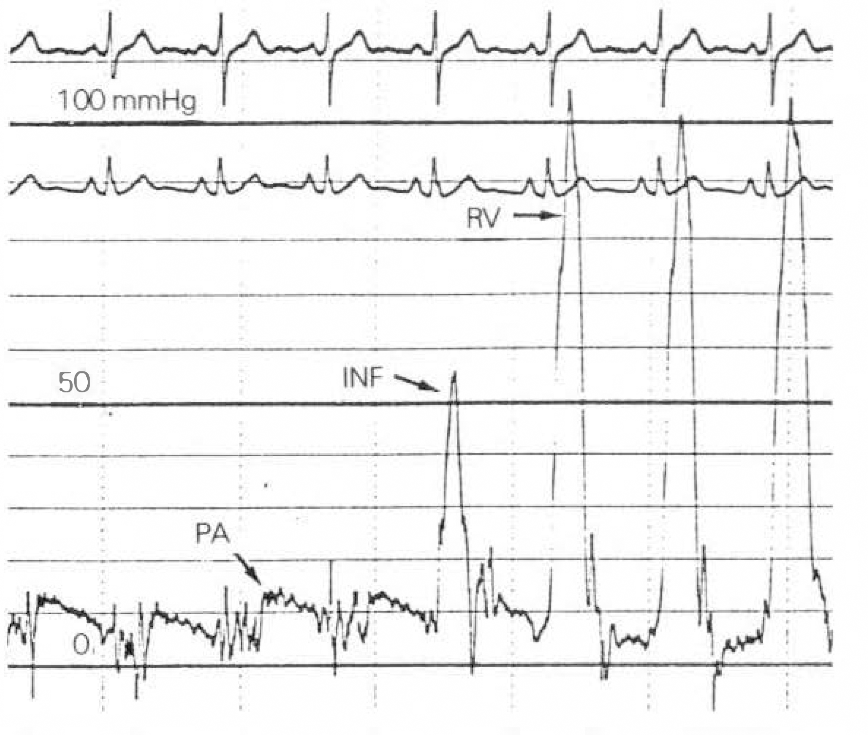
Fig. 3.
Comparison of infundibular pressure gradient by doppler echocardiography and catheterization.
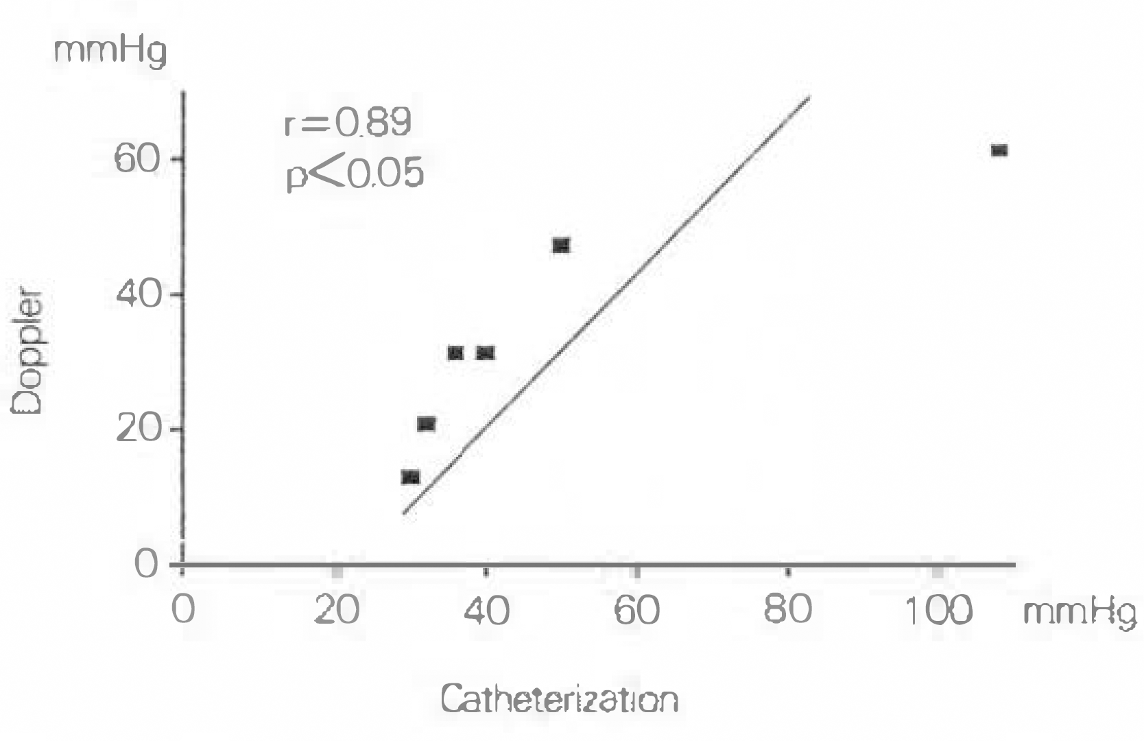
Table 1.
Comparison of doppler velocity spectrum in valvular and infundibular PS
| Valvular PS | Infundibular PS | |
|---|---|---|
| Peak velocity(m/sec) | 4.33 | 2.84∗ |
| AT/ET | 0.51 | 0.73∗ |
Table 2.
Doppler, catheterization and operation findings of study patients
 XML Download