Abstract
Background
Dynamic echoes in the left atrium, spontaneous echo contrast(SEC), represents a marker for thromboembolic risk in patients(pts) with mitral stenosis(MS). The aims of this study were to determine the factors associated with SEC in pts with MS and to observe the immediate effect of percutaneous mitral, valvuloplasty(PMV) on SEC.
Methods
Biplane transesophageal echocardiography(TEE) including Doppler measurement of left atrial appendage flow was performed before and immediately after PMV in 50 pts with MS[32 in normal sinus rhythm(NSR) and 18 in atrial fibrillation(AF)]. Hemodynamic data of left atrial pressure, transmitral pressure gradient, mitral valve area by Gorlin's method (MVA) and cardiac output(CO) by thermodilution method were obtained before and after successful PMV.
Results
Before PMV(MVA of 0.9 ± 0.3cm2), SEC was observed in 60% (30/50) of tight MS (13/32 in NSR, 17/18 in AF). The presence of AF(p = 0.001). increased left atrial dimension(p= 0.001), decreased appendage peak positive velocity(APPV, p = 0.03). decreased MVA(p = 0.01) and reduced CO(p = 0.001) were positive predictive factors for SEC: AF was the most powerful factor among them. In pts with NSR MVA(p=0.01) was the only factor for SEC before PMV. After successful PMV(MVA of 2.0 ± 0.4cm2) SEC was still observed in 6 pts(12%) with AF. AF(p=0.001). increased left atrial dimension(p=0.06) and decreased APPV(p = 0.001) were favorable factors for persistence of SEC after PMV. but hemodynamic indices were not associated with SEC after PMV. New development of mitral regurgitation after PMV was the only predictive factor for disappearance of SEC(p = 0.04). In pts with NSR PMV promptly normalized the APPV with disappearance of SEC.
Conclusion
In pts with tight MS. different factors may be associated with SEC according to the rhythm. PMV is an effective method to abolish SEC with hemodynamic improvement. Despite the similar MVA and hemodynamic indices, possible preventive effect of thromboembolism after PMV may be more prominent in pts with NSR compared to those with AF.
Go to : 
References
1). Daniel GD, Nellessen U, Schroder E, Nonnast-Daniel B, Bednarski P, Nikutta P, Lichtlen PR. Left atrial spontaneous echo contrast in mitral valve disease: an indicator for an increased thromboembolic risk. J Am Coll Cardiol. 11:1204–1211. 1988.

2). Castello R, Pearson AC, Labovitz AJ. Prevalence and clinical implications of atrial spontaneous contrast in patients undergoing transesophageal echocardiography. Am J Cardiol. 65:1149–1153. 1990.

3). Black IW, Hopkins AP, Lee LCL, Walsh WF, Jacobson BM. Left atrial spontaneous echo contrast: A clinical and echocardiographic analysis. J Am Coll Cardiol. 18:398–404. 1991.

4). Pozzoli M, Febo O, Torbicki A, Tramarin R, Calsamiglia G, Cobbeli F, Specchia G, Roelandt JRTC. Left atrial appendage dysfunction: A cause of thrombosis ? Evidence by transesophageal echocardiography-Doppler studies. J Am Soc Echocardiogr. 4:435–41. 1991.
5). Pollick C, Taylor D. Assessment of left atrial appendage function by transesophageal echocardiography: implications for the development of thrombus. Circulation. 84:223–231. 1991.

6). Garcia-Fernandez M, Torrecilla EG, Roman DS, Azevedo J, Bueno H, Moreno MM, Delcan JL. Left atrial appendage Doppler flow patterns: implications on thrombus formation. Am Heart J. 124:955–961. 1992.
7). 송재관· 박숭정 • 박성욱 • 김원호 • 두영철 • 김재 중 • 이종구. 중중의 숭모판협착증 환자에서의 화 심방이혈류; 정상인과의 비교및 경피적 풍선성형 술의 효과. 순환기. 23:549–560. 1993.
8). Park SJ, Kim JJ, Park SW, Song JK, Doo YC, Lee SJK. Immediate and one-year results of percutaneous mitral balloon valvuloplasty using Inoue and double-balloon techniques. Am J Cardiol. 71:938–943. 1993.

9). Acar J, Michel PL, Cormier B, Vahanian A, lung B. Features of patients with severe mitral stenosis with respect to atrial rhythm: atrial fibrillation in predominant and tight mitral stenosis. Acta Cardiologica. 47:115–124. 1992.
10). 김철호 · 이명묵 • 이영우. 숭모판협착중에서 경식 도초음파로 발견된 자발에코영상(Spontaneous Echo Contrast)의 의 미. 22:389–395. 1992.
11). Vigna C, de Rito V, Criconia GM, Russo A, Testa M, Fanelli R, Loperfido F. Left atrial thrombus and spontaneous echo-contrast in nonanticoagulated mitral stenosis: A transesophageal echocardiographic study. Chest. 103:348–352. 1993.
12). Iliceto S, Antonelli G, Sorino M, Biasco G, Rizzon P. Dynamic intracavitary left atrial echoes in mitral stenosis. Am J Cardiol. 55:603–606. 1985.

13). Beppu S, Nimura Y, Sakakibarra H, Nagata S, Park YD, Izumi S. Smoke-like echo in the left atrial cavity in mitral valve disease: its features and significance. J Am Coll Cardiol. 6:744–749. 1985.

Go to : 
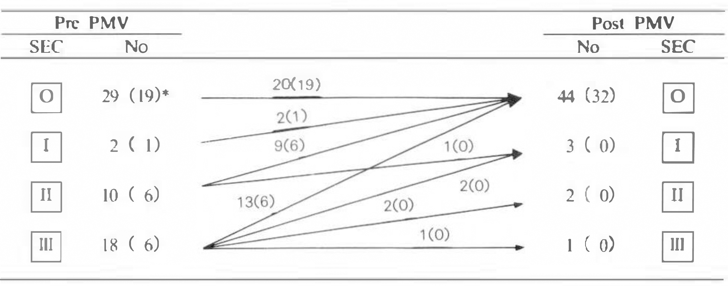
 | Fig. 1.Changes of spontaneous echo contrast(SEC) after percutaneous mitral valvuloplasty(PMV).
∗Number within parenthesis is the patients with sinus rhythm.
|
Table 1.
Factors correlated with spontaneous echo contrast(SEC) before percutaneous mitral valvuloplasty (PMV): Univariate analysis
| SEC(+) (N = 30) | SEC(–) (N = 20) | P-value | |
|---|---|---|---|
| 1. Rhythm | |||
| Atrial fibrllation | 17 | 1 | |
| Sinus rhythm | 13 | 19 | 0.001 |
| 2. Echo variables | |||
| Left atrial dimension(mm) | 50 ± 7 | 42 ± 11 | 0.001 |
| Appendage diameter(mm) | 22 ± 4 | 17 ± 8 | NS |
| Appendage area(mm2) | 56 ± 17 | 42 ± 24 | NS |
| APPV∗ (cm/sec) | 10 ± 8 | 25 ± 19 | 0.03 |
| 3. Hemodynamic variables | |||
| Left atrial pressure(mmHg) | 27 ± 10 | 24 ± 12 | NS |
| Mitral gradient(mmHg) | 18 ± 8 | 15 ± 7 | NS |
| Mitral valve area(cm2) | 0.8 ± 0.2 | 0.9 ± 0.4 | 0.01 |
| Cardiac output(L/min) | 3.4 ± 1.8 | 4.2 ± 2.1 | 0.001 |
Table 2.
Factors correlated with spontaneous echo contrast(SEC) in patients with sinus rhythm before PMV
| SEC(+) (N=13) | SEC(–) (N=19) | P-value | |
|---|---|---|---|
| 1. Echo variables | |||
| Left atrial dimension (mm) | 47 ± 6 | 41 ± 12 | NS |
| Appendage diameter(mm) | 20 ± 3 | 17 ± 9 | NS |
| Appendage area(mm2) | 52 ± 10 | 42 ± 25 | NS |
| APPV∗ (cm/sec) | 16 ± 7 | 26 ± 19 | NS |
| 2. Hemodynamic variables | |||
| Left atrial pressure(mmHg) | 32 ± 7 | 24 ± 12 | NS |
| Mitral gradient(mmHg) | 22 ± 6 | 15 ± 9 | NS |
| Mitral valve area(cm2) | 0.8 ± 0.2 | 0.9 ± 0.4 | 0.01 |
| Cardiac output(L/min) | 3.2 ± 2.1 | 4.2 ± 2.2 | NS |
Table 3.
Factors correlated with spontaneous echo contrast(SEC) after PMV
| SEC(+) (N = 6) | SEC(–) (N = 44) | P-value | |
|---|---|---|---|
| 1. Rhythm | |||
| Atrial fibrillation | 6 | 12 | |
| Sinus rhythm | 0 | 32 | 0.001 |
| 2. Echo variables | |||
| Left atrial dimension(mm) | 49 ± 8 | 41 ± 10 | NS |
| Appendage diameter(mm) | 22 ± 2 | 18 ± 6 | NS |
| Appendage area(mm2) | 53 ± 11 | 45 ± 22 | NS |
| APPV∗ (cm/sec) | 9 ± 7 | 34 ± 26 | 0.001 |
| 3. Hemodynamic variables | |||
| Left atrial pressure(mmHg) | 16 ± 3 | 14 ± 7 | NS |
| Mitral gradient(mmHg) | 6 ± 3 | 6 ± 3 | NS |
| Mitral valve area(cm2) | 1.9 ± 0.5 | 1.9 ± 0.5 | NS |
| Cardiac output(L/min) | 4.7 ± 1.5 | 4.2 ± 2.1 | NS |
| 4. Mitral regurgitation ≥ 1 | 0 | 20 | 0.04 |
Table 4.
Factors correlated with spontaneous echo contrast(SEC) in patients with atrial fibrillation after PMV
| SEC(+) (N = 6) | SEC(–) (N=12) | P-value | |
|---|---|---|---|
| 1. Echo variables | |||
| Left atrial dimension (mm) | 49 ± 8 | 49 ± 5 | NS |
| Appendage diameter(mm) | 22 ± 2 | 23 ± 7 | NS |
| Appendage area(mm2) | 53 ± 11 | 58 ± 30 | NS |
| APPV∗ (cm/sec) | 9 ± 7 | 6 ± 10 | NS |
| 2. Hemodynamic variables | |||
| Left atrial pressure(mmHg) | 16 ± 3 | 13 ± 8 | NS |
| Mitral gradient(mmHg) | 6 ± 3 | 5 ± 4 | NS |
| Mitral valve area(cm2) | 1.9 ± 0.5 | 1.8 ± 0.2 | NS |
| Cardiac output(L/min) | 4.7 ± 1.5 | 4.2 ± 2.1 | NS |
| 3. Mitral regurgitation ≥ 1 | 0 | 8 | 0.01 |


 PDF
PDF ePub
ePub Citation
 Print
Print


 XML Download