Abstract
Background
The evaluation of valvular regurgitation is a long standing clinical problem. While many ways to evaluate the severity of valvular insufficiency have been tried, none allows precise quantification of regurgitation volume. Invasive angiographic grading of mitral regurgitation is semiquantitative and subjective.
Recent studies have shown that using color Doppler flow mapping can identify a blue-red aliasing radius, corresponding to a proximal isovelocity surface area(PISA), proximal to a regurgitant orifice. Thus regurgitant volume across the mitral orifice can be calculated.
Methods
The clinical applicability of the PISA method was evaluated prospectively in 23 patients with mitral regurgitation and also the regurgitant volume calculated by using the time-velocity integral (TVI) method. The regurgitant jet area were compared to regurgitant volume calculated from the PISA method.
Results
1) Regurgitant volume calculated by using the PISA method correlated well with the regurgitant volume calculated by using the TVI method(r=0.73, P = 0.0001).
2) Regurgitant volume calculated by using the PISA method did not correlate with the regurgitant jet to left atrial ratio(r=–0.02, P = 0.94).
3) Eccentricity of regurgitant flow did not influence the result of PISA method.
Go to : 
References
1). Helmcke F, Nanda NC, Hsiung MC, Soto B, Adey CK, Goyal RG, Gatewood RP. Color Doppler assessment of mitral regurgitation with orthogonal planes. Circulation. 75:175–183. 1987.

2). Jones M, Hoit B, Eidbo E, et al. Variability of Doppler color flow mapping imaging of regurgitant jets in an animal model of mitral insufficiency. (Abstract) J Am Coll Cardiol. 9:64A. 1987.
3). Miyatake K, Izumi S, Okamoto M, Kinoshita N, Asonuma H, Nakagawa H, Yamamoto K, Takamiya M, Sakakibara H, Nimura Y. Semiquantitative grading of severity of mitral regurgitation by realtime two-dimensional Doppler flow imaging technique. J Am Coll Cardiol. 7:82–88. 1986.

4). Spain MG, Smith M, Harrison M, et al. Hemodynamic implications of jet size by color Doppler flow imaging in patients with mitral regurgitation. (Abstract) J Am Coll Cardiol. 9:67A. 1987.
5). Spain MG, Smith MD, Grayburn PA, Harlamert EA, DeMaria A, O'Brien M, Kwan OL. Quantitative assessment of mitral regurgitation by Doppler color flow imaging: Angiographic and hemodynamic correlations. J Am Coll Cardiol. 13:585–590. 1989.

6). Yoshida K, Yoshikava J, Yamaura Y, Hozumi T, Shakudo M, Akasaka T, Kato H. Value of acceleration flows and regurgitant jet direction by color Doppler flow mapping in the evaluation of mitral valve prolapse. Circulation. 81:879–885. 1990.

7). Yoshida K, Yoshikawa J, Yamamura Y, Hozumi T, Akasaka T, Iutaya T. Assessment of mitral regurgitation by biplane transesophageal color Doppler flow mapping circulation. 82:1121–26. 1990.
8). Jenni R, Ritter M, Eherli F, Grimn J, Krayenbuhl HP. Quantification of mitral regurgitation with amplitude-weighted mean velocity form continuous wave Doppler spertra. Circulation. 79:1294–1299. 1989.
9). Sahn DJ, Chung KJ, Tamura T, et al. Factors affecting jet visualization by color flow mapping Doppler echo: In vitro studies. (Abstract) Circulation. 74(II):11–271. 1986.
10). Sahn DJ, Simpson IA, Murillo A, Valdes-Cruz LM. Observations of acceleration proximal to restrictive orifices in congenital heart diseases: Important clues for the interpretation of Doppler color flow maps. Circulation. 78(Suppl II):11–649. 1988.
11). Sahn DJ. Instrumentation and physical factors related to visualization of stenotic and regurgitant jets by Doppler color flow mapping. J Am Coll Cardiol. 12:1354–1365. 1988.

12). Simpson LA, Sahn DJ. Hydrodynamic investigation of a hemodynamic problem: A review of the in vitro evaluation of mitral insufficiency by color Doppler flow mapping. J Am Soc Echocardiogr. 2:67–74. 1989.

13). Switzer DF, Yoganathan AP, Nanda NC, Woo Y-R, Ridgway AJ. Calibration of color Doppler flow mapping during extreme hemodynamic conditions in vitro: A foundation for a reliable quantitative grading system for aortic incompetence. Circulation. 75:837–846. 1987.

14). Chen C, Flachskampf FA, Rodriguez LL, Liu CM, Thomas JD. Impinging wall jets are much smaller than free jets: An in vitro color Doppler study (abstract). Circulation. 80(Suppl II):11–570. 1989.
15). Moises V, Shandas R, Maciel B, Beltran M, Recusani F, Sahn DJ. Consistency of color flow Doppler estimation of regurgitant flow rate across varying size orifices and multiple orifice communications using flow convergence concepts: Studies in an “invitro” model (abstract). J Am Coll Cardiol. 13:22A. 1989.
16). Bolger AF, Eigler NL, Maurer G. Quantifying valvular regurgitation: Limitations and inherent assumptions of Doppler techniques. Circulation. 78:1316–1318. 1988.

17). Bolger AF, Eigler NL, Pfaff JM, Resser KJ, Maurer G. Computer analysis of Doppler color flow mapping images for quantitative assessment of in vitro fluid jets. J Am Coll Cardiol. 12:450–457. 1988.

18). Simpson IA, Valdes-Cruz LM, Sahn DJ, Murillo A, Tamura T, Chung KJ. Doppler color flow mapping of simulated in vitro regurgitant jets: Evaluation of the effects of orifice size and hemodynamic variables. J Am Coll Cardiol. 13:1195–1207. 1989.

19). Bargiggia GS, Bertucci C, Raisaro A, Setta S, Angoli L, Recusani F, Gallati M, Tronconi L. Quantitative assessment of mitral regurgitation by color Doppler analysis of flow convergence region: Usefulness of continuity equation. Proceedings of the Sixth International Congress on Cardiac Doppler. 140. 1988.
20). Bargiggia GS, Recusani F, Yoganathan AP, Valdes-Cruz LM, Raisaro A, Simpson IA, Sung HW, Tronconi L, Sahn DJ. Color flow Doppler quantitation of regurgitant flow rate using the flow convergence region proximal to the orifice of a regurgitant jet (abstract). Circulation. 78(suppl II):11–609. 1988.
21). Bargiggia GS, Bertucci C, Raisaro A, et al. Quantitative assessment of mitral regurgitation by color Doppler analysis of flow convergence region: Usefulness of continuity equation. (Abstract) Proceedings of Sixth International Congress on Cardiac Doppler. 140:1988.
22). Bargiggia GS, Bertucci C, Raisaro A, Barba F, Gallati M, Recusani F, Tronconi L. A new method for the assessment of mitral regurgitation based on color doppler analysis of the flow convergence region (abstract). Eur Heart J. 10:405. 1989.
23). Bargiggia GS, Raisaro A, Bertucci C, Bramucci E, Recusani F, Montemartini C. Color Doppler analysis of proximal flow convergence region in patient with mitral regurgitation. Cardiovascular Imaging. 2:137–141. 1990.
24). Bargiggia GS, Tronconi L, Sahn DJ, Recusani F, Raisaro A, discrvi S, Valdes-Crux LM, Montemartini C. A new method for quantitation of mitral regurgitation based on color flow Doppler imaging of flow convergence proximal to regurgitant orifice. Circulation. 84:1481–89. 1991.

25). Gardin J. Doppler color flow “proximal isovelocity surface area(PISA)”: An alternative method for estimating volume flow across narrowed orifice regurgitant valves and intracardiac shunt lesion. Echocardiography. 9:39–42. 1992.
26). Giesler M, Scaugch M. Color Doppler determination of regurgitant flow: from proximal isovelocity surface areas to proximal velocity profiles. Echocardiography. 9:51–62. 1992.
27). Giesler M, et al. Color Doppler echocardiographic determination of mitral regurgitant flow from the proximal velocity profile of the flow convergence region. Am J Cardiol. 71:217–24. 1993.

28). Okamoto M, Morichika N, Nakagawa H, et al. Assessment of supra – valvular abnormal signal with color Doppler flow mapping in patients with aortic regurgitation. Am Heart J. 119:339. 1990.
29). Recusani F, Bargiggia GS, Yoganathan AP, Raisaro A, Valdes-Cruz LM, Sung HW, Bertucci C, Gallati M, Moises VA, Simpson IA, Tronconi L, Sahn DJ. A new method for quantification of regurgitant flow rate using color Doppler flow imaging of the flow convergence region proximal to a discrete orifice: An in vitro study. Circulation. 83:594–604. 1991.

30). Utsunomiya T, Ogawa T, Tang HA, Henry WL, Gardin JM. Doppler color flow mapping of the “proximal isovelocity surface area”: A new method for measuring volume blood flow across an orifice (abstract). J Am Coll Cardiol. 13:225A. 1989.
31). Utsunomiya T, Ogawa T, Patel D, Nguyen D, Gardin JM. Color flow Doppler “proximal isovelocity surface area” method for estimating volume flow. Independence from machine factors (abstract). Circulation. 80(Suppl II):11–577. 1989.
32). Utsunomiya T, Quan M, Doshi R, et al. Effect of flow rate, orifice size and aliasing calculation using Doppler color proximal isovelocity surface area method. (Abstract) J Am Coll Cardiol. 15:89A. 1990.
33). Utsunomiya T, Mehta K, Nguyen D, et al. Accuracy of flow rate calculation in funnel orifices by color Doppler zero-shift “proximal isovelocity surface area” method. (Abstract) J Am Coll Cardiol. 17:148A. 1991.

34). Utsunomiya T, Ogawa T, Patel D, et al. Doppler color flow mapping of the “proximal isovelocity surface area”. A new method for measuring volume flow across an orifice. J Am Soc Echocardiogr. 4:338–348. 1991.
35). Utsunomiya T, Ogawa T, Doshi R, et al. Doppler color flow “proximal isovelocity surface area” method for estimating volume flow rate. Effects of orifice shape and machine factors. J Am Coll Cardiol. 17:1103. 1991.

36). Rouse H. Fluid Mechanics for Hydraulic Engineers. New York: Dover Publications Inc.;p. 83–95. 1961.
37). Utsunomiya T, Doshi R, Patel D, Mehta K, Nguyen D, Henry WL, Gardin JM. Calculation of volume flow rate by the proximal isovelocity surface area method: Simplified approach using color Doppler zero baseline shift. J Am Coll Cardiol. 22:277–82. 1993.

38). Utsunomiya T, Doshi R, Patel D, Nguren D, Mehta K, Gardin J. Regurgitant volume estimation in patient with mitral regurgitation initial studies using the color Doppler “proximal isovelocity surface area” method. Echocardiography. 9:63–70. 1992.
39). Rokey R, Sterling LL, Zoghbi WA, et al. Determination of regurgitant fraction inisolated mitral or aortic regurgitation by pulsed Doppler two-dimensional echocardiography. J Am Coll Cardiol. 7:1273–1278. 1986.
40). 김삼수 • 김학중: Echocardiogγ'llm에 관한 연구. 대한내과학회지. 15:679. 1972.
41). 박옥규. 판막 질환의 비관혈적 평가. 순환기. 18–3(부록):10–16. 1986.
42). 배종화 · Wong M, Shah PM. 연가양 숭모판의 특징적 도플러 십초음파도 소견. 순환기. 233–241. 1986.
43). 김권삼 · 김명식 • 송정상 • 배종화. 한국인 정상 성인에서 연속따 Doppler 심초음파도로 측정한 판막 혈류방향과 최대속도 순환기. 17:95–101. 1987.
44). 배 종화 · Wong M, Vijayaraghavan G, Shah PM. Doppler 초음파에 의한 협착구 전후의 압력 및 혈류속도의 상관관계에 관한 실험적 연구, 순환기. 25:37–44. 1985.
45). 이문호 · 이기영 • 조정휘 • 김권삼 · 김병식 • 송 정상 • 배종화. 숭모판 일탈출증 에서 Doppler 섬초음파를 이용한 숭모판 폐쇄부정중의 빈도와 중동도 에 대한 관찰 대한 내과학회잡지. 35:336–342. 1987.
46). Sandler H, Dodge HT, Hay RE, Rackley CE. Quantitation of valvular insufficiency in man by angiocardiography. Am Heart J. 65:501–513. 1963.

47). Croft CH, Lipscomb K, Mathis K, Firth BG, Nicod P, Filton G, Winniford MD, Hillis LD. Limitation of quantitative angiographic grading in aortic and mitral regurgitation. Am J Cardiol. 53:1593–1598. 1984.
48). Chen C, Thomas JD, Anconina J, Harrigan P, Mueller L, Picard MH, Levine RA, Weyman AE. Impact of impinging wall jets on color Doppler quantification of mitral regurgitation. Circulation. 84:712–720. 1991.
49). Krabill KA, Sung H-W, Tamura T, Chung KJ, Yoganathan AP, Sahn DJ. Factors influencing the structure and shape of stenotic and regurgitant jets: An in vitro investigation using Doppler color flow mapping and optical flow visualization. J Am Coll Cardiol. 13:1672–1681. 1989.

50). Maciel BC, Moises VA, Shandas R, Simpson IA, Beltran M, Valdes-Cruz LM, Sahn DT. Effects of pressure and volume of the receiving chamber on the spatial distribution of regurgitant jet as imaged by color Doppler flow mapping. Circulation. 83:605–613. 1991.
51). Enriquez-sarano M, Jajik AJ, Bailey KR, Seward JB. Color folw imaging compared with quantitative Doppler assessment of seventy of mitral regurgitation: Influence of eccentricity of jet and mechanism of regurgitation. J Am Coll Cardiol. 21:1211–1219. 1993.
Go to : 
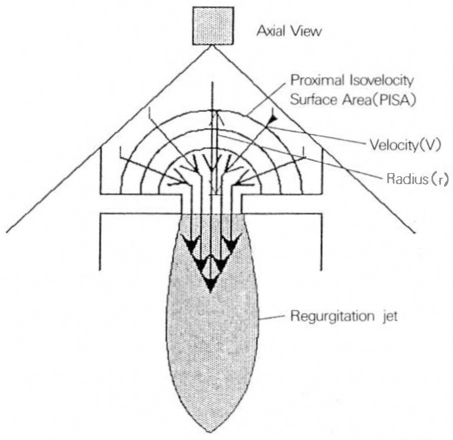
 | Fig. 1.PISA의 원리: 좁은 줄구로 황하는 혈류는 점차빠른 속도로 집종되어 반구형태의 돌속면올이룬다. 이 동속면상으| flow rate(PISA × velocity)는 좁은 줄구를 통한 flow rate(Regurgitantfow)와 같다 |
 | Fig. 2.Color Doppler systolic image in a patient with severe mitral regurgitation. Proximal isovelocity surface area (arrow) is clearly visible proximal to the mitral valve orifice. |
 | Fig. 3-1.Illustration of quantitative Doppler measurements by time-velocty interal method.
Pannel A: Parasternal long-axis view: measurement of the length at the left ventricular outflow tract (LVOT), Pannel B: Apical long-axis view: position of the sample volume(closed box) at LVOT, Pannel C: Apical four-chamber view: measurement of the length at the mitral annulus, Pannel D: Apical four-chamber view: position of the sample volume(closed box) at the mid portion of mitral annulus.
|
 | Fig. 3-2.Continued Figure 3-1.
Pannel A: Pulsed-wave Doppler image at the LVOT Pannel B: Pulsed-wave Doppler image at the mitral annulus.
|
Table 1.
Clinical characteristics of the patients
Table 2.
Results of time–velocity integral(TVI) method
| Variable | Mitral | Aortic |
|---|---|---|
| Annular diameter(cm) | 3.4 ± 0.6 | 2.1 ± 0.3 |
| TVI(cm) | 18.9 ± 9.2 | 19.6 ± 6.0 |
| Flow volume(ml) | 161.4 ± 43.6 | 69.7 ± 26.2 |
Table 3.
Results of maximal regurgitant jet to left atrial ratio
Table 4.
Results of proximal isovelocity surface area method
| Variable | Data |
|---|---|
| Maximal radius(cm) | 0.9 ± 0.2 |
| Velocity(cm/sec) | 50.5 ± 7.9 |
| MRTVI(cm) | 141.1 ± 30.0 |
| MRPFV(m/sec) | 4.6 ± 0.4 |
| MR volume(ml) | 101.6 ± 39.0 |
Table 5.
Comparison of Regurgitant volumes measured by PISA method and TVI method
Table 6.
Correlation between the regurgitant jet to left atrium ratio and the regurgitant volume measured by PISA method and TVI method
| TVI method | Jet dimension | Jet area | ||
|---|---|---|---|---|
| r | p-value | r | p-value | |
| Total | –0.09 | 0.67 | –0.10 | 0.65 |
| Sinus | 0.17 | 0.57 | 0.20 | 0.49 |
| AF | –0.61 | 0.08 | –0.66 | 0.05 |
| Central | 0.26 | 0.36 | 0.07 | 0.82 |
| Eccentric | 0.49 | 0.18 | –0.27 | 0.48 |


 PDF
PDF ePub
ePub Citation
 Print
Print


 XML Download