Abstract
Background
The pattern of left ventricular filling as depicted by Doppler echocardiographic transmitral flow velocities has been used to left ventricular diastolic properties and altered transmitral and aortic flow by abnormal myocardial wall motion and left ventricular function in ischemic heart disease was predicted during exercise stress echocardiography.
Methods
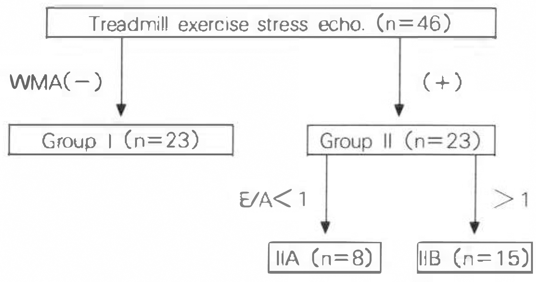
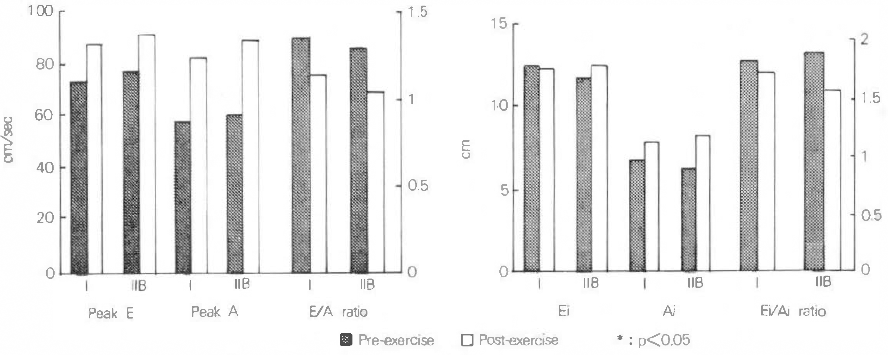
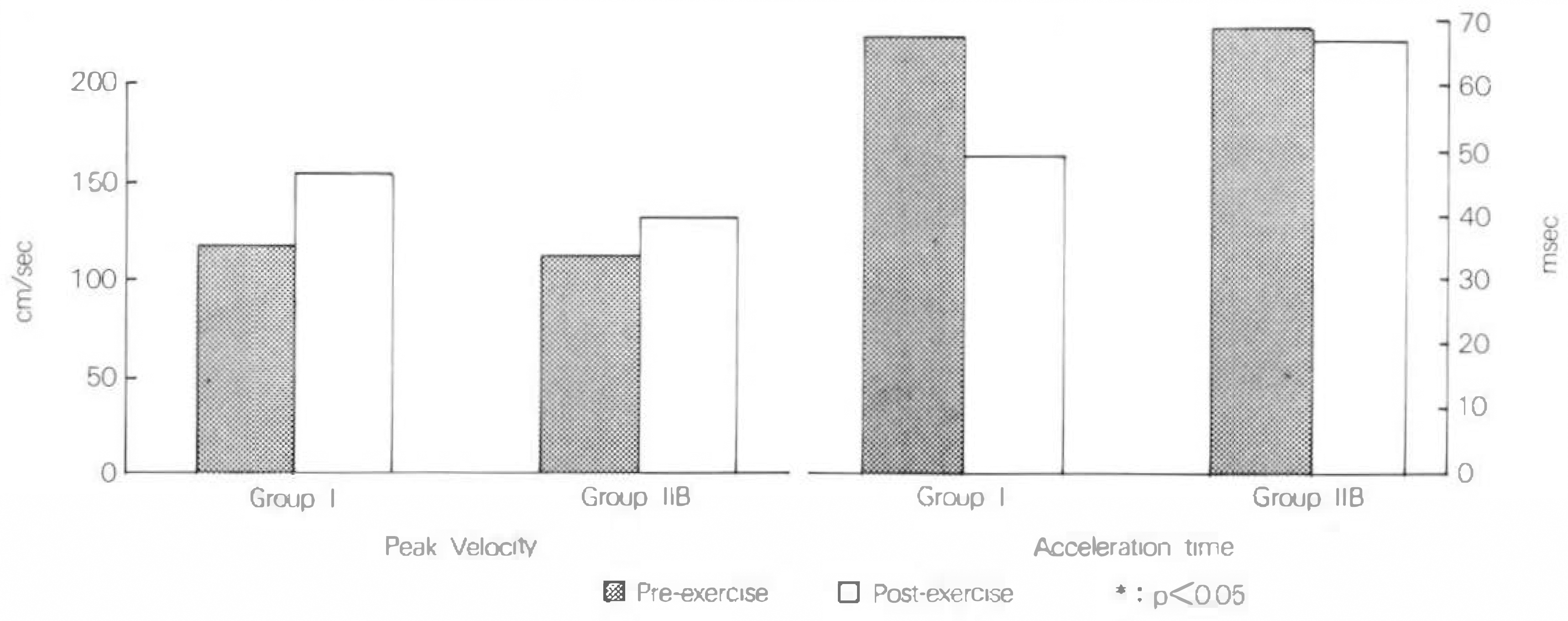
To determine the effects of altered wall motion on Doppler indexes of mitral and aortic flow, we studied 46 angina pectoris patients and divided into 3 groups(Group I: No wall motion abnormality before and after exercise. Group IIA: Wall motion abnormality after exercise and E/A ratio<1 at rest Group IIB: Wall motion abnormality after exercise and E/A ratio>1 at rest). Transmitral flow measurements comprised peak and integrated early passive(E) and late atrial(A) filling velocities and ratio. Aortic flow measurements comprised peak velocity and acceleration time.
Results
Heart rate in group I was 63.1/min at rest and 88.6/min at Doppler echocardiography after exercise. Group IIB was 63.6/min. 83.9/min, respectively. In group IIB E/A ratio was significantly decreased after exercise(p<0.001) and acceleration time of aortic flow was not significantly decreased.
References
1). 박노훈 · 김신애 · 김기업 · 신숭호 • 김순깅 · 서세 동· 김성구· 권영주;협심중 환자에서 운동부하 도플러 심초음파륜 이용한 승모판 혈류변화에 관한 연구. 순환기학회지. 22:580. 1992.
2). Agati L, Arata L, Luongo R, lacoboni C, Renzi M. Assessment of severity of coronary narrowings by quantitative exercise echocardiography and comparison with quantitative arteriography. Am J Cardiol. 67:1201. 1991.

3). Crawford MH, Amon KW, Vance WS. Exercise 2-dimensional echocardiography: Quantitation of left ventricular performance in patients with severe angina pectoris. Am J Cardiol. 51:1. 1983.
4). Miki S, Murakami T, Iwase T, Tomita T, Suzuki Y. Dependence of Doppler echocardiographic transmitral early peak velocity on left ventricular systolic function in coronary artery disease. Am J Cardiol. 67:470. 1991.

5). Morganroth J, Chen CC, David D, Sawin HS, Naito M. Exercise cross-sectional echocardiographic diagnosis of coronary artery disease. Am J Cardiol. 47:20. 1981.

6). Crouse LJ, Harbrecht JJ, Vacek JL, Rosamond TL, Kramer PH. Exercise echocardiography as a screening test for coronary artery disease and correlation with coronary arteriography. Am J Cardiol. 67:1213. 1991.

7). Chen C, Rodriguez L, Guerrero JL, Marshall S, Levine RA. Noninvasive estimation of the instantaneous first derivative of left ventricular pressure using continuous-wave Doppler echocardiography. Circulation. 83:2101. 1991.

8). Cuocolo A, Sax FL, Brush JE, Maron BJ, Bacharach SL. Left ventricular hypertrophy and impaired diastolic filling in essential hypertension: diastolic mechanisms for systolic dysfunction during exercise. Circulation. 81:978. 1990.

9). Crouse LJ, Harbrecht JJ, Vacek JL, Rosamond TL, Kramer PH. Exercise echocardiography as a screening test for coronary artery disease and correlation with coronary arteriography. Am J Cardiol. 67:1213. 1991.

10). Ishida Y, Meisner JS, Tsujioka K, Gallo JI, Yoran C. Left ventricular filling dynamics: influence of left ventricular relaxation and left atrial pressure. Circulation. 74:187. 1986.

11). Appleton CP, Hatle LK. The natural history of left ventricular filling abnormalities: Assessment by two-dimensional and Doppler echocardiography. Echocardiography. 9:437. 1992.

12). Danford DA, Huhta JC, Murphy DJ. Doppler echocardiographic approaches to ventricular diastolic function. Echocardiographiy. 3:33. 1986.

13). Xie GY, Berk MR, Smith MD, Gurley JC, DeMaria AN. Prognostic value of Doppler transmitral flow patterns in patients with congestive heart failure. J Am Coll Cardiol. 24:132. 1994.

14). Nishimura RA, Tajik AJ. Quantitative hemodynamics by Doppler echocardiography: A noninvasive alternative to cardiac catheterization. Pregress in Cardiovascular Diseases. 36:309. 1994.

15). Grodecki PV, Klein AL. Pitfalls in the echo-Doppler assessment of diastolic dysfunction. Echocardiography. 10:213. 1993.

16). Gardin JM, Dabestani A, Takenaka K, Rohan MK, Knoll M. Effect of imaging view and sample volume location on evaluation of mitral flow velocity by pulsed Doppler echocardiography. Am J Cardiol. 57:1335. 1986.

17). Herzog CA, et al. Effect of atrial pacing on left ventricular diastolic filling measured by pulsed Doppler echocardiography. J Am Coll Cardiol. 9(Suppl. A):197A. 1987.
18). Harrison MR, Clifton GD, Pennell AT, DeMaria AN, Cater A. Effect of heart rate on left ventricular diastolic transmitral flow velocity patterns assessed by Doppler echocardiography in normal subjects. Am J Cardiol. 67:622. 1991.

19). Downes TR, Nomeir AM, Smith KM, Stewart KP, Little WC. Mechanism of altered pattern of left ventricular filling with aging in subjects without cardiac disease. Am J Cardiol. 64:523. 1989.

20). Arrighi JA, Dilsizian V, Perrone-Filardi P, Diodati JG, Bacharach SL, Bonow RO. Improvement of the age-related impairment in left ventricular diastolic filling with verapamil in the normal human heart. Circulation. 90:213. 1994.

21). Stoddard MF, Pearson AC, Kern MJ, Ratcliff J, Mrosek DG. Influence of alteration in preload on the pattern of left ventricular diastolic filling as assessed by Doppler echocardiography in humans. Circulation. 79:1226. 1989.

22). Courtois M, Vered Z, Barzilai B, Ricciotti NA, Prez JE. The transmitral pressure-flow velocity relation: Effect of abrupt preload recution. Circulation. 78:1459. 1988.
23). Gardin JM, Kozlowski J, Dabestani A, Murphy M, Kusnick C, Allfie A, Russell D, Henry WL. Studies of Doppler aortic flow velocity during supine bicycle exercise. Am J Cardiol. 57:327. 1986.

24). Harrison MR, Clifton GD, Sublett KL, DeMaria AN. Effect of heart rate on Doppler indexes of systolic function in humans.
Fig. 1.
Classification of subjects by treadmill exercise echocardiography.
WMA: wall motion abnormality

Table 1.
Doppler data of group l(n = 23)
Table 2.
Doppler data of group IIA(n = 8)
Table 3.
Doppler data of group IIB(n = 15)


 PDF
PDF ePub
ePub Citation
 Print
Print


 XML Download