

 PDF
PDF ePub
ePub Citation
 Print
Print


INTRODUCTION
The success of aneurysm surgeries lie in the complete clipping of the aneurysm neck and in the preservation of branching and perforating arteries. Injury to perforating arteries has always been one of the major causes of postoperative morbidity in aneurysm clipping. This is of serious concern especially in specific locations such as the basilar bifurcation and anterior choroidal artery region. If possible, no neurosurgeon would like to include a perforator inside the clip to achieve satisfactory clipping even when only a small area of infarction can be seen on a postoperative follow-up computed tomography (CT) scan.
To avoid these complications, a variety of intraoperative technical advancements have been employed to immediately readjust an inappropriate and imperfect aneurysm clipping intraoperatively; intraoperative digital subtraction angiography (DSA),123 microvascular Doppler ultrasonography (MDU),4 electrophysiological monitoring,456 intraoperative indocyanine green angiography (ICGA) and neuroendoscopic techniques,7891011 have been used alone or in combination to provide sufficient intraoperative neuromonitoring during cerebrovascular surgery.
In the past, intraoperative DSA was considered the gold standard for detecting aneurysm neck remnants and unintended branch occlusions. However, this method is time consuming, expensive, difficult to access, and associated with not a low complication rate.12312 MDU is a safe, uncomplicated, and repeatable procedure with a low cost and low rate of morbidity. Even in deep surgical fields, MDU evaluation of vessels is possible and is not greatly influenced by other adverse occurrences such as micro-bleedings. However, MDU may be limited in the assessment of small perforating arteries and neck remnants.4 Intraoperative monitoring, including motor-evoked potential (MEP) and somatosensory-evoked potential (SSEP) monitoring, is an indirect method for the verification of safe clip placement. However, those two methods have some limitations in the identification of cerebral ischemia, especially outside the pyramidal tract.13 Gruber et al.14 reported a comprehensive prospective comparison and analysis of intraoperative monitoring and imaging modalities performed. They were able to accurately identify the specific advantages and limitations of various modalities used in neurovascular surgery. Although it is difficult, inconvenient, and time-consuming to use all these modalities on a routine basis, a neurovascular surgeon should be familiar with how to use them and be comfortable with the multimodal assessment of the intraoperative evaluation of aneurysm clipping outcomes. Moreover, they must be prepared to use all available adjuncts to optimize surgical results and minimize complications. This review is intended for neurosurgeons who are subspecializing in cerebrovascular surgery. The purpose of this study is to review practical and general surgical tips, preoperative planning, and minimization of complications in the microsurgical dissection and clipping of intracranial aneurysms (IAs).
LOCATIONS OF THE PERFORATING ARTERY OR PERFORATORS
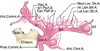
Most commonly, saccular, cerebral bifurcation aneurysms occur at the distal terminus or origin of the following arteries: basilar, internal carotid, main stem of the middle cerebral artery (MCA), and anterior communicating artery (AcomA) region (Fig. 1).15 With the introduction of new innovative devices and techniques, cerebral aneurysms are microscopically clipped more easily these days. However, identifying the origin of perforators and small branches in relation to the aneurysm neck still remains crucial for successful surgery. Pritz16 carefully reviewed their personal series of 142 saccular aneurysms that were treated microsurgically and in which preoperative imaging and intraoperative recordings were conducted. The authors found that perforators were present in 7% of basilar artery(BA) bifurcations, 17% of internal carotid artery (ICA) bifurcation aneurysms, 12% of middle cerebral aneurysms, and 11% of anterior communicating aneurysms.
Five different major locations of aneurysm with many perforators will be subsequently discussed. Table 1 presents the location, blood supply area, and clinical symptoms related to each perforator.
1. Perforators in basilar bifurcation aneurysm
Basilar apex aneurysms comprise 5% to 8% of all intracranial aneurysms.17 The direct surgical treatment of this kind of lesion remains a major challenge for the vascular neurosurgeon because of technical difficulties and the inherent risks of deep location. Many studies have reported the existence of these small braches. Tulleken and Luiten reported five cases of perforators arising from the basilar apex.18 However, no perforating branches arose from the tip of the basilar artery.19
Caruso et al.17 investigated the patterns of perforators in 50 fixed human cadaver brains. They found that perforators arose from the P1 segment in all specimens with no vertical perforators. Pritz16 also analyzed the relationship between perforators and secondary branch origin and the aneurysm neck in their 142 clinical series. They found a single perforator from the aneurysm neck in four cases, and P1 segment perforators in two cases. These branches may adhere closely to basilar bifurcation aneurysms. During the clipping process, it is common for contralateral P1 segment perforator branches to be included between the clip blades. All the perforators should be preserved regardless of their caliber, because it is impossible during neurosurgical procedures to judge their functional importance.
Angiograms should be used to identify the direction of the aneurysm fundus, the angle of bifurcation, and the relationship with the dorsum sellae for the appropriate surgical approach such as the pterional, subtemporal, and orbitozygomatic. Preoperative knowledge of some anatomical characteristics such as the height of the basilar apex and the angle between both P1 segments can help in deciding on a neurosurgical approach and may improve operative results in the management of basilar bifurcation aneurysm.
2. Perforators in internal carotid artery (ICA) bifurcation aneurysm
ICA bifurcation aneurysms represent 2% to 9% of all IAs.2021222324 ICA bifurcation aneurysms are located at the bifurcation of the ICA where it divides into the A1 segment of the anterior cerebral artery (ACA) and the M1 segment of the MCA. Anatomically, ICA bifurcation is usually the highest point of the Circle of Willis.
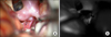
The exposure and clipping of ICA bifurcation aneurysms is highly demanding due to the following:25 (1) high position from the skull base, (2) adherence of the aneurysmal dome to the frontal lobe base, (3) numerous perforators in the base/or dome of the aneurysm, and (4) the high risk of intraoperative premature rupture during dissection. Consequently, meticulous dissection of surrounding perforators demands not only exceptional microsurgical skills, but also patience to work on the aneurysmal base for a sharp dissection. The orientation of the aneurysm dome, in particular, is very important for aneurysm clipping because the posterior direction is the most difficult to clip without perforator injury. They are heavily involved with the perforators, posing the greatest risk of perforator injury. The dome is partially obstructed by the ICA so that good visualization of the perforators, even for a small aneurysm dome, is particularly difficult. Furthermore, after clipping, an aneurysm remnant may be easily left unnoticed at the dead angle behind the ICA trunk. To overcome this surgical difficulty, Lehecka et al. have recommended that a tentative clip be inserted at first, avoiding the perforators, and then the dome be further dissected. Finally, the previous clip should be exchanged for a small-as- and light-as-possible final clip.25 The perforators must then be checked again by manipulating and shifting the clipped aneurysm (Fig. 2).
There are a large number of perforators behind the ICA bifurcation. These perforators originate from: (1) the choroidal segment of the ICA, (2) the anterior choroidal artery (AchA), (3) the recurrent artery of Hubner (RAH), (4) the medial lenticulostriate artery (MLA), and (5) the lateral lenticulostriate artery (LLA).262728293031 These small braches may be attached and/or stretched to varying degrees by ICA bifurcation aneurysms, or arise from the base of the aneurysm. Extreme care should be taken in every stage of dissection and clipping. Dashti et al. opined that several vascular anomalies of both the M1 and A1 segments should be considered before surgery.2627 It is most crucial to identify the point of origin and course of all the perforators, which may adhere to the posterior wall of the aneurysm, and to preserve them during the various stages of dissection, temporary, or final clipping, since most of them will be on the posterior portion of the aneurysm.
If the aneurysm is obstructing the surgical view of the perforators hidden behind the dome, it may be necessary to place the tentative clip at the aneurysm base at first to secure the space and then continue dissection for the perforators. A small cottonoid or cellulose material may be useful to protect and mobilize perforators during the final dissection at this stage. Occasionally, operators encounter situations where the aneurysm dome is partially or fully buried in the frontal base. The pia mater can then be opened around the aneurysm dome, and the subpial tissue can be resected to isolate the aneurysm.
3. Perforators in proximal middle cerebral artery (MCA) aneurysm
Proximal M1 aneurysms are located in the main trunk of the MCA, between the bifurcation of the ICA and the main bifurcation of M1.27 Proximal MCA aneurysms are microsurgically and hemodynamically challenging. Proximal MCA aneurysms are located deep and proximally in the sylvian cistern in a complex cisternal and vascular anatomy, where anatomical variations may affect the surgical outcome. These aneurysms are closely connected to one or more branching arteries of M1 and are often small and thin-walled, which make their precise clipping difficult. Proximal MCA aneurysms are uncommon. Rinne et al.32 reported that proximal M1 aneurysms represented 6% of all aneurysms and 16% of MCA aneurysms. Proximal MCA aneurysms are, in particular, often associated with other aneurysms in 72% in their series.32
Preservation of M1 branches is pertinent in the treatment for M1 aneurysms. Türe et al.31 classified M1 branches into: (1) the cortical branches (often named as temporopolar, frontotemporal, and orbitofrontal branches), and (2) the lateral lenticulostriate artery (LLA) branches. LLAs enter the brain via the central and lateral parts of the anterior perforating substance and supply the substantia innominata, putamen, globus pallidus, head and body of the caudate nucleus, internal capsule and adjacent corona radiata, and the central portion of the anterior commissure.31
During dissection and clipping of M1 aneurysms, the portion and pattern of exit of LLAs are of special concern because proximal M1 aneurysms may involve these perforators at their branching sites. In these cases, the LLAs may be stretched, distorted, and compressed by M1 aneurysmal clipping.
Proximal MCA aneurysms are often wide-necked and occasionally, are closely connected to a M1 branch at its origin on M1; these characteristics favor open microsurgery rather than endovascular surgery. The direction of the parent and branching arteries and the orientation of the aneurysm fundus are the most important factors affecting the efficacy and safety of clipping. Therefore, neurosurgeons should have a clear understanding of these three-dimensional (3D) relationships before surgery.
4. Perforators in proximal anterior cerebral artery (ACA) aneurysm
Proximal A1 aneurysms are located in the proximal segment (A1) of the ACA, between the bifurcation of the ICA and the junction of the A1 and A2 segments at the AcomA complex. The ICA bifurcates just below the anterior perforating substance into 2 terminal branches, the M1 and the A1. The A1 arises from the ICA in the carotid cistern and, with a medial and somewhat anterior course, enters the cistern of lamina terminalis. A group of thick arachnoid bands extending from the olfactory triangle to the lateral side of the optic nerve encase the A1 segment at this point. This is important to note when dissection and mobilization of the A1 segment is required.26 The course of the A1 segment varies considerably according to its length and dominance under the frontal lobe.33 Vascular anomalies are frequently seen with proximal A1 aneurysms. Anomalies of the A1 segment include hypoplasia, aplasia, duplication, fenestration, and infraoptic course.34
Proximal A1 aneurysms are rare, forming less than 1% of all IAs.3536373839 These aneurysms usually arise at the origin of perforators on the A1 segment and may be adherent to the perforators.3536373839 Microsurgical clipping is preferable to endovascular treatment due to its small size and involvement of perforating arteries. Sometimes, the Proximal A1 aneurysm is of a fusiform or dissecting nature, which makes aneurysmal clipping challenging or even impossible.4041424344 Many studies suggest that proximal A1 aneurysms, generally rupture at smaller sizes than other IAs in general.3536373839 Consequently, unruptured A1 segment aneurysms may require therapy even though they are relatively small.
The perforators of the A1 segment can be divided into two parts: (1) the MLA, and (2) RAH. The number of MLAs varies highly, with a mean of 8 (range, 2-15).3445 Most MLAs arise from the proximal half of the A1 trunk. MLAs are different from the LLAs that arise from the M1 segment.2627 MLAs supply the septum pellucidum, the medial part of the anterior commissure and pallidum, the pillars of the fornix, the paraolfactory area, the anterior limb of the internal capsule, the anterior-inferior part of the striatum, and the anterior hypothalamus.344546 Knowing the spectrum of neurologic symptoms that may occur if these perforators are injured is important.
In general, the RAH arises from the first few millimeters of the A2 segment or the A1-A2 junction, and in only about 10% of cases, it originates from the distal A1 segment.34454748 However, the point of origin of the RAH and its course in relation to the A1 trunk varies considerably. Therefore, an understanding of this variability is of primary importance when dissecting and placing temporary clips on the A1 segment during dissection. The RAH supplies the anterior inferior striatum, anterior limb of the internal capsule, olfactory region, anterior hypothalamus, frontobasal cortex, and subcortical white matter.34454647 The point of origin and the course of the RAH should be identified to prevent damage during further dissection toward AcomA complex and temporary clipping on the A1 segment.
Preservation of all perforators might be impossible in fusiform and dissecting aneurysms because a long segment of the A1 segment is involved with several perforators. In these cases, an encircling Sundt clip is used to clip the affected segment although there still is the probable risk of perforator injuries. Simple trapping involving segments without revascularization may cause cerebral infarction if the contralateral A1 is hypoplastic or aplastic.
5. Perforators in anterior choroidal artery (AchA) aneurysm
AchA aneurysms represent 2%-5% of all intracranial aneurysms, arising from the posterolateral wall of the supraclinoid ICA at the origin of the AchA.495051 The point of origint of the AchA is frequently incorporated in the base of the aneurysm and may be distorted or hidden by the aneurysm dome.5051 Occlusion of the AchA causes severe ischemia in the posterior limb of the internal capsule, resulting in clinical conditions such as hemiparesis, hemisensory loss, and homonymous hemianopsia.
The AchA is divided into two segments: (1) the cisternal segment, starting from the origin of the AchA and extending into the choroidal fissure, and (2) the choroidal segment, formed by several branches passing through the choroidal fissure to the choroid plexus of the temporal horn.5253
After its origin, the AchA has an initial lateral course inside the carotid cistern and then follows a posteromedial course to enter the crural cistern. Passing through the wing of the ambient cistern, it travels the lateral and inferior to the optic tract, crosses the optic tract first from the 0lateral to medial, and then from the medial to lateral to enter the choroidal fissure.525354
Branches of the AchA supply the uncus, the posteromedial amygdala, the anterior hippocampus and the dentate gyrus, the tail of caudate nucleus, the inferior aspect of the optic chiasm, posterior two-thirds of the optic tract, the optic radiation, medial segments of the globus pallidus, the genu and posterior limb of the internal capsule, the middle one-third of the cerebral peduncle, the substantia nigra, the upper part of the red nucleus, a portion of the subthalamus, the lateral thalamic nuclei, the lateral geniculate body, and the choroid plexus of the lateral ventricle.535556 The vascular territory of the AchA varies reciprocally with the supply from the posterior communicating artery, the ICA, and the MCA.3353 To identify and preserve the AchA during different stages of dissection or temporary clipping, the neurosurgeon should be aware of these possible anatomic variations.
THE ROLE OF ADJUVANT MONITORING DEVICES
In recent years, MDU, intraoperative DSA, electrophysiological monitoring, neuroendoscope, and ICGA have been used alone or in combination to provide sufficient intraoperative neuromonitoring during cerebrovascular surgery.14
The ‘ultimate, all-in-one’ diagnostic tool has not yet been designed, but it should be simple, rapid, reliable, inexpensive, radiation free, noninvasive, integrated into the surgical work flow, repeatedly applicable, yet sensitive and specific for detecting aneurysm remnants on the hidden side of the vessel, even in calcified aneurysms, as well as diagnosing hemodynamically relevant major vessel stenosis or perforating artery occlusions. Due to the various situations described in many studies where one tool failed but another tool was able to diagnose a problem after clipping, it is more likely that a combination of tools will be more useful than even the best single tool. Thus far, there is no tool superior to intraoperative DSA for identifying a neck remnant in an incompletely clipped aneurysm. No tool is faster than MDU for checking whether a major arterial branch is occluded after a clip application. To make sure that the aneurysm sac is excluded from the circulation, nothing is more reliable than puncturing the sac. Inadvertent occlusion or kinking of perforating arteries may be best visualized using ICGA loop analysis, keeping in mind that the moment of ICGA inflow is the most diagnostically effective and that backflow may dye a vessel and prevent normal filling even after 1 to 2 seconds. Hidden proximal deep perforating arteries such as branches of the AchA or the posterior communicating artery are possibly best diagnosed by SSEP or MEP monitoring. Yet in some cases, neuroendoscopy can visualize compromised perforating or branching arteries or, as shown by the authors, an incompletely clipped aneurysm. However, for a correct interpretation of postclipping ICGA, it may be required to have information available about the normal flow delay of branching arteries caused by their different lengths until they come into the visual inspection field.
It is reasonable to assess the correct clip position by sequentially using the aforementioned methods. After clipping and surgical inspection, parent artery patency is rapidly confirmed sonographically and major cerebral perfusion impairment is ruled out. Aneurysm occlusion, perforator patency, and correct parent artery reconstruction are instantly evaluated by ICGA and intraoperative DSA, However, none of the monitoring tools used was shown to be absolutely reliable, and each device had distinct shortcomings when used as a stand-alone method without intraoperative correlation of the information generated.
In conclusion, we suggest that no one intraoperative monitoring or vascular imaging technique currently has the potential to replace all others, but the methods described are complementary in nature. Moreover, a high level of intraoperative safety can be provided when these techniques are used together.
1. Neuroendoscope
The neuroendoscope has been widely used in the field of neurosurgery, for both cerebrovascular surgery and tumor resection with various advantages. Particularly, it offers higher magnification, better observation, and additional illumination and, thus, provides information that may not be available in microscopic aneurysmal surgery. In general, the microscope only gives us a straight line of sight and results in inadequate observation of structures that may lie behind other structures.57 To overcome the above limitations, many neurosurgeons have recommended neuroendoscopy for aneurysm surgeries. Through this procedure, surgeons can detect hidden structures, dissect perforators at the back of aneurysm and check for complete clipping of aneurysms. Since Apuzzo et al. adopted the side-angled rigid endoscope for surgery,58 its usefulness has been reported in compensating for the shortcomings of the direct microscope.789596061626364 In summary, the advantages of the neuroendoscope are as follows: (1) increased light intensity while approaching an object, (2) clear depiction of details in close-up positions, and (3) extended viewing angles.6566 The position of the clip blades in relation to perforating branches in the vicinity of the aneurysm neck can be assessed endoscopically in selected cases.
The efficacy of endoscope-assisted aneurysmal clipping depends on the location of the aneurysm. The neuroendoscope is especially helpful in the identification of deep-seated aneurysms. However, several shortcomings of the neuroendoscope have been reported as follows:7 (1) the endoscope can cause aneurysmal rupture during entry or inspection, especially in cases of large or giant aneurysms, (2) the endoscope is helpless when there are blood clots in the operating field and clots have to be irrigated and removed, and (3) there is still a lack of sufficient equipment specially designed for endoscopic surgery.
2. Indocyanine green video angiography (ICGA)
Intraoperative ICG-VA has been widely used in aneurysm surgery to confirm the complete occlusion of the aneurysm and for the precise assessment of small perforating arteries in the vicinity of the aneurysm neck.456768697071 This method offers a real time assessment of the blood flow of vasculatures and reduces operative morbidity related to vascular occlusion after aneurysmal clipping, thus, increasing the number of positive outcomes for aneurysmal surgery.57 However, the major limitation of ICGA is that the microscope must have a direct line-of-sight to the region of interest, which might hamper the identification of pinched hidden arteries as well as remnants at the averted aneurysm neck.
Mielke et al.13 in their clinical series, applied a prototype endoscope for the visualization of ICG fluorescence in hidden regions of the microsurgical field and evaluated its potential usefulness compared with microscopic ICGA. They suggested that endoscopic ICGA was capable of emerging as a useful adjunct to aneurysm surgery and had the potential to further improve operative results. Yoshioka and Kinouchi57 found that endoscopic ICGA was especially useful in ICA-PcomA aneurysm cases for the observation of the blood flow at the origins of the perforators behind the parent arteries and aneurysms. Thus, combination endoscopy and microscopic guidance may reveal important information hidden from the microscope, and may be used to verify the status of the operative process under direct visualization.1357
3. Microvascular Doppler ultrasonography (MDU)
MDU extended the information derived from surgical exploration after aneurysm clipping and provided repeatable information about parent artery patency. Despite extensive effort to preserve patency at the time of the clip application, intraoperative 3D visual inspection of the entire vascular complex may not reveal arterial compromise or occlusion. An intraoperative MDU has been used for this purpose for the past few years.727374
The usefulness of MDU is for detecting the intravascular flow and patency, as well as the decrease or increase in CBF through the parent arteries for vascular compromise. For example, after clipping of the aneurysm with an atherosclerotic neck, the vessel contour itself looks normal and shows no compromise, microscopic ICGA can show the intravascular flow, and MEP/SSEP cannot detect a decrease in CBF. However, the patient can manifest ischemic symptoms. So, quantitative monitoring of flow patency is also important, and MDU can be useful in evaluating quantitative as well as qualitative intravascular flow. Intraoperative MDU has an advantage as it assists surgeons in making difficult intraoperative decisions, especially, when a compromise is necessary between complete obliteration of the aneurysm neck and maintenance of adequate parent and branching vessel patency. Therefore, MDS is a safe, feasible, and very reliable technique and should be used routinely in intracranial aneurysm surgery. The major limitation of this method is as follows: (1) inability to detect the persistence of a minimal blood flow after clipping, (2) aneurysm remnants or a residual neck may go unnoticed, and (3) Events after completing the IMD evaluation may potentially lead to a vessel stenosis and an ischemia.75
4. Electrophysiological monitoring
The many advances in motor system assessment achieved in the last two decades have undoubtedly improved monitoring efficacy without unduly compromising safety. Although the aforementioned intraoperative vascular imaging techniques are used to directly visualize the cerebral arteries of interest, electrophysiological neuromonitoring can be considered a method for permanent background surveillance. SSEP and MEP monitoring have provided functional information affecting the surgical strategy. This information is crucial and may change the surgical strategies employed. However, we still need to explore the usefulness and problems of intraoperative monitoring such as MEP and SSEP to evaluate brain dysfunction caused by an insufficiency of cerebral blood flow. Since it is difficult to predict the postoperative ischemic complications despite evaluating the motor function via MEP monitoring in some cases.76 Future studies and experience will likely clarify existing controversies and bring further advances.
CONCLUSIONS
To prevent kinking or occlusion of adjacent and/or perforating branches, the smallest possible while still sufficient final clip should be applied to prevent perforator injury. A proper selection of clips with different shapes and lengths of blades, and applicators, suiting each aneurysmal configuration, should be ready for use. Moreover, adjuvant monitoring devices and various types of dissectors for perforator preservation should be used appropriately.

 XML Download