Journal List > Chonnam Med J > v.53(1) > 1075005

Nguyen and Cho: Versatile Functions of Caveolin-1 in Aging-related Diseases

Abstract

Caveolin-1 (Cav-1) is a trans-membrane protein that is a major component of the caveolae structure on the plasma membrane. Cav-1 is involved in the regulation of various cellular processes, including cell growth, differentiation, endocytosis, and in particular it has been implied in cellular senescence. Here we review current knowledge about Cav-1 in cellular signaling and discuss the role of Cav-1 in aging-related diseases.

INTRODUCTION

Caveolae are structures with a diameter of 50-100 nm that have been identified by electron microscopy as flask-shaped invaginations in the membrane lipid rafts of various cell types and tissues, being especially abundant in endothelial cells and adipocytes.123 Although caveolae were discovered over 60 years ago, research on these structures has made slow progress until the discovery of caveolins, the structural proteins of caveolae.456 The identification of caveolins in caveolae structures has provided important insights into the differences between caveolae and lipid rafts.789 The caveolin family consists of 3 distinct proteins including caveolin-1 (Cav-1), caveolin-2 (Cav-2), and caveolin-3 (Cav-3).51011 Of these, Cav-1 and Cav-2 are highly co-expressed in non-muscle cells, whereas Cav-3 is mainly found in skeletal and cardiac muscle cells.101213 Among the caveolins, the structure and function of Cav-1 has been the most studied.1415 Cav-1 is well established as the major structural protein of caveolae, because these structures disappear from the lipid rafts of Cav-1-depleted cells.1617 Moreover, caveolae are composed of various molecules such as cholesterol, sphingolipids, cavins, and other trans-membrane proteins.101118
Various signaling molecules and different types of membrane receptors are enriched in caveolae and regulate their specific functions by binding with caveolins.19202122 Therefore, caveolae and structural caveolins, particularly Cav-1, have been implicated in various cellular events such as endocytosis, cellular signaling, membrane transport, and immunity, as well as in related diseases.518232425 However, much remains to be discovered about caveolae structure, formation, regulation, and function in different cell types and in pathology.52426 This review discusses caveolae and Cav-1 along with their functions, particularly in aging and aging-related diseases.

CAV-1/CAVEOLAE FUNCTIONS

1. Structure of Cav-1 and its function in caveolae formation

Cav-1 was the first protein identified in the caveolin family and is the most highly investigated among them.131415 Experimental results have indicated that Cav-1 is a trans-membrane protein, but it is different from other trans-membrane proteins in that both terminals are located in the cytoplasm.227 Normally, Cav-1 predominantly localizes on the plasma membrane as an oligomer, but it is also found in the Golgi apparatus in its monomer form.72829 The Cav-1 protein has two isoforms, Cav-1α and Cav-1β, which have different sizes due to differences in the translation initiation site of Cav-1 mRNA that results in Cav-1β lacking 32 amino acids at its N terminal. Therefore, Cav-1β, like Cav-1α, has one phosphorylation site at serine 80, which has been suggested as playing an important function in the regulation of the cholesterol tracking and secretory pathways. On the other hand, Cav-1α has an additional phosphorylation site at tyrosine 14, which has been proposed as having a vital role in the response to growth factors and other stimuli.18233031 In addition to these phosphorylation sites, the Cav-1 protein has three palmitoylation sites at cysteine 133, 143, and 156 that are required for oligomerization and cholesterol transport.732 The oligomerization domain is believed to be a central functional region of the Cav-1 protein because it is required for caveolae formation. Cav-1 is also known as a scaffolding protein that concentrates and functionally regulates various cellular signaling. Through the scaffolding domain, Cav-1 mainly interacts with different signaling molecules and regulates signaling cascade.183334 Furthermore, Cav-1 binds to cholesterol with high affinity, a crucial property for the stabilization of Cav-1 in caveolae.435 Taken together, based on the characteristics and properties of Cav-1, it is believed that this protein is not only a structural protein but is also involved in broad cellular physiological processes such as vesicular transport and regulation of signal transduction and differentiation as well as associated diseases.

2. Cav-1 in cellular signaling

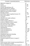
Cav-1 has been implicated as a specific regulator, which plays a role in signal transduction.1336 Using methods that isolate caveolae structures, many proteins and lipids associated with caveolae have been detected.4112037 As a consequence, many of these molecules are believed to be associated with the regulation of signal transduction by Cav-1 (Table 1). eNOS, a nitric oxide synthase targeted to the caveolae, is the best characterized Cav-1 binding partner, and the presence of Cav-1 inhibited eNOS signaling in both in vitro and in vivo experiments.1138 Moreover, Cav-1 has been proposed to attenuate the activity of other signal transduction proteins such as the epidermal growth factor (EGF) receptor (EGFR).4394041 In contrast, Cav-1 positively regulates several proteins such as the insulin and estrogen receptors.14203942 In most cases, signal proteins interact with the Cav-1 scaffolding domain via special binding motifs to activate signaling pathways.41134 However, in some cases, companion proteins that lack binding motifs still interact with Cav-1, such as a nerve growth factor receptor, a sterol carrier protein or a DNA-binding protein inhibitor.3443 Furthermore, Cav-1 is also involved in the regulation of cell fates such as cell proliferation, survival, and differentiation, leading to the development of associated diseases. For instance, down-regulation of Cav-1 leads to decreased E-cadherin expression but increased β-catenin transcription, subsequently enhancing the invasion of cancer cells.44 Along with this finding, the association of Cav-1 and metalloproteases also supports cancer invasion and metastasis.45 Moreover, Cav-1 also associates with different innate immune receptors such as Toll like receptors (TLRs), a class of proteins that play a key role in the innate immune systems.1946 Hence, Cav-1, the major protein of caveolae, is important in the regulation of cellular signaling.

3. Cav-1 gene regulation mechanisms

Cav-1 has various functions in the regulation of cells and tissues, and the mechanisms of Cav-1 gene regulation have been investigated. Initial data shows that both the levels of Cav-1 mRNA and protein are up-regulated during adipogenesis, and glucose transporter 4 (GLUT4) may play a role in this process, but the detailed mechanism is still unknown.47 Subsequently, Yamamoto investigated the function of the adenylyl cyclase pathway on Cav-1 gene expression and observed that Cav-1 mRNA levels decreased by activating this pathway.48 In addition to this finding, other data suggests that Myc, a transcription factor, and prolactin are involved in the negative regulation of Cav-1 mRNA.4950 Details about the underlying mechanisms have recently emerged, providing evidence that certain element and transcription factors are involved in Cav-1 gene regulation. For instance, sterol regulatory element-binding protein 1 inhibits Cav-1 gene transcription, resulting in the control of intracellular, free cholesterol transport.51 In addition to this finding, the transcription factor GATA-6, a GATA-binding factor 6, was shown to bind to the Cav-1 promoter and suppress gene expression.52 On the other hand, FOXO, the O subclass of the forkhead family of transcription factors, has been identified as a binding partner of the Cav-1 promoter and promotes Cav-1 gene expression.53 Moreover, epigenetic marks that control gene expression without modifying the DNA sequence have also been investigated as important factors that regulate Cav-1 expression.10 Epigenetic processes induce DNA promoter hypermethylation by adding methyl groups to cytosine residues by DNA methyltransferase on CpG dinucleotides that have a high density at promoter regions of the genome, named CpG islands, and this CpG island methylation inhibits the activation of transcriptional elements.54 To date, many studies have shown that epigenetics plays a crucial role in the methylation and histone modifications of Cav-1, and the majority of these reports have focused on the effects of epigenetics in various cancer types such as breast, prostate, lung, liver, ovarian, and colon cancer.1055

CAV-1 IN AGING-RELATED DISEASES

1. Aging

Cav-1 is well known as a gatekeeper of cellular senescence and is widely used to detect the aging phenotype.515253 Senescent human diploid fibroblasts (HDFs) show an increased level of Cav-1, and studies show that it colocalizes with EGFR.56 Cav-1 is also involved in senescence-associated morphological changes by modulation of focal adhesion kinase and actin stress fiber formation.5758 Moreover, overexpression of Cav-1 induces premature cellular senescence in primary cultures of murine fibroblasts.59 Another experiment with Cav-1 knockout (KO) mice showed that the presence of Cav-1 was important for maintaining good health.60 Cav-1 is also believed to be an important regulator of premature senescence of the lung fibroblasts of cigarette smokers.61 Interestingly, a reduction of the Cav-1 level in senescent HDF cells induced the restoration of the extracellular signal-regulated kinases (Erks) signaling pathway upon EGF stimulation, not only in terms of its phosphorylation but also its translocation into the nucleus and activation of transcriptional factors similar to young HDF cells.58 Reduction of Cav-1 in senescent cells can lead to morphological changes similar to young cells, causing them to become small and spindle-shaped.57 These results clearly point towards a central role of Cav-1 in suppressing mitogenic signaling and regulating morphological changes, leading to the promotion of cellular senescence.
Increased Cav-1 expression affects tissue aging by inducing tissue-specific dysfunctions and age-related diseases. Moreover, in the skin of the elderly, Cav-1 upregulation contributes to skin aging by suppressing collagen synthesis via the transforming growth factor-β pathway in dermal fibroblasts.62 In vascular aging, it is well known that Cav-1 is essential for vascular function and is involved in senescence phenotypes and inflammatory responses.63 Cav-1 has also been associated with various age-related diseases, such as cancer, type II diabetes, atherosclerosis, and infectious diseases, as discussed in the following sections.

2. Cancer

Older tissues are considered to be fertile ground for cancer development since a number of cells in those tissues accumulate mutations leading to an increased cancer rate in the elderly.64 Much evidence has emerged about the functions of Cav-1 in cancer.11 Cav-1 was initially hypothesized to be a tumor suppressor, since Cav-1 maps to a tumor suppressor region of the genome.1165 Moreover, experimental results showed that Cav-1 was deleted or down-regulated in many oncogene-transformed cells and that oncogene-transformed phenotypes could be reversed by the re-introduction of Cav-1 expression.11 However, Cav-1 does not only function as a tumor suppressor in cancer, since different explanations of Cav-1 expression levels have been proposed, and Cav-1 mutations are diverse depending on the cancer type.6667 For instance, Cav-1 expression levels are down-regulated in various cancers such as ovarian, lung, and mammary carcinomas, but are up-regulated in other cancers such as bladder, esophagus, and prostate carcinomas.65 Contradictory roles of Cav-1 in various cancers have been shown in animal models and human patients. Specifically, Cav-1 increases in metastatic breast and prostate cancers compared with that in primary tumors68 and promotes cell survival through Akt, a serine/threonine-specific protein kinsase, signaling and by regulating metastatic cancer microenvironments in patients with metastatic prostate cancers. Cav-1 is also secreted during prostate cancer development, indicating its potential as a diagnostic and therapeutic target.69
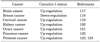
These results suggest the two faces of Cav-1, since it may be lost, promoting the growth of cancer cells in early stages, but enhances expression in late stages of cancer, provoking tumors to become aggressive and metastasize (Table 2).

3. Type II diabetes

The rate of type II diabetes is increased in the elderly, but the underlying mechanisms remain partly unknown.70 Recent evidence shows that Cav-1 may play a role as a mediator in insulin signaling and a defect of Cav-1 is involved in non-obese type II diabetes development. The first clues about the function of Cav-1 in type II diabetes came from experimental results in Cav-1 KO mice that showed these mice to be insulin resistant with attenuated insulin signaling.7172 Additionally, other studies have determined that the level of Cav-1 in skeletal muscle was associated with age-dependent type II diabetes.73 During aging, Cav-1 directly binds to insulin receptors and mediates the ensuing signaling pathways, leading to increased glucose absorption in the muscle. Cav-1 downregulation in old mice caused severe impairments of glucose and insulin tolerance, whereas Cav-1 upregulation in aged diabetic JYD mice, non-diabetic C57BL/6 , DBA/2 mice, develop insulin resistance and type 2 diabetes when they grow old, significantly improved insulin sensitivity, with a concomitant increase in glucose uptake by the skeletal muscle. In addition, sex hormones are implicated in the regulation of Cav-1 expression, which subsequently affects insulin resistance and type II diabetes in aged, male, diabetic JYD mice.74 Moreover, the mechanistic links between Cav-1 and type II diabetes have become better understood, and Cav-1 expression is associated with the RhoA/Rho kinase, a small GTPases protein of Ras family, pathway that controls diabetes.75 This research suggests various roles of Cav-1 in type II diabetes. In addition, increased Cav-1 expression in the smooth muscle upregulates the RhoA/Rho kinase pathway, leading to an increase in sustained muscle contractions in diabetic mice and patients with hyperglycemia. However, most studies on Cav-1 were limited to muscle tissues, and the roles Cav-1 in type II diabetes need to be further studied in liver, adipose tissues, and other related tissues.

4. Atherosclerosis

Atherosclerosis is a disease that is classified as a universal form of vascular aging in which lipids and cellular debris build up inside blood vessels, and Cav-1 has been shown to have an important function in the development of this disease.76 Cav-1 is abundantly expressed in endothelial cells as the main transporter of cholesterol and low-density lipoprotein (LDL) and is therefore involved in key events leading to atherosclerosis development.77 Some lipoproteins involved in lipid metabolism are also enriched and regulated in caveolae structures,78 and Cav-1 expression was sufficient to induce atherosclerosis in a mouse model.79 Furthermore, Cav-1 KO mice displayed selective loss of endothelial functions, leading to pulmonary and cardiac defects, atherosclerosis, and tissue ischemia. Analysis of lipoproteins in the plasma from Cav-1 KO mice displays a shift in their distribution towards an atherogenic profile characteristic of hypertriglyceridemia (elevated VLDL/chylomicrons), with intolerance in the clearance of oral fat load independent of lipoprotein and hepatic lipase activities.80 Moreover, it has been proposed that Cav-1 has two different functions in accelerating atherosclerosis. In endothelial cells, Cav-1 regulates low-density protein transcytosis in the intima as well as, it also involves in development of atherosclerosis by amplifying inflammation in macrophages, subsequently enhancing plaque formation.2881 Therefore, this data suggests that caveolae and caveolin-1 must plays an important role in the regulation of endothelial function and in the development of atherosclerosis.

5. Infectious diseases

The elderly population is more susceptible to infection by age-associated changes in physiology and anatomy as well as the weakening of the immune system.8283 Along with aging, epithelial cells experience changes in their signaling pathways and functions, subsequently affecting the ability of physical barriers such as the skin, bladder, and bronchial and digestive systems to prevent infection.83 Interestingly, caveolae-dependent endocytosis is elevated in senescent host cells, and this pathway may serve as a gateway toward increased infection in the elderly.84 Caveolae-dependent infection is divided into 2 different categories, infection by caveolae-dependent endocytosis and Cav-1-dependent actin rearrangement. The ability of caveolae, with their flask-shaped invagination structures, to exit the plasma membrane as enclosed vesicles first gave rise to the idea of an endocytotic function of these structures in 1975.85 Additionally, it has been established that caveolae have an advantage over other endocytotic pathways, since they ferry most cargo directly to the Golgi apparatus or endoplasmic reticulum without lysosome fusion.848687 Hence, the caveolae-mediated endocytosis pathway protects cargo from degradation by lysosome enzymes and maintains them in the cytoplasm. Therefore, many viruses and bacteria use this as a smart pathway to escape the lysosomal degradation in host cells and cause disease.84888990 On the other hand, Cav-1 is highly expressed and is important for enhancing Salmonella typhimurium (S. typhimurium) infection in M-like cells and senescent fibroblasts. More importantly, the mechanism of Cav-1-mediated invasion of S. typhimurium has been identified. Unlike other pathogens, Cav-1 is involved in the early phagocytotic processing of S. typhimurium into host cells through binding with SopE, a bacterial effector protein delivered into host cells via the type III secretion system. After injecting into host cells, SopE binds and activates cellular Rac1 protein, leading to cellular responses such as the rearrangement of actin fibers for facilitating infection. The expression of Cav-1 recruits and induces association of Rac1 and SopE in caveolae for invasion of S. typhimurium into host cells.8284 M cells are found in peyer's patch which is known as a main gate of S. typhimurium infection. Interestingly, during differentiation of M cells from epithelial precursor cells, Cav-1 remarkably increased and finally mediated the transcytosis of S. typhimurium. These results indicate that up-regulated Cav-1 in senescent host cells or M cells may explain the high susceptibility to microbial infections that occurs with age.

6. Immunosenescence

Recent evidence has been used to outline the function of Cav-1 in both adaptive and innate immunity. Cav-1 is believed to have an important function in the activation of lymphocytes since it induces T lymphocyte proliferation and cytokine production9192 and stimulates B lymphocytes to develop an immune response against thymus-independent antigens.93 Moreover, Cav-1 KO mice show defects in innate immunity, resulting in insignificant decrease in survival by infection or increased production of inflammatory cytokines.94 Along with this finding, several papers have reported an association between Cav-1 and Toll-like receptors (TLRs), receptors that recognize the conserved structures of microbes early in infection. The initial evidence was provided by Lei and Morrison,95 who showed that Cav-1 expression was altered in lipopolysaccharide (LPS)-activated murine macrophages, and they suggested that Cav-1 levels might be correlated with LPS signaling. More interestingly, a new function of Cav-1 in age-associated immunity (immunosenescence) has recently emerged, since there is evidence of a direct interaction between Cav-1 and TLR5.46 This interaction is important to maintain TLR5 expression and signaling during the aging process.46 Experimental results also support Cav-1 dependent TLR5 preservation in aging. Overexpressed Cav-1 in vitro enhances TLR5 mRNA through the mitogen-activated protein kinases (MAPKs) pathway and prolonged TLR5 protein half-life through direct interaction.96 KO mice research also demonstrate that TLR5/MyD88, an adaptor protein of TLRs, signaling is regulated by Cav-1 and induces the production of pro-inflammatory cytokines by stimulation of a TLR5 agonist flagellin.46 Moreover, it also proved that flagellin-conjugated antigens induce a significantly higher level of specific antibodies in response to the antigen and better protection against lethal challenges.46 It means that Cav-1/TLR5 signaling plays a key role in age-associated innate immune responses and that stimulation of Cav-1/TLR5 may be a good model of a mucosal vaccine adjuvant against infection via overcoming immunosenescence in the elderly.

CONCLUSIONS

Cav-1/caveolae have emerged as having vital functions in aging and aging-related diseases. The different density of Cav-1/caveolae in different cell types or tissues may associate with particular functions (Table 1). In aging, Cav-1 is up-regulated, causing cellular senescence with changing in cellular morphology, weakening the proliferation rate as well as cellular signaling. However, Cav-1 up-regulation also has a vital role in maintaining and stabilizing TLR5 against immunosenescence. These results significantly contribute to our understanding of innate immunity in aging and support the need to extend Cav-1 research in immunity. Moreover, Cav-1 expression level changes in various types of cancer, subsequently affecting cancer growth rates, are considered to be a valuable prognostic marker for cancer. Furthermore, the function of Cav-1/caveolae in other aging related-diseases such as type II diabetes and atherosclerosis is also well established and suggests that Cav-1/caveolae may serve as a therapeutic target for these diseases. Taken together, Cav-1/caveolae research in the aging field is important to more deeply understand the underlying mechanisms of Cav-1-dependent aging processes as well as efficiently develop therapies against aging-related diseases.

Figures and Tables

TABLE 1

Summary of caveolin-1 binding partners

cmj-53-28-i001
TABLE 2

Caveolin-1 status in various cancer

cmj-53-28-i002

Notes

CONFLICT OF INTEREST STATEMENT None declared.

References

1. Thomas CM, Smart EJ. Caveolae structure and function. J Cell Mol Med. 2008; 12:796–809.
crossref
2. Liu P, Rudick M, Anderson RG. Multiple functions of caveolin-1. J Biol Chem. 2002; 277:41295–41298.
crossref
3. Anderson RG. The caveolae membrane system. Annu Rev Biochem. 1998; 67:199–225.
crossref
4. Boscher C, Nabi IR. Caveolin-1: role in cell signaling. In : Jasmin JF, Frank PG, Lisanti MP, editors. Caveolins and Caveolae. New York: Springer;2012. p. 29–50.
5. Parton RG, Simons K. The multiple faces of caveolae. Nat Rev Mol Cell Biol. 2007; 8:185–194.
crossref
6. Schlegel A, Lisanti MP. A molecular dissection of caveolin-1 membrane attachment and oligomerization Two separate regions of the caveolin-1 C-terminal domain mediate membrane binding and oligomer/oligomer interactions in vivo. J Biol Chem. 2000; 275:21605–21617.
crossref
7. Razani B, Woodman SE, Lisanti MP. Caveolae: from cell biology to animal physiology. Pharmacol Rev. 2002; 54:431–467.
crossref
8. Sowa G, Pypaert M, Sessa WC. Distinction between signaling mechanisms in lipid rafts vs. caveolae. Proc Natl Acad Sci U S A. 2001; 98:14072–14077.
crossref
9. Patel HH, Murray F, Insel PA. Caveolae as organizers of pharmacologically relevant signal transduction molecules. Annu Rev Pharmacol Toxicol. 2008; 48:359–391.
crossref
10. Low JY, Nicholson HD. Epigenetic modifications of caveolae associated proteins in health and disease. BMC Genet. 2015; 16:71.
crossref
11. Quest AF, Gutierrez-Pajares JL, Torres VA. Caveolin-1: an ambiguous partner in cell signalling and cancer. J Cell Mol Med. 2008; 12:1130–1150.
crossref
12. van Helmond ZK, Miners JS, Bednall E, Chalmers KA, Zhang Y, Wilcock GK, et al. Caveolin-1 and -2 and their relationship to cerebral amyloid angiopathy in Alzheimer's disease. Neuropathol Appl Neurobiol. 2007; 33:317–327.
crossref
13. Williams TM, Lisanti MP. The caveolin proteins. Genome Biol. 2004; 5:214.
14. Yun JH, Park SJ, Jo A, Kang JL, Jou I, Park JS, et al. Caveolin-1 is involved in reactive oxygen species-induced SHP-2 activation in astrocytes. Exp Mol Med. 2011; 43:660–668.
crossref
15. Stahlhut M, van Deurs B. Identification of filamin as a novel ligand for caveolin-1: evidence for the organization of caveolin-1-associated membrane domains by the actin cytoskeleton. Mol Biol Cell. 2000; 11:325–337.
crossref
16. González-Muñoz E, López-Iglesias C, Calvo M, Palacín M, Zorzano A, Camps M. Caveolin-1 loss of function accelerates glucose transporter 4 and insulin receptor degradation in 3T3-L1 adipocytes. Endocrinology. 2009; 150:3493–3502.
crossref
17. Miyawaki-Shimizu K, Predescu D, Shimizu J, Broman M, Predescu S, Malik AB. siRNA-induced caveolin-1 knockdown in mice increases lung vascular permeability via the junctional pathway. Am J Physiol Lung Cell Mol Physiol. 2006; 290:L405–L413.
crossref
18. Parton RG, del Pozo MA. Caveolae as plasma membrane sensors, protectors and organizers. Nat Rev Mol Cell Biol. 2013; 14:98–112.
crossref
19. Wang XM, Kim HP, Nakahira K, Ryter SW, Choi AM. The heme oxygenase-1/carbon monoxide pathway suppresses TLR4 signaling by regulating the interaction of TLR4 with caveolin-1. J Immunol. 2009; 182:3809–3818.
crossref
20. Pike LJ. Growth factor receptors, lipid rafts and caveolae: an evolving story. Biochim Biophys Acta. 2005; 1746:260–273.
crossref
21. Gratton JP, Bernatchez P, Sessa WC. Caveolae and caveolins in the cardiovascular system. Circ Res. 2004; 94:1408–1417.
crossref
22. Smith RM, Harada S, Smith JA, Zhang S, Jarett L. Insulin-induced protein tyrosine phosphorylation cascade and signalling molecules are localized in a caveolin-enriched cell membrane domain. Cell Signal. 1998; 10:355–362.
crossref
23. Lajoie P, Nabi IR. Lipid rafts, caveolae, and their endocytosis. In : Kwang WJ, editor. International Review of Cell and Molecular Biology. AmsterdamL: Academic Press;2010. p. 135–163.
24. Bastiani M, Parton RG. Caveolae at a glance. J Cell Sci. 2010; 123:3831–3836.
crossref
25. Quest AF, Leyton L, Párraga M. Caveolins, caveolae, and lipid rafts in cellular transport, signaling, and disease. Biochem Cell Biol. 2004; 82:129–144.
crossref
26. Cheng JPX, Nichols BJ. Caveolae: one function or many? Trends Cell Biol. 2016; 26:177–189.
crossref
27. Levine TB, Levine AB. Metabolic Syndrome and Cardiovascular Disease. New York: John Wiley & Sons;2012.
28. Jasmin JF, Frank PG, Lisanti MP. Caveolins and caveolae: roles in signaling and disease mechanisms. New York: Springer;2012.
29. Lin YC, Lin CH, Kuo CY, Yang VC. ABCA1 modulates the oligomerization and Golgi exit of caveolin-1 during HDL-mediated cholesterol efflux in aortic endothelial cells. Biochem Biophys Res Commun. 2009; 382:189–195.
crossref
30. Boettcher JP, Kirchner M, Churin Y, Kaushansky A, Pompaiah M, Thorn H, et al. Tyrosine-phosphorylated caveolin-1 blocks bacterial uptake by inducing Vav2-RhoA-mediated cytoskeletal rearrangements. PLoS Biol. 2010; 8:DOI: 10.1371/journal.pbio.1000457.
crossref
31. Schlegel A, Arvan P, Lisanti MP. Caveolin-1 binding to endoplasmic reticulum membranes and entry into the regulated secretory pathway are regulated by serine phosphorylation. Protein sorting at the level of the endoplasmic reticulum. J Biol Chem. 2001; 276:4398–4408.
crossref
32. Qin H, Bollag WB. The caveolin-1 scaffolding domain peptide decreases phosphatidylglycerol levels and inhibits calcium-induced differentiation in mouse keratinocytes. PLoS One. 2013; 8:e80946.
crossref
33. Collins BM, Davis MJ, Hancock JF, Parton RG. Structure-based reassessment of the caveolin signaling model: do caveolae regulate signaling through caveolin-protein interactions? Dev Cell. 2012; 23:11–20.
crossref
34. Byrne DP, Dart C, Rigden DJ. Evaluating caveolin interactions: do proteins interact with the caveolin scaffolding domain through a widespread aromatic residue-rich motif? PLoS One. 2012; 7:e44879.
crossref
35. Le Lay S, Rodriguez M, Jessup W, Rentero C, Li Q, Cartland S, et al. Caveolin-1-mediated apolipoprotein A-I membrane binding sites are not required for cholesterol efflux. PLoS One. 2011; 6:e23353.
crossref
36. Joshi B, Bastiani M, Strugnell SS, Boscher C, Parton RG, Nabi IR. Phosphocaveolin-1 is a mechanotransducer that induces caveola biogenesis via Egr1 transcriptional regulation. J Cell Biol. 2012; 199:425–435.
crossref
37. Cameron PL, Ruffin JW, Bollag R, Rasmussen H, Cameron RS. Identification of caveolin and caveolin-related proteins in the brain. J Neurosci. 1997; 17:9520–9535.
crossref
38. Chen Z, Bakhshi FR, Shajahan AN, Sharma T, Mao M, Trane A, et al. Nitric oxide-dependent Src activation and resultant caveolin-1 phosphorylation promote eNOS/caveolin-1 binding and eNOS inhibition. Mol Biol Cell. 2012; 23:1388–1398.
crossref
39. Chen X, Whiting C, Borza C, Hu W, Mont S, Bulus N, et al. Integrin alpha1beta1 regulates epidermal growth factor receptor activation by controlling peroxisome proliferator-activated receptor gamma-dependent caveolin-1 expression. Mol Cell Biol. 2010; 30:3048–3058.
crossref
40. Lajoie P, Partridge EA, Guay G, Goetz JG, Pawling J, Lagana A, et al. Plasma membrane domain organization regulates EGFR signaling in tumor cells. J Cell Biol. 2007; 179:341–356.
crossref
41. Labrecque L, Nyalendo C, Langlois S, Durocher Y, Roghi C, Murphy G, et al. Src-mediated tyrosine phosphorylation of caveolin-1 induces its association with membrane type 1 matrix metalloproteinase. J Biol Chem. 2004; 279:52132–52140.
crossref
42. Schlegel A, Wang C, Katzenellenbogen BS, Pestell RG, Lisanti MP. Caveolin-1 potentiates estrogen receptor alpha (ERalpha) signaling. caveolin-1 drives ligand-independent nuclear translocation and activation of ERalpha. J Biol Chem. 1999; 274:33551–33556.
43. Zhang X, Ling MT, Wang Q, Lau CK, Leung SC, Lee TK, et al. Identification of a novel inhibitor of differentiation-1 (ID-1) binding partner, caveolin-1, and its role in epithelial-mesenchymal transition and resistance to apoptosis in prostate cancer cells. J Biol Chem. 2007; 282:33284–33294.
crossref
44. Lu Z, Ghosh S, Wang Z, Hunter T. Downregulation of caveolin-1 function by EGF leads to the loss of E-cadherin, increased transcriptional activity of beta-catenin, and enhanced tumor cell invasion. Cancer Cell. 2003; 4:499–515.
crossref
45. Williams TM, Lisanti MP. Caveolin-1 in oncogenic transformation, cancer, and metastasis. Am J Physiol Cell Physiol. 2005; 288:C494–C506.
crossref
46. Lim JS, Nguyen KC, Nguyen CT, Jang IS, Han JM, Fabian C, et al. Flagellin-dependent TLR5/caveolin-1 as a promising immune activator in immunosenescence. Aging Cell. 2015; 14:907–915.
crossref
47. Scherer PE, Lisanti MP, Baldini G, Sargiacomo M, Mastick CC, Lodish HF. Induction of caveolin during adipogenesis and association of GLUT4 with caveolin-rich vesicles. J Cell Biol. 1994; 127:1233–1243.
crossref
48. Yamamoto M, Okumura S, Oka N, Schwencke C, Ishikawa Y. Downregulation of caveolin expression by cAMP signal. Life Sci. 1999; 64:1349–1357.
crossref
49. Park WY, Cho KA, Park JS, Kim DI, Park SC. Attenuation of EGF signaling in senescent cells by caveolin. Ann N Y Acad Sci. 2001; 928:79–84.
crossref
50. Park DS, Razani B, Lasorella A, Schreiber-Agus N, Pestell RG, Iavarone A, et al. Evidence that Myc isoforms transcriptionally repress caveolin-1 gene expression via an INR-dependent mechanism. Biochemistry. 2001; 40:3354–3362.
crossref
51. Bist A, Fielding PE, Fielding CJ. Two sterol regulatory element-like sequences mediate up-regulation of caveolin gene transcription in response to low density lipoprotein free cholesterol. Proc Natl Acad Sci U S A. 1997; 94:10693–10698.
crossref
52. Boopathi E, Gomes CM, Goldfarb R, John M, Srinivasan VG, Alanzi J, et al. Transcriptional repression of Caveolin-1 (CAV1) gene expression by GATA-6 in bladder smooth muscle hypertrophy in mice and human beings. Am J Pathol. 2011; 178:2236–2251.
crossref
53. van den Heuvel AP, Schulze A, Burgering BM. Direct control of caveolin-1 expression by FOXO transcription factors. Biochem J. 2005; 385:795–802.
crossref
54. Verma M, Srivastava S. Epigenetics in cancer: implications for early detection and prevention. Lancet Oncol. 2002; 3:755–763.
crossref
55. Deb M, Sengupta D, Kar S, Rath SK, Roy S, Das G, et al. Epigenetic drift towards histone modifications regulates CAV1 gene expression in colon cancer. Gene. 2016; 581:75–84.
crossref
56. Park WY, Park JS, Cho KA, Kim DI, Ko YG, Seo JS, et al. Up-regulation of caveolin attenuates epidermal growth factor signaling in senescent cells. J Biol Chem. 2000; 275:20847–20852.
crossref
57. Cho KA, Ryu SJ, Oh YS, Park JH, Lee JW, Kim HP, et al. Morphological adjustment of senescent cells by modulating caveolin-1 status. J Biol Chem. 2004; 279:42270–42278.
crossref
58. Cho KA, Ryu SJ, Park JS, Jang IS, Ahn JS, Kim KT, et al. Senescent phenotype can be reversed by reduction of caveolin status. J Biol Chem. 2003; 278:27789–27795.
crossref
59. Volonte D, Zhang K, Lisanti MP, Galbiati F. Expression of caveolin-1 induces premature cellular senescence in primary cultures of murine fibroblasts. Mol Biol Cell. 2002; 13:2502–2517.
crossref
60. Head BP, Peart JN, Panneerselvam M, Yokoyama T, Pearn ML, Niesman IR, et al. Loss of caveolin-1 accelerates neurodegeneration and aging. PLoS One. 2010; 5:e15697.
crossref
61. Volonte D, Kahkonen B, Shapiro S, Di Y, Galbiati F. Caveolin-1 expression is required for the development of pulmonary emphysema through activation of the ATM-p53-p21 pathway. J Biol Chem. 2009; 284:5462–5466.
crossref
62. Lee JA, Choi DI, Choi JY, Kim SO, Cho KA, Lee JB, et al. Methyl-β-cyclodextrin up-regulates collagen I expression in chronologically-aged skin via its anti-caveolin-1 activity. Oncotarget. 2015; 6:1942–1953.
crossref
63. Powter EE, Coleman PR, Tran MH, Lay AJ, Bertolino P, Parton RG, et al. Caveolae control the anti-inflammatory phenotype of senescent endothelial cells. Aging Cell. 2015; 14:102–111.
crossref
64. de Magalhães JP. How ageing processes influence cancer. Nat Rev Cancer. 2013; 13:357–365.
crossref
65. Williams TM, Lisanti MP. Caveolin-1 in oncogenic transformation, cancer, and metastasis. Am J Physiol Cell Physiol. 2005; 288:C494–C506.
crossref
66. Martinez-Outschoorn UE, Sotgia F, Lisanti MP. Caveolae and signalling in cancer. Nat Rev Cancer. 2015; 15:225–237.
crossref
67. Boscher C, Nabi IR. Caveolin-1: role in cell signaling. In : Jasmin JF, Frank PG, Lisanti MP, editors. Caveolins and Caveolae: Roles in Signaling and Disease Mechanisms. New York, NY: Springer;2012. p. 29–50.
68. Yang G, Truong LD, Timme TL, Ren C, Wheeler TM, Park SH, et al. Elevated expression of caveolin is associated with prostate and breast cancer. Clin Cancer Res. 1998; 4:1873–1880.
69. Li L, Ittmann MM, Ayala G, Tsai MJ, Amato RJ, Wheeler TM, et al. The emerging role of the PI3-K-Akt pathway in prostate cancer progression. Prostate Cancer Prostatic Dis. 2005; 8:108–118.
crossref
70. Gunasekaran U, Gannon M. Type 2 diabetes and the aging pancreatic beta cell. Aging (Albany NY). 2011; 3:565–575.
crossref
71. Cohen AW, Razani B, Wang XB, Combs TP, Williams TM, Scherer PE, et al. Caveolin-1-deficient mice show insulin resistance and defective insulin receptor protein expression in adipose tissue. Am J Physiol Cell Physiol. 2003; 285:C222–C235.
crossref
72. Razani B, Combs TP, Wang XB, Frank PG, Park DS, Russell RG, et al. Caveolin-1-deficient mice are lean, resistant to diet-induced obesity, and show hypertriglyceridemia with adipocyte abnormalities. J Biol Chem. 2002; 277:8635–8647.
crossref
73. Oh YS, Khil LY, Cho KA, Ryu SJ, Ha MK, Cheon GJ, et al. A potential role for skeletal muscle caveolin-1 as an insulin sensitivity modulator in ageing-dependent non-obese type 2 diabetes: studies in a new mouse model. Diabetologia. 2008; 51:1025–1034.
crossref
74. Oh YS, Lee TS, Cheon GJ, Jang IS, Jun HS, Park SC. Modulation of insulin sensitivity and caveolin-1 expression by orchidectomy in a nonobese type 2 diabetes animal model. Mol Med. 2011; 17:4–11.
crossref
75. Mahavadi S, Nalli A, Kumar D, Bhattacharya S, Zhou R, Grider J, et al. Increased expression of caveolin-1 is associated with up-regulation of the RhoA/Rho kinase pathway and smooth muscle contraction in diabetes (1110.11). FASEB J. 2014; 28.
crossref
76. Drab M, Verkade P, Elger M, Kasper M, Lohn M, Lauterbach B, et al. Loss of caveolae, vascular dysfunction, and pulmonary defects in caveolin-1 gene-disrupted mice. Science. 2001; 293:2449–2452.
crossref
77. Ross R. Atherosclerosis--an inflammatory disease. N Engl J Med. 1999; 340:115–126.
78. Frank PG, Woodman SE, Park DS, Lisanti MP. Caveolin, caveolae, and endothelial cell function. Arterioscler Thromb Vasc Biol. 2003; 23:1161–1168.
crossref
79. Fernández-Hernando C, Yu J, Dávalos A, Prendergast J, Sessa WC. Endothelial-specific overexpression of caveolin-1 accelerates atherosclerosis in apolipoprotein E-deficient mice. Am J Pathol. 2010; 177:998–1003.
crossref
80. Razani B, Combs TP, Wang XB, Frank PG, Park DS, Russell RG, et al. Caveolin-1-deficient mice are lean, resistant to diet-induced obesity, and show hypertriglyceridemia with adipocyte abnormalities. J Biol Chem. 2002; 277:8635–8647.
crossref
81. Frank PG. Endothelial caveolae and caveolin-1 as key regulators of atherosclerosis. Am J Pathol. 2010; 177:544–546.
crossref
82. Lim JS, Shin M, Kim HJ, Kim KS, Choy HE, Cho KA. Caveolin-1 mediates Salmonella invasion via the regulation of SopE-dependent Rac1 activation and actin reorganization. J Infect Dis. 2014; 210:793–802.
crossref
83. Gavazzi G, Krause KH. Ageing and infection. Lancet Infect Dis. 2002; 2:659–666.
crossref
84. Lim JS, Choy HE, Park SC, Han JM, Jang IS, Cho KA. Caveolae-mediated entry of Salmonella typhimurium into senescent nonphagocytotic host cells. Aging Cell. 2010; 9:243–251.
crossref
85. Simionescu N, Siminoescu M, Palade GE. Permeability of muscle capillaries to small heme-peptides. Evidence for the existence of patent transendothelial channels. J Cell Biol. 1975; 64:586–607.
crossref
86. Kiss AL, Botos E. Endocytosis via caveolae: alternative pathway with distinct cellular compartments to avoid lysosomal degradation? J Cell Mol Med. 2009; 13:1228–1237.
crossref
87. Gruenberg J, van der Goot FG. Mechanisms of pathogen entry through the endosomal compartments. Nat Rev Mol Cell Biol. 2006; 7:495–504.
crossref
88. Pang H, Le PU, Nabi IR. Ganglioside GM1 levels are a determinant of the extent of caveolae/raft-dependent endocytosis of cholera toxin to the Golgi apparatus. J Cell Sci. 2004; 117:1421–1430.
crossref
89. Ferrari A, Pellegrini V, Arcangeli C, Fittipaldi A, Giacca M, Beltram F. Caveolae-mediated internalization of extracellular HIV-1 tat fusion proteins visualized in real time. Mol Ther. 2003; 8:284–294.
crossref
90. Schubert W, Frank PG, Razani B, Park DS, Chow CW, Lisanti MP. Caveolae-deficient endothelial cells show defects in the uptake and transport of albumin in vivo. J Biol Chem. 2001; 276:48619–48622.
crossref
91. Tomassian T, Humphries LA, Liu SD, Silva O, Brooks DG, Miceli MC. Caveolin-1 orchestrates TCR synaptic polarity, signal specificity, and function in CD8 T cells. J Immunol. 2011; 187:2993–3002.
crossref
92. Ohnuma K, Uchiyama M, Yamochi T, Nishibashi K, Hosono O, Takahashi N, et al. Caveolin-1 triggers T-cell activation via CD26 in association with CARMA1. J Biol Chem. 2007; 282:10117–10131.
crossref
93. Medina FA, Williams TM, Sotgia F, Tanowitz HB, Lisanti MP. A novel role for caveolin-1 in B lymphocyte function and the development of thymus-independent immune responses. Cell Cycle. 2006; 5:1865–1871.
crossref
94. Medina FA, de Almeida CJ, Dew E, Li J, Bonuccelli G, Williams TM, et al. Caveolin-1-deficient mice show defects in innate immunity and inflammatory immune response during Salmonella enterica serovar Typhimurium infection. Infect Immun. 2006; 74:6665–6674.
crossref
95. Lei MG, Morrison DC. Differential expression of caveolin-1 in lipopolysaccharide-activated murine macrophages. Infect Immun. 2000; 68:5084–5089.
crossref
96. Lim JS, Nguyen KC, Han JM, Jang IS, Fabian C, Cho KA. Direct Regulation of TLR5 Expression by Caveolin-1. Mol Cells. 2015; 38:1111–1117.
crossref
97. Escriche M, Burgueño J, Ciruela F, Canela EI, Mallol J, Enrich C, et al. Ligand-induced caveolae-mediated internalization of A1 adenosine receptors: morphological evidence of endosomal sorting and receptor recycling. Exp Cell Res. 2003; 285:72–90.
crossref
98. Liu L, Xu HX, Wang WQ, Wu CT, Chen T, Qin Y, et al. Cavin-1 is essential for the tumor-promoting effect of caveolin-1 and enhances its prognostic potency in pancreatic cancer. Oncogene. 2014; 33:2728–2736.
crossref
99. Yang G, Xu H, Li Z, Li F. Interactions of caveolin-1 scaffolding and intramembrane regions containing a CRAC motif with cholesterol in lipid bilayers. Biochim Biophys Acta. 2014; 1838:2588–2599.
crossref
100. Engelman JA, Chu C, Lin A, Jo H, Ikezu T, Okamoto T, et al. Caveolin-mediated regulation of signaling along the p42/44 MAP kinase cascade in vivo. A role for the caveolin-scaffolding domain. FEBS Lett. 1998; 428:205–211.
crossref
101. Sato M, Hutchinson DS, Halls ML, Furness SG, Bengtsson T, Evans BA, et al. Interaction with caveolin-1 modulates G protein coupling of mouse β3-adrenoceptor. J Biol Chem. 2012; 287:20674–20688.
crossref
102. Calizo RC, Scarlata S. A role for G-proteins in directing G-protein-coupled receptor-caveolae localization. Biochemistry. 2012; 51:9513–9523.
crossref
103. Shen J, Lee W, Li Y, Lau CF, Ng KM, Fung ML, et al. Interaction of caveolin-1, nitric oxide, and nitric oxide synthases in hypoxic human SK-N-MC neuroblastoma cells. J Neurochem. 2008; 107:478–487.
crossref
104. Kabayama K, Sato T, Saito K, Loberto N, Prinetti A, Sonnino S, et al. Dissociation of the insulin receptor and caveolin-1 complex by ganglioside GM3 in the state of insulin resistance. Proc Natl Acad Sci U S A. 2007; 104:13678–13683.
crossref
105. Sato Y, Sagami I, Shimizu T. Identification of caveolin-1-interacting sites in neuronal nitric-oxide synthase. Molecular mechanism for inhibition of NO formation. J Biol Chem. 2004; 279:8827–8836.
crossref
106. Volonte D, Liu Z, Musille PM, Stoppani E, Wakabayashi N, Di YP, et al. Inhibition of nuclear factor-erythroid 2-related factor (Nrf2) by caveolin-1 promotes stress-induced premature senescence. Mol Biol Cell. 2013; 24:1852–1862.
crossref
107. Li L, Ren C, Yang G, Goltsov AA, Tabata K, Thompson TC. Caveolin-1 promotes autoregulatory, Akt-mediated induction of cancer-promoting growth factors in prostate cancer cells. Mol Cancer Res. 2009; 7:1781–1791.
crossref
108. Mercier I, Jasmin JF, Lisanti MP. Caveolins in Cancer Pathogenesis, Prevention and Therapy. New York: Springer;2011.
109. Razani B, Lisanti MP. Two distinct caveolin-1 domains mediate the functional interaction of caveolin-1 with protein kinase A. Am J Physiol Cell Physiol. 2001; 281:C1241–C1250.
crossref
110. Jiao J, Garg V, Yang B, Elton TS, Hu K. Protein kinase C-epsilon induces caveolin-dependent internalization of vascular adenosine 5'-triphosphate-sensitive K+ channels. Hypertension. 2008; 52:499–506.
crossref
111. Conde-Perez A, Gros G, Longvert C, Pedersen M, Petit V, Aktary Z, et al. A caveolin-dependent and PI3K/AKT-independent role of PTEN in β-catenin transcriptional activity. Nat Commun. 2015; 6:8093.
crossref
112. Senetta R, Stella G, Pozzi E, Sturli N, Massi D, Cassoni P. Caveolin-1 as a promoter of tumour spreading: when, how, where and why. J Cell Mol Med. 2013; 17:325–336.
crossref
113. Razani B, Zhang XL, Bitzer M, von Gersdorff G, Böttinger EP, Lisanti MP. Caveolin-1 regulates transforming growth factor (TGF)-beta/SMAD signaling through an interaction with the TGF-beta type I receptor. J Biol Chem. 2001; 276:6727–6738.
crossref
114. Lim JS, Nguyen KC, Han JM, Jang IS, Fabian C, Cho KA. Direct Regulation of TLR5 Expression by Caveolin-1. Mol Cells. 2015; 38:1111–1117.
crossref
115. Czikora I, Feher A, Lucas R, Fulton DJ, Bagi Z. Caveolin-1 prevents sustained angiotensin II-induced resistance artery constriction and obesity-induced high blood pressure. Am J Physiol Heart Circ Physiol. 2015; 308:H376–H385.
crossref
116. Tahir SA, Park S, Thompson TC. Caveolin-1 regulates VEGF-stimulated angiogenic activities in prostate cancer and endothelial cells. Cancer Biol Ther. 2009; 8:2286–2296.
crossref
117. Hehlgans S, Cordes N. Caveolin-1: an essential modulator of cancer cell radio-and chemoresistance. Am J Cancer Res. 2011; 1:521–530.
118. Chiu WT, Lee HT, Huang FJ, Aldape KD, Yao J, Steeg PS, et al. Caveolin-1 upregulation mediates suppression of primary breast tumor growth and brain metastases by stat3 inhibition. Cancer Res. 2011; 71:4932–4943.
crossref
119. Suprynowicz FA, Disbrow GL, Krawczyk E, Simic V, Lantzky K, Schlegel R. HPV-16 E5 oncoprotein upregulates lipid raft components caveolin-1 and ganglioside GM1 at the plasma membrane of cervical cells. Oncogene. 2008; 27:1071–1078.
crossref
120. Park J, Bae E, Lee C, Yoon SS, Chae YS, Ahn KS, et al. RNA interference-directed caveolin-1 knockdown sensitizes SN12CPM6 cells to doxorubicin-induced apoptosis and reduces lung metastasis. Tumour Biol. 2010; 31:643–650.
crossref
121. Diaz-Valdivia N, Bravo D, Huerta H, Henriquez S, Gabler F, Vega M, et al. Enhanced caveolin-1 expression increases migration, anchorage-independent growth and invasion of endometrial adenocarcinoma cells. BMC Cancer. 2015; 15:463.
crossref
122. Chatterjee M, Ben-Josef E, Thomas DG, Morgan MA, Zalupski MM, Khan G, et al. Caveolin-1 is associated with tumor progression and confers a multi-modality resistance phenotype in pancreatic cancer. Sci Rep. 2015; 5:10867.
crossref
123. Yang G, Goltsov AA, Ren C, Kurosaka S, Edamura K, Logothetis R, et al. Caveolin-1 upregulation contributes to c-Myc-induced high-grade prostatic intraepithelial neoplasia and prostate cancer. Mol Cancer Res. 2012; 10:218–229.
crossref
124. Thompson TC, Tahir SA, Li L, Watanabe M, Naruishi K, Yang G, et al. The role of caveolin-1 in prostate cancer: clinical implications. Prostate Cancer Prostatic Dis. 2010; 13:6–11.
crossref
TOOLS
Similar articles