

 PDF
PDF ePub
ePub Citation
 Print
Print


INTRODUCTION
Measurement of C-peptide levels is currently believed to be the best method to evaluate endogenous insulin secretion and may assist in the clinical management of diabetes mellitus (DM), especially in insulin-treated patients in whom the diabetes subtype is uncertain.1 Even though type 1 diabetes mellitus (T1DM) is characterized by a lack of insulin, some patients have significant residual beta-cell function. The progression rate of insulin deficiency, meaning the loss of beta-cell function, varies greatly among patients. Factors associated with significant residual beta-cell function include age at diagnosis, early diagnosis, and onset of DM after infection.2
Most children with positive, multiple autoantibodies develop T1DM, but progression rates vary widely from a few months to several years after seroconversion. Age at seroconversion, autoantibody levels, and affinity are factors associated with the progression of the disease among children with multiple autoantibodies who may develop T1DM later and show rapid disease progression (T1DM within three years after seroconversion) or slower progression (remaining non-diabetic for ≥10 years after seroconversion).34 Earlier studies have shown that the genotypes of non-human leukocyte antigen (HLA) T1DM susceptibility genes influence the likelihood and/or rate of diabetes development.4
Recently, there has been a resurgence of interest in the determination of C-peptide levels. The amount of residual insulin secretion is reflected by C-peptide level or by the hormone itself; accordingly, the risk of serious hypoglycemia or late microvascular complications decreases with slight residual insulin secretion.5 The heterogeneity of diabetes at clinical onset with the increased incidence in children makes C-peptide important since it may contribute to the classification of newly diagnosed children.6 This study aims to investigate the relationship between C-peptide levels and clinical/laboratory parameters of children with T1DM as measured at six-month intervals for the first three years after diagnosis.
MATERIALS AND METHODS
1. Subjects
In total, 109 children with T1DM who were newly diagnosed at the Pediatric Endocrinology Department of Chonbuk National University Children's Hospital between January 1, 2000 and December 31, 2014 were included in the current study. Exclusion criteria were as follows: (1) previously diagnosed T1DM (n=9); (2) change in subtype from T1DM to type 2 DM (T2DM) (n=5); and (3) follow-up measurements of C-peptide not performed (n=61). Finally, 34 children (19 girls, 15 boys), aged one to 19 years (mean age: 9.7±4.6 years) with T1DM were enrolled in the study. The decision as to whether each patient's disease progression was rapid or slow was based on their C-peptide level at 36 months, since we assumed that most of the C-peptide levels of T1DM patients would be fully decreased at three years after diagnosis. Therefore, all subjects were categorized into two groups based on their C-peptide levels at 36 months: 27 into a rapid progression group with a C-peptide <0.6 ng/mL at 36 months (Group A) and seven into a slow progression group with a C-peptide level ≥0.6 ng/mL at 36 months (Group B).
The study subjects were also subdivided into three groups based on their severity of symptoms at diagnosis as follows: asymptomatic patients with glucosuria only (5.9%), patients with polydipsia, polyuria or weight loss (67.6%), and patients with significant symptoms with complications such as diabetic ketoacidosis (DKA) (26.5%). DKA was diagnosed according to hyperglycemia with plasma glucose >200 mg/dL, acidosis (arterial potential hydrogen (pH) <7.3), and ketonemia and/or ketonuria with symptoms of DM. This study was approved by the Institutional Review Board of the Clinical Research Institute of Chonbuk National University Hospital (IRB No. CUH 2017-03-022-002).
2. Methods
We retrospectively reviewed the medical records of the 34 included patients and recorded their clinical characteristics and laboratory data. Laboratory findings at the time of diagnosis included serum fasting C-peptide; serum glucose; insulin; hemoglobin A1c (HbA1c); pancreatic autoantibodies (anti-glutamic acid decarboxylase, insulin autoantibody, islet antigen 2, and islet cell antibody 512); thyroid autoantibodies (antithyroid peroxidase antibody, antithyroglobulin antibody, antimicrosomal antibody, thyroid-stimulating hormone receptor antibody); and HLA typing. Overnight fasting serum C-peptide levels were measured using an immunofluorescence assay (Cobas E601 analyzer; Japan Hitachi, Tokyo, Japan). In this study, a cut-point at the C-peptide level ≥0.6 ng/mL was used to define “preserved C-peptide,” indicating residual beta-cell function.78
A diagnosis of DM is based on the following criteria: (1) symptoms of diabetes plus random glucose levels of ≥200 mg/dL, (2) fasting plasma glucose levels of ≥126 mg/dL, or (3) two-hour post-load glucose levels of ≥200 mg/dL. In this study, the initial classification of DM was done based on signs, clinical symptoms, and demographic features of the patient, and later confirmed by laboratory tests for autoantibodies associated with DM, since T1DM is defined by the presence of one or more of these autoimmune markers. HLA genotyping, C-peptide, family history, and the clinical course of the patient were also considered in the classification of DM.9
3. Statistical analysis
A statistical analysis was performed using SPSS ver. 18.0 (IBM Corp., Armonk, NY, USA). The Kolmogorov-Smirnov test was used to determine whether the data followed a normal distribution. The Mann-Whitney U test was used to compare patient age, serum C-peptide, insulin, HbA1c, autoantibody, pH, and follow-up C-peptide levels between the rapid and slow progression groups. A simple correlation analysis was used to compare correlations between age, body mass index (BMI), pH, insulin, and HbA1c and C-peptide levels at different time points. The results are expressed as means±standard deviation. Statistical significance was defined as p<0.05 for all clinical and laboratory data.
RESULTS
1. Subjects' demographic and clinical characteristics
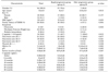
The demographic and clinical characteristics of the study subjects are shown in Table 1. Of the total 34 patients, 79.4% were part of the rapid progression group (Group A) and 20.6% were part of the slow progression group (Group B). Patients in Group A had a younger mean age at the time of diagnosis (A: 9±4.3 years vs. B: 13.6±3.6 years; p=0.013) and a lower mean BMI (A: 15.5±2.5 kg/m2 vs. B: 18.7±3.3 kg/m2; p=0.035), and showed severe symptoms with complication such as DKA (p=0.035) as compared with the patients in the slow progression group (Group B). In contrast, asymptomatic glucosuria was observed only in Group B (28.6%). Group A also showed a significant difference in initial C-peptide levels (A: 0.5±0.46 ng/mL vs. B: 1.87±1.08 ng/mL; p=0.001) and follow-up C-peptide levels (C-peptide at 6 months, p=0.000; 12 months, p=0.000; 18 months, p=0.000; 24 months, p=0.000; 30 months, p=0.000; 36 months, p=0.000). There were no significant intergroup differences with regards to patient sex, family history of T2DM, baseline HbA1c, pH, pancreatic autoantibodies, thyroid antibodies, or serum insulin.
2. C-peptide levels at diagnosis and a comparison of follow-up C-peptide levels
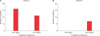
Among the 27 studied subjects of Group A who showed C-peptide levels of <0.6 ng/mL at 36 months after diagnosis, 16 patients showed C-peptide levels of <0.6 ng/mL and 11 patients showed C-peptide levels of ≥0.6 ng/mL at the time of T1DM diagnosis (Fig. 1). By contrast, among the subjects in Group B, who showed C-peptide levels of ≥0.6 ng/mL at 36 months after diagnosis, all seven patients showed C-peptide levels of ≥0.6 ng/mL at the time of T1DM diagnosis.
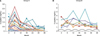
We examined follow-up C-peptide levels during the first three years after diagnosis in Groups A and B. Serum C-peptide levels in Group A decreased sequentially to <0.6 ng/mL (Fig. 2). In contrast, C-peptide levels in Group B remained ≥0.6 ng/mL over the three-year period.
3. Correlation (γ) of levels of C-peptide with clinical variables
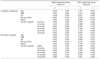
Simple correlation analyses showed that C-peptide levels at diagnosis and at 24 months post-diagnosis in the 34 patients were correlated with BMI (at diagnosis: p=0.009, γ=0.440; 24 months: p=0.002, γ=0.516; Table 2). C-peptide levels at 12 and 36 months post-diagnosis were correlated with age (12 months: p=0.046, γ=0.344; 36 months: p=0.014, γ=0.417) and BMI (12 months: p=0.030, γ=0.373; 36 months: p=0.009, γ=0.443). However, C-peptide levels were not correlated with the other laboratory variables studied, including pH, insulin, and HbA1c.
When evaluated separately, C-peptide levels at diagnosis in Group A were correlated with age (p=0.046, γ=0.387) and follow-up C-peptide levels (p=0.022, γ=0.438, Table 3). In Group B, C-peptide levels at diagnosis were correlated with HbA1c (p=0.046, γ=−0.756), but not with follow-up C-peptide levels. However, in Group B, the C-peptide levels at 36 months were not significantly correlated with HbA1c, but were correlated with the C-peptide levels at 24 months (p=0.034, γ=0.792).
DISCUSSION
The majority of patients with childhood-onset T1DM are relatively insulin deficient within two to three years after diagnosis as compared to those who have T2DM, in whom C-peptide levels tend to persist.10 However, it can be difficult to classify DM in some cases due to an overlapping of the presentation of typical laboratory findings or clinical symptoms between the different DM types. For example, pancreatic autoantibodies can be detected in some young patients with T2DM. Additionally, a rapid worldwide increase in childhood obesity means that even patients with T1DM can present with obesity.1112
Even very low residual beta-cell function is associated with enhanced glycemic control, a lower incidence of hypoglycemia, and considerable reductions in the development of vascular complications in T1DM patients.131415 The amount of endogenous insulin secretion associated with these clinical improvements is relatively small, and residual beta-cell function is present in many patients during the first few years after diagnosis.16 To evaluate residual beta-cell function, we examined the serum C-peptide levels of 34 subjects with T1DM at six-month intervals for the first three years after diagnosis. The serum C-peptide level corresponding with absolute insulin deficiency or absolute insulin requirement in the fasting state was <0.08 nmol/L (0.24 ng/mL).1 Although most of the serum C-peptide levels were low at diagnosis in the T1DM subjects, the average C-peptide level at 36 months in the current study was 0.35 ng/mL, which suggests the presence of residual insulin secretion function in the T1DM patients. From this result, we can speculate that therapies aimed at preserving the remaining beta-cell function in patients with T1DM are needed and may be beneficial.
In addition, the importance of initiating intensive diabetic management as early as possible after T1DM is diagnosed has emerged. The Diabetes Control and Complications Trial found that the use of aggressive treatment at diagnosis, with rapid and strict control of hyperglycemia, may result in improved beta-cell function and could help sustain endogenous insulin secretion.8 Shah et al.17 also suggested that the suppression of endogenous insulin by intensive, continuous insulin treatment for the first two weeks after diagnosis of T1DM may improve beta-cell function during the subsequent year. Many studies to date have investigated C-peptide levels at the time of DM diagnosis, whereas only a few have examined follow-up C-peptide levels regularly for several years; thus, our study has significance in this regard.181920 Furthermore, we tried to subdivide the 18 patients with initial C-peptide levels of ≥0.6 ng/mL at the time of T1DM diagnosis into rapid and slow progression groups, but found no significant difference between the two groups, probably due to the small number of patients studied.
A study in Japan showed that the time frames of early-stage T1DM onset and progression can vary.21 As in our study, the disease progresses rapidly with definite clinical symptoms (rapid-onset form) in some patients, while others are identified via the use of urine glucose screening at a pre-clinical period, but present with no symptoms of DM (slow-onset form). It is probable that genetic background as well as environmental factors influence the clinical features of the early phase of childhood-onset T1DM.22 Urakami et al.23 speculated that a change in the islet cell antibody titer indicates the slow destruction of the pancreatic beta cells by the immune system. It is also presumable that immunogenic factors could alter the clinical features of T1DM in children in the early disease phase. However, in the current study, there was no intergroup difference in pancreatic autoantibody titers. The term “latent autoimmune diabetes in adults” refers to adult diabetic patients who are initially non–insulin-requiring but who have immune markers of T1DM that can progress to insulin dependency.24 This term has been largely used in the last few years when referring to autoimmune forms of diabetes that do not initially require insulin; however, it is now known that these forms of diabetes are neither latent nor limited to occurring in adults.
Children with lower BMIs showed a tendency toward rapidly declining C-peptide levels in our study; in other words, they seemed to have a lower ability to preserve beta-cell function. Redondo et al.5 found that at the time of diagnosis of autoimmune T1DM, obese and overweight children were, respectively, 2.4 and >4 times more likely to have preserved C-peptide levels than lean children, independent of other factors such as age, sex, ethnicity, and puberty. In addition, as in our study, patients with a younger age at diagnosis also showed a rapid decline in C-peptide levels.52526 Patients in the rapid progression group showed more significant symptoms with complications like DKA in our study, and the findings of a previous study also suggest that C-peptide preservation is associated with the presentation of the disease in the absence of DKA.18 Insulin deficiency is the major factor of DKA; however, the metabolic effects of DKA may transiently reduce insulin secretion. Thus, children who present with DKA at diagnosis may have significantly lower C-peptide levels.
Therapies focusing on preserving residual beta-cell function will likely improve the patient's ability to achieve good glycemic control.27 However, HbA1c, the standard measure of glycemic control, is not suitable for predicting the primary outcomes in clinical trials of such therapies. Instead, the most appropriate measurement of endogenous insulin secretion and beta-cell function is the standardized measurement of C-peptide levels. Sensitive and reproducible assays for measuring C-peptide levels are highly accessible.28 Further, HbA1c did not show any correlation with C-peptide levels in our study. Based on a review of recent studies evaluating new therapies for T1DM, the worldwide group of experts assembled by the American Diabetes Association concluded that evaluating beta-cell function via C-peptide level measurement is the most suitable primary outcome for crucial intervention studies of therapies aimed at preserving beta-cell function in patients with T1DM.16 Our study showed that C-peptide levels at 36 months in Group A and B was correlated with only C-peptide level at 24 months, but not with C-peptide levels in other periods. Therefore, considering the honeymoon period and other variables, at least 24 months should be allowed to pass in order to predict C-peptide level in the long-term.
This study has some limitations. First, it was performed in a single center, and the number of included patients was very small (n=34). Additionally, since only seven patients were included in Group B, the intergroup results might have been affected by statistical errors. Therefore, a wider range of further joint research and epidemiologic studies with a larger number of patients included are required. Second, the retrospective design of this study based on medical record review may have resulted in errors. Third, the impact of factors that can affect C-peptide levels, such as the presence of infection and puberty, is unknown.29
In conclusion, patients who are younger, have a lower BMI, significant symptoms, and a low initial C-peptide level tend to show a rapid rate of decrease in C-peptide levels. Therefore, the early provision of intensive insulin therapy to preserve beta-cell function, in an effort to prevent several metabolic and cardiovascular complications, which carry social and economic costs, should be considered promptly in these groups with specific clinical findings. Future efforts should be focused on aspect of the relationship between C-peptide and residual insulin secretion in order to predict the clinical outcomes of newly diagnosed T1DM and to preserve residual beta-cell function for as long as possible.

 XML Download