

 PDF
PDF ePub
ePub Citation
 Print
Print


INTRODUCTION
The main characteristics of the culprit lesions in acute myocardial infarction (AMI) include a lower plaque volume with a higher thrombus burden. Healing patterns of the target lesions in AMI are different from those with stable angina. Delayed arterial healing at the culprit sites after drug-eluting stent (DES) placement for AMI is associated with increased risk of late stent thrombosis.
In the present review article, we describe the current use of DES in AMI in Korea and introduce novel DES uses in development for patients with AMI.
DES IN AMI
The Korea Acute Myocardial Infarction Registry (KAMIR) was established in commemoration of the 50th anniversary of Korea Circulation Society.1 We have registered more than 62,000 patients with AMI since 2005. Rates of percutaneous coronary intervention (PCI) were 96.7% in ST-elevation myocardial infarction (STEMI) and 82.7% in non-ST-elevation myocardial infarction (NSTEMI), and the success rates were 99.4% and 99.5%, respectively.2 Penetration rate of DES is higher than 90% (97% currently) in Korea.3 Stent thrombosis occurred in 1.1 % of patents after implantation of Cypher® (Cordis Corporation, Bridgewater, NJ, USA) and Taxus® (Boston Scientific, Natick, MA, USA) stents during a 6-month clinical follow-up.4 The outcome of DES was compared with that of bare metal stents (BMS) in patients with AMI. The event rates were lower in patients receiving DES compared to BMS with lower incidence of repeat intervention without increasing the risk of mortality, myocardial infarction (MI) and stent thrombosis.5 However, safety issues of DES still need to be addressed in real-world practice. Based on our clinical study using intravascular ultrasound (IVUS), we found that plaque prolapse occurred frequently in as many as 27% of patients with AMI after stent implantation. Predictors of plaque prolapse were long stent length and ruptured plaques, indicating plaque prolapse may be one of the risk factors for stent thrombosis in AMI.6 Virtual histology-IVUS examination in diabetic AMI patients showed multiple plaque rupture and 72% of thrombus in 60% and 72% of cases, respectively.7 No reflow developed in approximately 10 to 20% of AMI patients after stent implantation. IVUS predictors of no reflow after stenting were high density calcium and large necrotic cores at minimal luminal sites.8 High degrees of calcification and multiple plaque rupture were demonstrated in AMI patients with chronic kidney disease.9 Clinical, angiographic and IVUS predictors of early stent thrombosis were analyzed in patients with AMI. Tissue prolapse, high level of troponin and no reflow phenomenon were associated with early stent thrombosis in AMI after stent implantation.10
Current limitations of DES are development of late stent thrombosis and inflammation mediated by polymer-degradation.11 The cause of stent thrombosis after DES implantation is multifactorial, incomplete endothelial coverage being the most powerful histologic predictor in combination with other clinical and procedural risk factors playing a role.1213 We have developed new DES to prevent stent thrombosis and restenosis.14 We established the Korea Cardiovascular Stent Research Institute and Mangho Stent Company. We have developed the Chonnam National University Hospital (CNUH) stent, a coronary stent with a design of our own.15 The CNUH stent was more flexible than four commercialized stents tested which are the Promus Element® (Boston Scientific, Natick, MA, USA), Cypher® (Cordis Corporation, Bridgewater, NJ, USA), Resolute Integrity® (Medtronic, Santa Rosa, CA, USA), Xience Prime® (Abbott Vascular, Santa Clara, CA, USA) stents.15
DES WITH ANTI-THROMBOTIC DRUGS
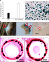
Heparin-coated stents inhibited stent restenosis and thrombosis in a porcine model. However, their clinical benefits could not be demonstrated.16 Platelet glycoprotein IIb/IIIa receptor blocker (ReoPro®)-coated stents inhibited platelet thrombi and restenosis in a porcine model and were associated with lower inflammation compared with Cypher® and Taxus® stents.17 ReoPro®-coated stents were feasible and significantly reduced in-stent neointimal hyperplasia in human coronary arteries, with potential therapeutic benefit in preventing stent restenosis (Fig. 1).18 Furthermore, ReoPro®-coated stent implantation was safe, and prevented stent thrombosis and neointimal hyperplasia on follow-up coronary angiograms and IVUS study in AMI patients.19 Target lesion revascularization and total major adverse cardiac events were lower in AMI patients who received ReoPro®-coated stents, compared with BMS.
DES WITH ANTI-OXIDANTS
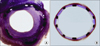
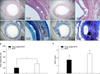
Anti-oxidants [carvedilol, probucol and alpha-lipoic acid (ALA)], were used for stent coating.202122 In a porcine model, carvedilol stents were more effective than probucol stents.21 No stent thrombosis was observed in clinical trials using carvedilol stents during a two-year clinical follow-up.23 Recently, we developed a ALA-coating stent, which inhibited neointimal cell proliferation by inhibiting phosphorylated extracellular signal-regulated kinases and phosphorylated signal transducers and activators of transcription 3, and promoted endothelial coverage in vitro study.22 ALA-coated stents inhibited Akt and vascular endothelial growth factor expression in a porcine model and may be effective for AMI patients with diabetes mellitus (Fig. 2).22
POLYMER-FREE DES
Non-polymer coating technology was developed to prevent stent thrombosis. New DES was developed using a titanium dioxide (TiO2) film by plasma enhanced chemical vapor deposition as a drug-combining matrix.24 Abciximab, ALA and heparin were effectively coated on the TiO2 film-coated stent surface, which was assessed by atomic focus microscopic imaging studies and still observed even after a one-month washing test. This non-polymer coating technique was registered as US patent in 2015 (US patent: 8,999,456B2).
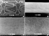
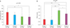
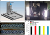
A dual-coating DES by grafting ALA with Abciximab was prepared on a BMS using non-polymer coating technology.25 Polymer-free TiO2 stent with abciximab or ALA was compared with biolimus-eluting stent (BES) in a porcine model. Percent area stenosis was not different between TiO2 stents with Abciximab or ALA and BES. However, fibrin and inflammation scores were lower in Abciximab and ALA-coated stents than in BES.26 We introduced nitrogen-doped TiO2 film as drug-binding matrices for the preparation of DES. Scanning electron microscope (SEM) findings revealed inhibition of platelet adhesion on heparin-, ALA- and Abciximab-grafted stent surfaces. Abciximab, heparin and ALA were released slowly from the stent surface over 1 month on released kinetics.27 Polymer-free DESs coated with everolimus using nitrogen-doped TiO2 film deposition were tested in vitro and in vivo in a porcine restenosis model.28 Electro-spinning coating with TiO2 film was used and this coating technique showed a rate of cumulative drug release to that of bioabsorbable polymer [poly lactic-co-glycolic acid (PLGA)] coating on the release kinetics. Coating thickness of everolimus-coated TiO2 film was 50 nm, which is the thinnest coating technology so far developed (Fig. 3). In an animal study, percent area stenosis of everolimus nitrogen doped TiO2 stent was comparable with Xience Prime® stents but the fibrin score was significantly lower in everolimus nitrogen doped TiO2 stents than Xience Prime® stents (Fig. 4).
DES WITH BIOCOMPATIBLE POLYMERS
Dextran is a natural polymers. An in vitro and in vivo comparison of dextran-based sirolimu-eluting stents and polylactic acid-based sirolimus stents was performed.29 The contact angle and the inflammation score of dextran-based DESs were lower than those of polylactic acid-based DESs. Immunofluorescence analysis revealed enhanced re-endothelialization and reduced inflammation in the dextran-based DES.
Biocompatible co-polymer systems using PLGA-polyethylene glycol (PEG) co-polymer coronary stents were evaluated in a porcine model.30 Novel coronary artery stents with co-polymers showed great potential for future clinical applications and may be applied in the development of novel coronary artery stents with biodegradable polymers.
Tacrolimus-eluting stents using a biodegradable polymer-coating technique were compared with sirolimus- and everolimus-eluting stents.31 The CNUH stents were used for the stent platform of 3 types of DESs. Inhibitory effects on smooth muscle cells (SMC) and human umbilical vein endothelial (HUVEC) proliferation were dose-dependent in all the 3 types of DESs.
DES WITH IMPROVED ENDOTHELIALIZATION
The effect of a novel peptide, WKYMVm- and sirolimus-coated stents on re-endothelialization and anti-restenosis was assessed in an animal model.32 The WKYMVm peptide, specially synthesized for homing endothelial colony-forming cells, was coated onto a BMS with hyaluronic acid. Thereafter, sirolimus was consecutively coated. The peptide was effectively attached to the surface of the stents and the sirolimus coating rendered the surface smooth. The release pattern of sirolimus was similar to that of commercial sirolimus-coated stents. Endothelial-cell proliferation was enhanced, whereas the proliferation of SMC was inhibited. In an animal study, the restenosis rates were lower than those of BMS and the endothelial healing was promoted, suggesting that consecutive coatings of WKYMVm and sirolimus onto BMS may have a potential role in re-endothelialization and neointimal suppression (Fig. 5).32 This innovation was registered as a US patent (US Patent: 9603891).
Endothelial progenitor cell (EPC)-capturing Aptamer stents improved re-endothelialization compared with BMS and Taxus® Stent.33 EPC-capturing Aptamer stents showed lower re-endothelialization scores and thrombus appearance rates compared to Taxus® stent.
DES WITH NATURAL BINDING TECHNIQUES
A natural binding technique was applied on the stent surface. A natural product, fucoidan, was used as a coating material onto BMS with in vivo evaluation. Fucoidan is derived from sea tangle and has anti-oxidant and mitogen-associated kinase inhibitory activities. Three coating technologies, multi-layer, single-layer and dual coating methods, were tested in Korea Cardiovascular Research Institute. Dual-coating stents with PLGA in the outer coating and fucoidan in the inner coating layers reduced percent area stenosis.34
Dopamine is a major component of mussels. Thromboresistant and endothelialization effects of dopamine-mediated heparin coating on a stent material surface were tested.35 Dopamine-mediated heparin coating improved endothelialization and inhibited platelet deposition. This innovation was registered as a Japanese patent (Japan patent: 5576441).
Phytoncide, a natural product derived from trees and plants was utilized for the coating of DES.36 Phytoncide-coated stents compared favorably with sirolimus eluting stents in terms of neointimal area, inflammation and fibrin scores.
DES WITH DUAL THERAPY SYSTEM
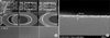
A novel DES with a dual therapy system was developed consisting of an abluminal and luminal coating technique.37 The abluminal coating layer contains anti-proliferative agent such as sirolimus, everolimus, paclitaxel and the luminal coating layer contains a re-endothelialization agent such as the WPP peptide or the CD34 antibody. In order to verify the bidirectional coating of materials, various morphologic analyses were performed using optical microscopy, SEM and fluorescence microscopy. The proliferation of SMC was inhibited by sirolimus, whereas the proliferation of HUVEC cells was enhanced by WKYMVm.
DES WITH GENE DELIVERY
Gene delivery stents using TiO2 and drug coating were developed.38 Plasmids were attached on the TiO2 film drug-coated stent surface and gene delivery was demonstrated in the porcine coronary artery SMC using stents and in the rat abdominal wall using gene-coated plate. a follow-up coronary angiogram revealed a patent stent without restenosis and with a clean surface of the stented artery. Histopathologic findings showed a small area of neotintima formation (Fig. 6).38 This innovation was registered as a Japanese patent in 2016 (Japan patent: 5922647). Suppression of post-angioplasty restenosis with an Akt1 siRNA-embedded coronary stent was tested in a rabbit model.39 On micro-computed tomography scans, the neointimal area was smaller in Akt1 siRNA-stents than those of BMS and hyaluronic acid-coated stents.
NEW BIOABSORBABLE VASCULAR SCAFFOLD
A bio-absorbable drug-coated stent was developed using a 3D-printing system with characterization and in vivo evaluation (Fig. 7).40 The fabricated polycaprolactone stent was coated with sirolimus mixed with PLGA and PEG via a spraying method for slow drug release. The engineered, drug-eluting, bioabsorbable vascular stent (BVS) proved effective in reducing neointimal hyperplasia in an animal study (Fig. 8).40
New BVSs using magnesium were developed and a smart stent with a micro-sensor for coronary pressure measurements is in development. Femtosecond laser stents are one of the new concepts of non-polymeric drug coating system, where femtosecond laser holes may increase biocompatibility and regulate cell proliferation with alignment.
LIMITATIONS
The present study described experiences from a single center on novel DESs, and thus may be limited in scope to reflect the real-world status of DES development. In addition, a number of DES models including stents with anti-oxidants, natural binding techniques, and gene-and-drug delivery capabilities are investigational and have yet to receive regulatory approval for clinical trials in Korea.
CONCLUSIONS
In conclusion, DESs are deemed safe and effective for the prevention of restenosis. However, problems such as late or very late stent thrombosis and late catch-up remain be to be solved. Currently, longer-term, dual anti-platelet therapy is recommended after DES implantation, especially in patients with AMI. Novel DES using non-polymer coating, natural binding technology, peptide coating, gene-and-drug delivery as well as bioabsorbable stents with new biology may improve long-term clinical outcomes and shorten the duration of dual antiplatelet therapy in AMI patients.

 XML Download