

 PDF
PDF ePub
ePub Citation
 Print
Print


Pulmonary arteriovenous malformation (PAVM) is a rare vascular communication in the lungs. The goals of occlusion treatments are to eliminate right-to-left shunt and treat complications.1 The success rate of transcatheter closure is unknown in infants with a huge PAVM. We report a 1-year-old girl, with a giant PAVM, who underwent transcatheter occlusion using Amplatzer vascular plugs, followed by additional Amplatzer vascular plugs with five Interlocking detachable coils when the PAVM recurred.
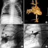
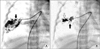
A 1-year-old girl was referred with cyanosis and clubbing fingers. In family history, her mother underwent a lobectomy for PAVM when she was 20 years old. Her maternal uncle underwent PAVM embolization with coils 5 years ago. Her oxygen saturation was 81% of the room air. On physical examination,her skin was normal, with no telangiectasia or hemangioma. There was a faint bruit with a mass-like density on the chest x-ray (Fig. 1A). Computed tomography revealed a tortuous vascular mass, measuring 3.8×2.5 cm (Fig. 1B). Percutaneous angiogram with intubation and intravenous sedation showed a giant PAVM with a huge major feeding vessel which originated from the pulmonary artery (Fig. 1C). Using an 8-mm Amplatzer vascular plug, the major feeding vessel was embolized. A minor feeding vessel was embolized with a 4-mm Amplatzer vascular plug and a proximal feeding vessel was embolized with a 6-mm Amplatzer vascular plug with successful occlusion of the PAVM (Fig. 1D). On the 4th day after the procedure, she was discharged with 94% oxygen saturation. Eight months later, her cyanosis reappeared. Following an angiogram, new collateral vessels were found feeding off a recurrent PAVM adjacent to the previous occlusion site (Fig. 2A). The proximal part of the feeding artery was occluded with an 8-mm Amplatzer vascular plug, but dye passed into a smaller distal part of the occluded major vessel. A residual, small shunt was occluded by five 6 × 20 cm Interlocking detachable coils. Finally, we confirmed complete occlusion of the recurrent PAVM (Fig. 2B). She was well, with 100% oxygen saturation during the 6-months follow-up.
PAVM is an abnormal, direct communication between the pulmonary arteries and veins which can cause serious complications such as septic embolism. PAVM can occur as isolated entities or be associated with hereditary hemorrhagic telangiectasia, as Rendu-Osler-Weber syndrome. Our patient had no telangiectasia, but her family history suggested a genetic component. No genetic analysis was performed. In the future, the patient's first-degree family will require screening for PAVM.
The past treatment of choice for PAVM was a surgical ligation of vessels and resection of the involved lung segment. Recently, percutaneous embolization with coils or detachable devices has replaced surgical ligation.2 Use of the Amplazter vascular plug can safely occlude a large PAVM in adult or a small child.34 We report here regarding our experience using Amplatzer vascular plugs for transcatheter embolization of multiple feeding vessels in a huge PAVM and recurrent PAVM that was finally occluded by combining an Amplazter vascular plug and multiple Interlocking detachable coils in a 1-year-old girl.
 XML Download