

 PDF
PDF ePub
ePub Citation
 Print
Print


INTRODUCTION
Recent data from the The Cancer Genome Atlas (TCGA) data revealed that Epstein-Barr virus (EBV)-associated tumors showed a PIK3CA mutation, PD-L1/2 overexpression, EBV-CIMP, CDKN2A silencing, and immune cell signaling.1 Moreover, EBV infection can cause tumorigenesis and malignant transformation of host cells through the activation of oncogenic pathways.23 Various evidence sources have also indicated that gene alterations may play an important role in the pathogenesis of EBV infection, including the promotion of pathological changes seen in gastric cancer.45 Some representative examples are p53, beta-catenin, and human epidermal growth factor receptor-2 (HER2), which are already known to be associated with the pathogenesis of EBVaGC. Therefore, studies of molecular alterations could provide potential biomarkers as therapeutic targets for EBV-associated gastric cancer (EBVaGC).
The TP53 gene is a tumor suppressor gene that is composed of 11 exons and encodes a nuclear p53 protein, which acts as a potent transcription factor with a key role in the maintenance of genetic stability.6 Thus, genetic alterations of the TP53 gene could be involved in gastric carcinogenesis. Previous studies have already demonstrated that a TP53 mutation is one of the most prevalent genetic alterations in gastric cancer.37 Plus, the association of p53 expression with tumor biological behavior and prognosis has also been reported in the gastric cancer patients.8 Interestingly, EBV infection has been associated with abnormal p53 expression, which may play an important role in inhibiting cell apoptosis and the tumorigenesis of EBVaGC.479 Meanwhile, beta-catenin is an important mediator of the Wnt signaling pathway, and mutation of beta-catenin has been identified in various human malignancies, including gastric cancer.10 Furthermore, Wnt signaling, induced by activated beta-catenin, regulates T cell responses.11 Therefore, these findings point to the possibility of beta-catenin as a prognostic and predictive marker in gastric cancer patients with EBV tumors, suggesting a pivotal role for the immune mechanism in these subsets of gastric cancer. In the ToGA trial, human epidermal growth factor receptor-2 (HER2)-positive gastric cancer patients who received first-line treatment with trastuzumab, an antibody targeting HER2, showed an improved overall survival.12 Several recent clinical studies have also investigated the link between HER2 and EBVaGC, where Zhang et al. reported that HER2 expression was inversely correlated with EBV infection.1314 Notwithstanding, the relationship between EBVaGC and HER2 remains controversial.1516
Accordingly, given that p53 protein, beta-catenin, and HER2 may be potential biomarkers as therapeutic targets for EBVaGC, this study assessed the expression of the p53 protein, beta-catenin, and HER2 and their prognostic implication in patients with EBVaGC.
PATIENTS AND METHODS
1. Study population
All the tissue samples used in this study were obtained from patients included in a previous study of EBVaGC, the inclusion criteria and results for which have already been reported.5 In brief, the study included 117 patients with histologically confirmed adenocarcinomas of the stomach that were identified as EBV-positive using EBV-encoded RNA in situ hybridization. Among the 117 patients, p53 expression was examined in 105 tumor tissues and beta-catenin in 57 tumor tissues, respectively. The baseline characteristics, including age, gender, TNM stage according to the American Joint Committee on Cancer (AJCC) staging 7th edition, and tumor histologic differentiation were all collected from the patients' medical records and surgical pathologic reports. This study was approved by the institutional review board at Kyungpook National University Medical Center (KNUMC) and informed consent was obtained from all the patients included in this study.
2. Immunohistochemistry and scoring
Immunohistochemical staining was performed using a Ventana Benchmark XT autoimmunostainer (Ventana Medical Systems, Tucson, AZ, USA) according to the manufacturer's instructions. Antibodies for the following molecules were used in this study: p53 (1:800; DAKO, Denmark), beta-catenin (1:200; DAKO, Denmark), and HER2 (pre-dilution; Ventana Medical Systems, Tucson, AZ, USA). The immunohistochemistry results were interpreted as follows: strong p53 nuclear expression in at least 50% of the tumor nuclei was interpreted as a positive result, strong beta-catenin expression in at least 10% of the cytoplasmic nuclei was interpreted as a positive result, and moderate or strong complete or basolateral membrane staining in 10% of the tumor cells was interpreted as a positive result for HER2. The histopathologic and immunohistochemistry analyses were conducted by two pathologists (A.N.S and H.I.B) at KNUMC.
3. Statistical analysis
The descriptive statistics are reported as proportions and medians. The categorical variables were evaluated using a χ2 test and Fisher's exact test, as appropriate. Disease-free survival (DFS) was defined as the time between diagnosis to tumor recurrence or death from any cause. Overall survival (OS) was calculated from the date of diagnosis to death from any cause. Data was censored if patients were free of recurrence or alive at the last follow-up. The Kaplan-Meier method was used to estimate the DFS and OS. The survival curves were compared using a log rank test according to the expression differences. Multivariate survival analyses were carried out using the Cox proportional hazard regression model. A p-value <0.05 was considered statistically significant. The statistical analyses were performed using SPSS for Windows (version 19.0, SPSS Inc., Chicago, Ill., USA).
RESULTS
1. Patient characteristics
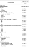
The patient characteristics are shown in Table 1. Most of the patients were male (79.5%) and the median age was 62 years (range 32-80). Most of the tumors (70.1%) were located in the body of the stomach. At the initial resection, 74 (63.2%) tumors were classified as stage I, 25 (21.4%) as stage II, 18 (15.4%) as stage III. Plus, according to the Lauren classification, 25 (21.4%) patients' tumors were intestinal, 81 (69.2%) were diffuse, and 11 (9.4%) were mixed type. Among the 117 patients, 57 (48.7%) were classified as having gastric carcinoma with lymphoid stroma (GCLC).
2. Immunohistochemistry analysis for p53, beta-catenin and HER2
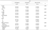
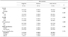
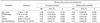
Immunohistochemical staining for p53 was performed on 105 tumor tissues, among which 25 tissues (23.8%) tested positive (Table 2). Positive expression of p53 was significantly associated with a high T stage (p=0.006). More tumors with lymph node metastasis were p53-positive (p=0.013). Beta-catenin expression was examined in 57 tumor tissues (Table 3). No difference in characteristics was noted according to the positive or negative expression of beta-catenin. Plus, no significant difference in tumor-infiltrating lymphocytes (TILs) was found according to the positive or negative expression of beta-catenin (p=0.735). Positive HER2 expression was only observed in 10 tumor tissues.
3. Survival according to p53 expression and beta-catenin
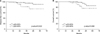
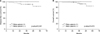
With a median follow-up duration of 36.8 months (range 6.5-60.6), the estimated 5-year DFS and OS rates were 83.9% and 81.0%, respectively. In the univariate analysis, the p53-positive EBVaGC patients showed an reduced DFS when compared with p53-negative EBVaGC patients (p=0.022), although the p53 status was only marginally associated with OS (p=0.080; Fig. 1). However, p53 expression showed no prognostic significance on DFS in the multivariate analysis (Table 4). Moreover, beta-catenin expression was not significantly associated with DFS (p=0.341) or OS (p=0.472; Fig. 2). Similarly, HER2 expression was not found to be associated with DFS or OS in the survival analysis (data not shown).
DISCUSSION
This study investigated the clinical significance of the p53 protein, beta-catenin, and HER2 expression in 117 patients with curatively resected EBVaGC. As a result, the overexpression of p53 was found to be associated with a significantly higher T stage and increased lymph node metastasis, suggesting that impaired p53 activity may be involved in the tumor progression of EBVaGC. Regarding the relationship between p53 overexpression and prognosis, while a trend was indicated, this needs to be elucidated in further studies.
EBV is a gammaherpesvirus whose latent infection is reported to be associated with approximately 10% of all gastric cancers.17 EBV can infect cells in a latent or lytic manner.18 EBVaGC also belongs to latency type I, which can include the expression of the viral genes EBV nuclear antigen 1 (EBNA1), latent membrane protein 2A (LMP2A), Bam HI-A rightward transcripts (BARTs), and EBV-encoded small RNA (EBER1/2).19 In particular, the expression of latency genes fulfils a relevant task in the initiation and neoplastic progression of EBV-associated epithelial cancers, including EBVaGC.20 Interestingly, these viral factors are known to interact with various cellular proteins, such as p53, which maintains genomic integrity in multicellular organisms by activating DNA repair, arresting cells at the G1/S checkpoint, and inducing apoptosis.21 For instance, EBNA1 expression decreases p53 activation and apoptosis in response to DNA damage, resulting in increased cell survival in gastric cancer.22 Plus, EBNA3C can down regulate p53 transcriptional activity in EBV-infected cells.23 Moreover, cumulative studies reported that the p53 protein is accumulated and phosphorylated in nasopharyngeal cancer, mediated by LMP1.242526 The present study found an association between p53 overexpression and tumor progression. Similar to the current results, Chen et al. reported that p53 mutation was associated with the initiation stages of gastric cancer.27 Although the exact mechanism remains unclear, the presence of clonal EBV in every tumor cell suggests that EBV infection precedes the final transforming event giving rise to a tumor.28 Yet, despite the association between advanced stage and p53 overexpression in EBVaGC tissue, no prognostic significance of p53 for DFS and OS was demonstrated. To date, the prognostic role of p53 expression detected by immunohistochemistry in gastric cancer remains controversial.729 Some studies have linked the absence of p53 expression with longer survival, implying that p53 is a negative prognostic factor.303132 Conversely, other studies have found no relation between p53 expression and survival.333435 Therefore, its prognostic relevance should be evaluated in further studies aimed at elucidating the biological significance of p53 expression in EBVaGC.
Beta-catenin has already been shown to be an important mediator of the Wnt signaling cascades.36 As a central molecule in the Wnt signaling pathway, beta-catenin expression is localized in the membrane, cytoplasm and nucleus. Cytoplasmic and nuclear beta-catenin are mainly involved in regulation of the Wnt signaling pathway.37 Beta-catenin accumulates in the nucleus and activates a target gene expression program with loss of E-cadherin, linking Epithelial-mesencymal transition to Wnt signaling.38 Recent studies demonstrated that aberrant activation of the Wnt/beta-catenin signaling pathway is involved in the development and progression of a significant proportion of gastric cancer cases.103940 In particular, activation of the Wnt/beta-catenin signaling pathway is involved in the immune system, including lymphocytes and immunosuppressive cytokines.41 Therefore, beta-catenin could be an important biomarker in EBVaGC, as EBV-positive tumors often show immune signaling activation.1
However, the present study found no statistically significant prognostic impact of beta-catenin in EBVaGC. Plus, no significant correlations were observed between beta-catenin and TILs. There are several possible reasons for this finding. First, the sample size was too small to make conclusions about the prognostic significance of beta-catenin. Second, the methodology used to interpret the expression of beta-catenin and the cutoff values need to be standardized. In this study, strong staining in 10% or more of the cytoplasmic-nuclei was interpreted as positive expression of beta-catenin. However, a recent study by Retterspitz et al. regarded that widespread staining of at least 35% of tumor nuclei indicated a positive expression.40 Thus, different sensitivities have been used to interpret the expression degree of beta-catenin.
HER2, a known proto-oncogene located on the long arm of human chromosome 17 (17q12), is a member of the epidermal growth factor receptor (EGFR) superfamily associated with tumor cell proliferation, apoptosis, adhesion, migration, and differentiation.4243 In gastric cancer, HER2 overexpression is associated with poor outcomes and aggressive disease.44 Thus, due to its pivotal role in tumor growth and relationship with therapeutic efficacy in gastric cancer, HER2 expression in EBVaGC was also evaluated in the current study. Clinical studies already reported that HER2 overexpression was only found in 1.6-5.6% of EBVaGC cases, which is consistent with the present result in terms of the frequency of HER2.1645 Interestingly, Zhang et al. reported a significantly reduced expression of HER2 in EBVaGC, possibly induced by LMP2A, a EBV latent protein.14 EBVaGC has distinct pathological features, presenting a generally diffuse-type histology.946 Thus, HER2 overexpression is relatively rare, making it difficult to analyze the relationship between HER2 and survival in EBVaGC.
In conclusion, the current study found a significant correlation between p53 expression and tumor progression in patients with EBVaGC. However, tissue expression of p53, beta-catenin, and HER2 was not identified as an independent prognostic marker in EBVaGC. Therefore, further investigation is needed to clarify the precise biologic significance of p53 in EBVaGC.
 XML Download