

 PDF
PDF ePub
ePub Citation
 Print
Print


INTRODUCTION
Extracorporeal membrane oxygenation (ECMO) is a life-saving salvage therapy for critically ill patients. ECMO use has increased over the past few decades, and according to the international summary of Extracorporeal Life Support Organization (ELSO) registry report in Jan 2017, patients on ECMO consist of 68% neonate and pediatric patients (Neonates: 44.8%, Pediatric: 24.1% and Adults: 31.1%).
While cardiopulmonary bypass (CPB) is used to assist patients undergoing open heart surgery, ECMO or extracorporeal life support (ECLS) provides a simplified version of CPB that is used temporarily to support refractory cardiac and respiratory failure. Depending on the cannulation type, it can assist both the cardiac and the pulmonary functions (venoarterial ECMO), or only the pulmonary function (venovenous ECMO). Regardless of this difference, an ECMO system is composed of a circuit, a pump, and an oxygenator.
Significant improvements in medical materials, components, and techniques over the past 50-60 years have allowed ECMO to be utilized not only in the operating room but also in the intensive care unit. A ventricular assist device (VAD) is an implantable mechanical device that pumps blood from the ventricles to the rest of the body without oxygenator. A VAD is usually used in patients with heart failure as a bridge to transplant or destination therapy. The characteristics of mechanical circulatory support systems are shown in Table 1.
Although ECMO is becoming more popular, hemorrhagic and thromboembolic complications remain the major threat of ECMO treatment, as they are the most frequent causes of death.1 Recently, the Extracorporeal Life Support Organization (ELSO) registry has reported that thrombotic complications occur in up to 20% of patients with ECMO support.2 These complications commence upon blood contact with artificial surfaces of the circuit, blood pump, and oxygenator system. Therefore, anticoagulation therapy is required in most cases to prevent these problems. Systemic anticoagulation therapy may aggravate a life-threatening hemorrhage, such as intracranial hemorrhage and infarction.3 Here, we describe the blood-surface interactions and anticoagulation therapies in pediatric patients compared with those in adults.
BLOOD INTERACTIONS WITH ARTIFICIAL SURFACES DURING ECMO
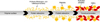
Because the blood is exposed to foreign non-endothelial materials (components of the circuit and oxygenator), all mechanical circulatory support systems can trigger acute inflammatory responses and coagulation (Fig. 1).4 However, these interactions trigger inflammatory and coagulative reactions that cannot be completely controlled to this day. The severity of the action may depend on the duration of support, the anticoagulant used, individual patient characteristics, and the type of artificial material employed. Contact with artificial surfaces activates the complement system through an alternative pathway that releases the anaphylatoxins C3a (alternative pathway) and C5a (terminal pathway).5 Activated complements induce the synthesis of cytokines, which belong to the subgroups of proinflammatory (interleukin-6 and interleukin-8, tumor necrosis factor-a) and anti-inflammatory (interleukin-10) cytokines.678
Shear stress induced by roller pumps induces red blood cell hemolysis. The recent development of centrifugal pumps has rendered hemolysis less problematic.9 Blood cells are injured by contact with artificial materials and by shear stress, turbulence, cavitation, and osmotic forces, triggering very high hemolysis rates.10 Plasma proteins and lipoproteins become progressively denatured during ECMO,1112 increasing plasma viscosity and reducing protein solubility. The levels of plasma immunoglobulins (Ig) G, A, and M as well as albumin decrease as well.13
HEMORRHAGIC AND THROMBOTIC COMPLICATIONS DURING ECMO
Hemorrhagic complications develop in 12%-52% of patients on ECMO.23913 A central nervous system (CNS) hemorrhage may be especially severe and is associated with high-levels of in-hospital mortality and morbidity. However, few studies have compared the prevalence of hemorrhage in neonates and infants. Some authors have grouped hemorrhagic complications in pediatric cardiac patients on ECMO according to age group and diagnostic category.3 Pediatric ECMO patients who had undergone cardiac surgery were found to be more prone to surgical site and CNS bleeding than other pediatric ECMO patients; the mortality rate was double the usual rate seen in ECMO.3 Thrombotic complications also occurred: clots in the circuit were reported in 15% of pediatric patients under ECMO, clots in the oxygenation system in 12%, and intracranial infarctions in 4%.14 There are many contributors to the risks of bleeding and thromboembolic events. Important factors of pro-hemorrhagic conditions include excessive heparin use for systemic anticoagulation, consumption of coagulation factors, low fibrinogen levels, thrombocytopenia, platelet dysfunction, hyperfibrinolysis, and other coagulation disorders. On the other hand, pro-thrombotic factors indicate inadequate use of heparin for systemic anticoagulation, acquired thrombin deficiency, consumption of Protein C, S, consumption of tissue factor pathway inhibitor (TFPI), endothelial dysfunction, heparin-induced thrombocytopenia (HIT), and blood stasis in the cardiac chambers.15
BASELINE LABORATORY DATA AND TRANSFUSION STRATEGY
If a patient is considered to be a candidate for ECMO, baseline laboratory data is essential. These may already be available for critically ill patients, but clinicians must review any incident of coagulopathy prior to ECMO. The required baseline laboratory values include the complete blood count (CBC) including platelet count, the prothrombin time, the activated partial thromboplastin time (aPTT), the fibrinogen, d-dimer levels, the activated clotting time (ACT), the level of antithrombin activity, and thromboelastographic/thromboelastometric data (if available).
The ELSO guidelines state that blood products such as platelets, frozen plasma, or cryoprecipitate can be administered if significant coagulopathy is evident prior to ECMO initiation.13 Hemoglobin levels should be maintained at a minimum value of 8 g/dL, depending on the clinical situation. If the level is lower than 8 g/dL, packed red blood cells (PRBC) should be administered. The platelet count should be above 80,000 cells/mm3; platelet concentrates are often needed, especially in patients who bleed actively or have a high risk of bleeding. Lower platelet counts (lower than normal but above 45,000 cells/mm3) may be acceptable for patients with a low risk of bleeding.16 Usually, fibrinogen progressively increases during ECMO; however, in postcardiotomy ECMO, within the first hours after ECMO implantation, the level of fibrinogen can be very low.17 The values of fibrinogen should be at a minimal level of 100 g/dL. If the values are lower than that, fibrinogen concentrate is available in most centers for supplementation. If fibrinogen concentrate is unavailable, cryoprecipitates can be administered.16
PRIMING OF THE ECMO CIRCUIT
Various combinations of blood products are used to prepare priming solutions. For adults, many centers use only crystalloid-based solutions, most likely due to the lack of time when treating a critically ill patient. For children, combinations of banked blood and other colloid solutions are most commonly used.13 Unfractionated heparin (UFH) 50-100 units per 1 unit of PRBC may also be added. In more urgent situations, priming can be performed with crystalloids while waiting for cross match results. Other blood products may be added when they become available. ECMO priming represents a massive transfusion especially for pediatric patients. To prevent cardiac arrest during this process, freshly irradiated and washed RBCs should be used in every new priming cycle.18
MANAGEMENT OF ANTICOAGULATION THERAPY
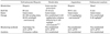
Anticoagulation therapy is an imperfect science, being especially difficult in neonates and children.19 No definitive anticoagulation therapies for neonates and young children on ECMO have yet emerged. Anticoagulation is important to maintain a clot-free oxygenator circuit, and to stabilize the patient hemostatically. It is especially important for patients with cardiac failure who are on ECMO, because left ventricular (LV) stasis can trigger LV thrombosis followed by fatal intracranial infarction.20 UFH is the most widely used and best-studied gold standard anticoagulant for pediatric patients; several other anticoagulants are also employed. The characteristics of the major anticoagulants are described in Table 2.
1. Unfractionated heparin
UFH is the anticoagulant most commonly used during ECMO in both adults and children. UFH interacts with antithrombin (AT) and the tissue factor pathway inhibitor (TFPI). The UFH-AT complex immediately inhibits coagulation.21 Once bound together, the UFH-AT complex has much greater effects of inhibition on coagulatory protein activity compared with AT alone. The UFH-AT complex potentiates the inhibitory effects of activated proteins, factors Xa, IXa, XIa, XIIa, and IIa (thrombin). The thrombin inhibition requires binding of both AT and thrombin to UFH, on the other hand, factor Xa inhibition requires that only AT binds to UFH. UFH inhibits thrombin only after its formation, and does not inhibit either thrombin formation or thrombin already bound to fibrin. UFH increases the effect of TFPI by increasing its affinity to factor Xa.2223 UFH has a short half-life and its action is reversible by protamine sulfate. The aPTT, ACT, and an anti-Xa assay are routinely used to monitor coagulation status. UFH is given as a bolus infusion (50-100 units/kg of body weight) to both adults and children after cannulation prior to initiation of ECMO. UFH is then infused continuously during ECMO support. Intermittent bolus infusions can be given depending on the clinical situation and the results of monitoring tests. In adults, lower doses may be appropriate; however, in neonates and pediatric patients, higher doses should be given when no hemorrhage is apparent.13 The target ACT level of 180-220 is typically achieved with an infusion rate of 20-50 units/kg/h. However, this rate varies among most neonatal and pediatric patients because of greater complexity of their condition. Low molecular weight heparin (LMWH) can also be administered subcutaneously in adult patients during ECMO, but is used less commonly. There are limited reports on LMWH in pediatric patients on ECMO.
2. Antithrombin replacement
Antithrombin (AT) is an inhibitor of serine proteases produced by the liver. AT acts as an anticoagulant principally by inhibiting thrombin and factor Xa.21 Although the optimal level of AT in ECMO patients is unknown, an AT deficiency should be suspected in neonates and children whose requirements for UFH increases to over 35-40 units/kg/h. A relative deficiency of AT contributes significantly to the increased UFH requirements of neonates and pediatric patients, and may render anticoagulation therapy inadequate. Neonates and infants have lower AT activities than older children and adults. Some centers prophylactically replace AT to the target ranges of 50%-100%.24 On the other hand, some centers replace AT only when a reduced effect of UFH is evident.23 Some authors recommend that the target AT level should be >50% in all patients; others prefer target levels of over 80% in neonates and over 100% in infants and children. AT concentrates (plasma-derived or recombinant) are commonly available and are given when AT activity falls below 30%-80%. AT replacement is preferred over infusion of frozen plasma treatments because frozen plasma contains only 1 U/mL AT and plasma would need to be given at over 20 mL/kg to reach an optimal level.19 After AT infusion, the UFH dose can be decreased by up to 50%. The ACT should be calculated or an anti-Xa assay performed 30-60 min after AT infusion.19
3. Alternative anticoagulants
Alternative anticoagulants include direct thrombin inhibitors (e.g., bivalirudin), factor Xa inhibitors (e.g., rivaroxaban, argatroban), factor XIIa inhibitors, and nafamostat mesilate. No large-scale studies on these alternatives have been performed in the pediatric population.
1) Direct thrombin inhibitors
Direct thrombin inhibitors (DTIs) inhibit both circulating and bound thrombin. DTIs are advantageous because they have a lower inter-individual variability as they bind directly to the thrombin, forming complexes. These effects are independent of AT as the levels are not predictable in pediatric population, especially in critically ill patients.25 Additionally, DTIs have more predictable pharmacokinetics compared to heparin because they are independent of plasma proteins. DTIs can be used as an alternative anticoagulant in children with heparin-induced thrombocytopenia (HIT).2627 The significant limitation of DTIs is the lack of an antidote. Although several studies demonstrated that recombinant activated clotting factor VII,2829 further studies are needed. There are limited reports on the use of DTIs in pediatric populations, especially those on ECMO support where further studies are needed.
Major DITs are bivalirubin, argatroban, and leupirudin. In children, bivalirudin and argatroban are frequently used and lepirudin is reported to increase the bleeding risk.30
Bivalirudin is a potential DTI that can be used as an initial anticoagulant in ECMO patients but can also be secondarily used in patients with HIT or UFH resistance.3132 Since bivalirudin metabolizes through intravascular proteolysis, its use can be considered in critically ill patients with hepatic and renal insufficiencies. Very limited data is available for neonates and pediatric patients, particularly those on ECMO. In 2013, Nagle et al. reported on 12 patients with UFH resistance, UFH failure, or HIT who switched from heparin to bivalirudin where anticoagulation were successful.33 Although pediatric dosages of bivalirudin have not yet been established, a pilot study demonstrated an effective and safe dose of 0.125 mg/kg bolus infusion followed continuously by 0.125 mg/kg/h.3435 The target therapeutic aPTT range is 1.5-2.0-times the normal value; a target of 1.5-2.5-times that of normal values is better when it is sought to maintain optimal circuit mechanics.36 The extent of thromboembolic complications was also found to be similar in ECMO patients on heparin and bivalirudin.3637
Argatroban is another synthetic DTI, derived from L-arginine, which binds reversibly to the thrombin active site. It is the first pediatric labeled anticoagulant in the United States for treatment of thrombosis in patients with HIT.29 In general treatment of thrombosis in children (not ECMO patients), it is administered at 0.75 mcg/kg/min continuously and the target aPTT is maintained at 1.5-3 times that of normal levels, which should be checked 2 h after the bolus administration.38 The dose can be titrated in step sizes of 0.25 mcg/kg/min according to the level of aPTT.39 Dose reduction should be considered in critically ill patients with impaired hepatic function because argatroban is hepatically eliminated.40 There were limited studies including ECMO patients. In 2006, Hursting et al. reviewed 12 patients aged 1 week to 16 years who received argatroban simultaneously with ECMO or VAD. Argatroban was administered during circuit priming, and both the ACT and aPTT were monitored. An oxygenator clot developed in one patient, disseminated intravascular coagulation in another, and gastrointestinal bleeding in a third.4142
2) Nafamostat mesilate
Nafamostat mesilate (NM) is a synthetic serine-protease inhibitor used for acute pancreatitis and shock. Recently, it has been used to reduce hemorrhagic complications of hemodialysis and plasmapheresis with its characteristics of a very short half-life. In several studies, NM has been reported to reduce urinary excretion of potassium and cause hyperkalemia. The drug and its metabolites inhibit amiloride-sensitive sodium conductance in the renal cortical collecting ducts leading to impaired urinary potassium excretion.4344
The results of its use during ECMO application were controversial, although most reported results were limited to Asian countries, mostly from Korea and Japan. Lim et al. found that NM was associated with a higher risk of bleeding than heparin in ECMO patients.45 In contrast, Park et al. and Han et al. reported that NM was a safe alternative anticoagulant for ECMO patients with a high risk of bleeding.4647 Nagaya et al. reported preliminary data on the use of NM in 12 neonates on ECMO who had a high risk of bleeding, and bleeding was well controlled in 8 of these 12 cases.48 The average dose used was 0.48±0.22 mg/kg/h and were monitored by measuring the activated clotting time (ACT).48 However, no large study of NM use has yet been reported, especially in the pediatric population. Even more, accurate dosage and titration method principles have not yet been established in the pediatric population and further studies are needed.
3) Other anticoagulants
Rivaroxaban is the first oral DTI that specifically inhibits Factor Xa and prevents thrombin formation. However, limited data on its use in pediatric patients are available.19 Rivaroxaban is used for treatment and prevention of deep vein thrombosis or pulmonary embolism in adult patients.
Factor XIIa inhibitors are new anticoagulants evaluated only in animal models of ECMO. A recombinant human FXIIa-neutralizing antibody prevented thrombosis in the extracorporeal circuit without increasing the risk of bleeding.49
MONITORING THE HEMOSTATIC SYSTEM DURING ECMO
Several in vitro whole-blood and plasma tests are available to measure UFH or other anticoagulants, and each has certain limitations when employed during ECMO. A lack of test standardization is a major limitation, especially when used to evaluate neonatal and pediatric populations. During ECMO employing UFH, use of a single test is not acceptable, but the need to perform multiple tests, several times daily can easily confuse team members. Each center must develop a monitoring method that works best in that center.
In addition, the ECMO circuit triggers more inflammation, in turn affecting cellular and plasma components in younger patients. Additionally, anticoagulation therapy and monitoring are more complicated in neonates and pediatric patients than in adults.
1. Activated clotting time (ACT)
The activated clotting time (ACT) is the most commonly used measure of UFH status during ECMO. This is an inexpensive, whole-blood bedside test. The ACT is the amount of time taken for blood to form a fibrin clot by adding various activators of coagulation (thus, the ACT is the time until the end of the clotting cascade).1319 However, the ACT does not only represent the effect of UFH, being affected by many factors including the platelet count, platelet functionality, levels of fibrinogen and coagulation factors, the patient's temperature, hemodilution status, and various technical parameters.50
A retrospective review of 600 pediatric ECMO patients given heparin revealed a modest correlation (r=0.48) between the ACT and the UFH dose, and concluded that the ACT did not accurately reflect UFH status.50 Other studies reported various correlations (from none to higher levels) between UFH status and the ACT.1319 There are limited studies in correlation of ACT and other anticoagulants.
2. Activated partial thromboplastin time (aPTT)
The aPTT guidelines were developed based on data from adults. The data correlated poorly with UFH status in neonatal and pediatric patients. The aPTT is more reliable in adults given modest doses of UFH; many adult ECMO centers prefer to use the aPTT rather than the ACT when monitoring and adjusting UFH doses. However, in infants, the baseline aPTT is prolonged, rendering the aPTT less reliable as a monitoring test.51 The UFH dose and the aPTT exhibit a linear relationship in normal pediatric subjects, but the aPTT varies unpredictably in those who are ill, limiting its utility when evaluating UFH status.1319 Compared with other monitoring tools, the aPTT correlated well with the anti-Xa activity levels in pediatric patients under cardiac catheterization and cardiac surgery.52 In addition, several studies report that the aPTT reflected UFH status better than the ACT in pediatric ECMO patients.53
Monitoring methods for other anticoagulants, such as DTIs have not yet been established, but aPTTs are most commonly used for monitoring. Further studies on monitoring various anticoagulants in the pediatric population are needed.
3. Anti-Xa assays
Many clinicians use anti-Xa assays as substitutes for the aPTT or ACT. These are plasma-based tests that are not affected by platelet activities. The assays measure UFH-AT complex levels, not thrombin inhibition. In a single-center study, the anti-Xa assay results correlated well with UFH status. Additional advantages of the assays include a reduced need for blood sampling, less transfusion of blood products, and fewer thrombotic/hemorrhagic complications.1617545556 However, anti-Xa assays are chromogenic. Thus, if the hemoglobin or bilirubin levels become elevated during ECMO, the assay may yield artificially low values; the data must thus be interpreted with caution.57 Newall et al. prospectively compared UFH concentrations (measured via protamine titration) and the effects of UFH (using the aPTT, an anti-Xa assay, and the thrombin clotting time) in pediatric patients under cardiac catheterization.58 No monitoring method yielded data that correlated well with protamine titration results. It was thus suggested that the current recommendations are inappropriate for pediatric populations; these require either a different monitoring test or an alternative anticoagulant.58 Despite these concerns, most ECMO centers use anti-Xa assay data to guide anticoagulation therapy. Some centers recommend that the daily ACT target range should be based on anti-Xa assay data and AT activity levels.5960
4. Viscoelastic testing
Viscoelastic tests (thromboelastography and rotational thromboelastometry) evaluate whole-blood coagulation and fibrinolysis. These are the only methods that evaluate clot strength, and can be performed bedside. They evaluate the time to initial fibrin formation, kinetics, clot establishment, the strength and stability of the clot, clot lysis, and the function of platelets.61 However, the tests are not yet widely available. One retrospective pilot study on children on ECMO revealed that qualitative platelet dysfunction was both frequent and severe upon thromboelastographic testing.62
CONCLUSION
Anticoagulation therapy is important to maintain the circuit and a clot-free oxygenator, and to stabilize the patient hemostatically, but it is especially complex in neonates and children. No definitive anticoagulation therapies for neonates and young children on ECMO have yet been established. Most anticoagulation studies are based on adult populations, and very few studies on the pediatric population on ECMO were found. Additionally, monitoring the levels of anticoagulation is more complicated in pediatric patients as ECMO circuit adds more complexity. To find the most acceptable anticoagulants for pediatric patients on ECMO, an effective dosage and monitoring method should be established based on newer anticoagulants. The greatest limitation of ECMO research, especially on pediatric populations, is the variabilities of ECMO protocols and algorithms among centers and countries. Many centers worldwide use non-standardized equipment (cannulae and oxygenators) to treat children and follow in-house management algorithms. Although most ECMO patients, especially pediatric patients, have their specific situation and individualized reasons for ECMO, following standardized algorithms is very important. Prospective, multicenter studies for newer anticoagulants in pediatric patients are urgently needed.

 XML Download