

 PDF
PDF ePub
ePub Citation
 Print
Print


INTRODUCTION
Vitamin D is a steroid hormone. It is mainly subdivided into vitamin D2 (ergocalciferol) and vitamin D3 (cholecalciferol).1 It can be produced in the skin with adequate ultraviolet exposure or ingested through diet and supplementation. 2 However, vitamin D deficiency has become a universal public health issue. Vitamin D deficiency is very common, and between 30 and 50% of the world's population suffers from it.3 Similarly, the Korea National Health and Nutrition Examination Survey data show that 50-60% of men and 60-70% of women experience severe vitamin D deficiency.45
Vitamin D deficiency is related to hypertension, diabetes mellitus, dyslipidemia, metabolic syndrome, stroke, myocardial infarction, and atherosclerosis.678910 Peripheral arterial disease (PAD) is a chronic occlusive arterial disease caused by atherosclerosis. The primary risk factors for PAD are increasing age, smoking, hypertension, diabetes mellitus, and dyslipidemia.11 It has not yet been determined, however, whether vitamin D deficiency or insufficiency is a risk factor for PAD. Some prior studies have reported that low serum 25(OH)D concentrations are associated with a higher prevalence of PAD,1213141516 whereas others have not.31718
Although several case–control studies have evaluated the association between vitamin D status and PAD19 in CVD patients, there have been few population-based studies, and those were performed in Western populations.20 Although ethnic differences could influence the relationship between serum 25(OH)D concentrations and the risk of PAD, to date, no studies have been performed in Asians. We therefore aimed to investigate whether low 25-hydroxyvitamin D (25(OH)D) levels were associated with increased risk of PAD in a Korean population.
MATERIALS AND METHODS
1. Subjects
The Dong-gu Study is an ongoing prospective study designed to investigate the prevalence and incidence of the risk factors for chronic diseases targeting the elderly population who live in Dong-gu.21 In total, 9,260 subjects aged 50 years and older were enrolled (3,711 males and 5,549 females) from 2007 to 2010. This cross-sectional study was conducted among 8,960 participants and excluded due to missing information on: vitamin D measurement (n=58), peripheral arterial disease (n=76), medication intake (n=166). All of the participants provided signed consent forms, and the study was performed according to the guidelines of the Declaration of Helsinki. The study protocol was approved by the Institutional Review Board (IRB) of Chonnam National University Hospital [No I-2008-05-056].
2. Anthropometric data and lifestyle factors
Demographic characteristics, medical history, and lifestyle were assessed by standardized questionnaires. Smoking status was divided into smokers (including ex-smokers) and non-smokers. Alcohol consumption was classified into current drinkers and non-drinkers. Education was categorized into middle school completion or less and high school completion or more. Exercise was classified as regular exercise or no regular exercise based on the weekly frequencies of recreational activities. Self-rated health status was classified as good (very good, good) or poor (average, poor, very poor). We examined whether participants had taken medications during the previous 2 weeks for hypertension, diabetes, or dyslipidemia. Waist circumference was measured at the midpoint between the ribs and pelvis, and body mass index (BMI) value was computed using height (m) and weight (kg) with the formula BMI=kg/m2. Blood pressure was measured using a mercury sphygmomanometer (Baumanometer; WA Baum Co, Inc., Copiague, NY, USA) placed around the right upper arm after 5 minutes of quiet sitting. Three consecutive readings of systolic and diastolic blood pressure were taken once per minute, and the average value was used for the analysis.
3. Laboratory measurements
Blood samples were drawn in the morning after a 12-h overnight fast. Levels of total cholesterol, triglycerides, high-density lipoprotein (HDL) cholesterol, and blood glucose were measured via enzymatic methods using an automatic analyzer (Model 7600; Hitachi, Ltd., Tokyo, Japan). High-sensitive C-reactive protein (CRP) levels were locally determined using a BN II nephelometer (Dade Behring, Marburg, Germany). Hemoglobin A1c (HbA1c) levels were measured using high-performance liquid chromatography (HPLC) using the Bio-Rad VARIANT II system (Bio-Rad, Hercules, CA, USA). Levels of 25(OH)D and parathyroid hormone (PTH) were determined by chemiluminescent microparticle immunoassay (CMIA) (ARCHTECT i2000, Abbott, Chicago, IL, USA).
4. Definition of peripheral arterial disease
PAD was defined as a ankle brachial blood pressure <0.9. ABI was provided automatically using an oscillometric method 22 with the VP-1000 system (Colin Co., Komaki, Japan). After 5 minutes of relaxation in the supine position, BP cuffs were placed around the subjects' arms and ankles. ABI was measured by the ratio of the ankle systolic blood pressure to the arm systolic blood pressure. If the ABI value was lower than 0.9, the patient was defined as having PAD.
5. Statistical analysis
Baseline characteristics were analyzed for PAD values and compared using the t-test and chi-squared test, as appropriate. Multivariate, logistic regression was used to evaluate the relationship between serum 25(OH)D levels and the risk of PAD, adjusting for age, sex, smoking, alcohol intake, medication use (hypertension, diabetes, and dyslipidemia), systolic and diastolic blood pressure, total cholesterol, HDL cholesterol, triglyceride, fasting glucose, and hs-CRP. We further adjusted for PTH to evaluate the mediating effect of PTH. Odds ratios (ORs) are expressed per standard deviation (SD) increase in 25(OH)D. All statistical tests were two-tailed, and the statistical significance level was set at p<0.05. Statistical analyses were performed using SPSS version 22.0 (SPSS, Inc., IBM, Chicago, IL, USA).
RESULTS
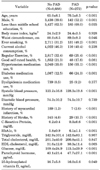
Among the 8,960 subjects, 272 (3.0%) had PAD. The mean ages of those with and without PAD were 70.1±8.5 and 65.0±8.1 years, respectively (p<0.001). Participants with PAD were more likely to be male, to have hadsmoked, to have increased systolic blood pressure, HbA1c, CRP, glucose, parathyroid hormone, to use anti-hypertensive medication and anti-diabetic medication, and to have a history of myocardial infarction and stroke, and they were less likely to perform regular exercise (Table 1).
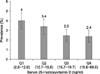
The prevalence of PAD according to the 25(OH)D quartile is presented in Fig. 1. The age and sex adjusted prevalence of PAD decreased with increasing 25(OH)D quartile. The prevalence of PAD according to the 25(OH)D quartile was 4.0%, 3.4%, 2.5%, and 2.4% for the first through fourth quartiles, respectively.
The association between serum 25(OH)D and the prevalence of PAD is presented in Table 2. When adjusted for age and sex, increased serum 25(OH)D levels were significantly associated with decreased risk of PAD (OR for one SD increase, 0.96; 95% CI, 0.94-0.99). Further adjustments for potential confounders attenuated this association (OR for one SD increase, 0.97; 95% CI, 0.95-1.0). When we further adjusted for parathyroid hormone to evaluate its mediating effect, the association was slightly attenuated (OR for one SD increase, 0.98; 95% CI, 0.95-1.0). Compared with the first quartile of 25(OH)D, the odds of PAD were 0.86 (95% CI, 0.62-1.21), 0.67 (95% CI, 0.46-0.97), and 0.71 (95% CI, 0.49-1.04) for the second, third, and fourth quartiles, respectively.
DISCUSSION
We found a significant, inverse relationship between serum 25(OH)D status and the risk of PAD in these Korean subjects aged 50 and older.
Three studies131518 have examined the relationship between vitamin D and PAD in the general population. Of those, two1315 were conducted using the US National Health and Nutrition Examination Survey (NHANES) 2001-2004 data. Melamed et al.13 reported that the prevalence rate ratio for the lowest quartile was 1.8 (95% CI, 1.19-2.74) compared with the highest quartile, which is in accordance with the present study. Reis et al.15 investigated whether there were racial differences in the association between vitamin D levels and the prevalence of PAD. They found that the prevalence of PAD was significantly lower in white than in black adults (5.3% and 8.5%, respectively); after adjusting for racial differences in 25(OH)D, the OR for PAD decreased by 31%, suggesting that racial differences in vitamin D status may explain racial difference in PAD prevalence. However, a prospective study performed in an older population of 1,568 Italian community-dwelling adults ≥ 65 years old with a mean follow-up duration of 4.4 years found no association between baseline serum 25(OH)D and an incident of PAD.18 This study had a small sample size and relatively short follow-up duration of 4.4 years. In addition, half of the study participants had sufficient levels of 25(OH)D (≥30 ng/mL), whereas only 7% had deficient levels of 25(OH)D (<10 ng/mL), which may have hindered the evaluation of the association between vitamin D status and PAD incidence.
The mechanism underlying the association between low serum 25(OH)D and risk of PAD can be explained as follows. Increased blood levels of 25(OH)D have been associated with decreased blood pressure, which could influence the risk of PAD. In the Health Professionals' Follow-up Study and the Nurses' Health Study, subjects whose 25(OH)D concentrations were less than 15 ng/mL had an 8.1 times and 2.7 times increased risk of hypertension, respectively.23 The mechanism linking vitamin D with hypertension may be explained by its role as a negative regulator of the renin-angiotensin system (RAS) and its influence on vascular endothelial function or vascular smooth muscle intracellular concentrations of calcium24. In addition, vitamin D inhibits the progression of inflammation and atherosclerosis by adjusting T helper cells, which in turn inhibit the development and progression of coronary artery disease and PAD.25
Although the present study had the advantage of being a large population-based study, it has several limitations. First, because the design was cross sectional, the established associations do not necessarily indicate a causal relationship. Second, levels of sunlight exposure and intakes for vitamin D were not measured, and both likely had effects on serum 25(OH)D levels. Third, a low ABI (<0.9) which was used in the present study may underestimate the prevalence of PAD.26 However, the ABI is a reliable screening test for PAD and a low ABI (≤0.9) independently predicts future cardiovascular events.27
In conclusion, low serum 25(OH)D levels were associated with the risk of PAD, independent of the traditional cardiovascular risk factors and parathyroid hormone. Our findings suggest that low vitamin D levels contribute to PAD in the Korean population.

 XML Download