

 PDF
PDF ePub
ePub Citation
 Print
Print


INTRODUCTION
Although the survival rate for advanced gastric cancer is steadily improving, the prognosis remains very poor.1 Gastric cancer is a particularly heterogeneous disease that is implicated in various gene-environment interactions resulting in the activation of several molecular pathways.2 MET is a proto-oncogene located on chromosome 7 (7q31) and encodes a tyrosine kinase membrane receptor for its physiologic ligand hepatocyte growth factor (HGF).3 The intracellular signaling cascades activated by the MET pathway include the phosphoinositide 3-kinase (PI3K) and mitogen-activated protein kinase (MEK) pathways. Activation of these signals leads to alterations in the transcription of various cellular genes and cross-signaling pathways such as the epidermal growth factor receptor (EGFR) and vascular endothelial growth factor receptor.4 Thus, MET and HGF play an essential role in tumor-related metastasis and angiogenesis as well as in the cellular proliferation and survival of tumors.5 Clinical studies have also shown that the alteration of MET or its family is associated with prognosis for various solid tumors.678910
Several recent reports have indicated that MET may be a prognostic marker and important target for cancer treatment using co-signal networks in the case of gastric cancer.11 For example, the expression of MET has been shown to be correlated with a poor prognosis for gastric cancer.1213 Toiyama et al. also reported that increased MET and HGF expression was significantly associated with poor prognosis and predicted peritoneal dissemination.14 In addition, the MET gene copy number appears to influence the survival of patients with gastric cancer.1516 Consequently, given these results, MET seems to play an important role in tumor growth and spread, thereby affecting the prognosis of gastric cancer.
Notwithstanding, relatively few published studies have investigated the MET gene copy number and its relationship to the clinical outcomes of resected gastric cancer. Accordingly, the present study analyzed the MET gene copy number and its impact on the survival of patients with curatively resected gastric cancer.
MATERIALS AND METHODS
1. Patients
Tissues were obtained from patients who participated in a pilot study of a combination regimen of cisplatin and S-1. The inclusion criteria and results of that study have been reported previously.17 In brief, the study included patients with histologically confirmed adenocarcinoma of the stomach who underwent curative surgery and received adjuvant chemotherapy with cisplatin and S-1. The pathologic staging was assessed according to TMN classifications from the 6th edition of the American Joint Committee on Cancer (AJCC). This pilot study was approved by the institutional review board (IRB) at Kyungpook National University Hospital (KNUH).
2. Analysis of MET gene copy number
Written informed consent for quantitative polymerase chain reaction (qPCR) was received from the patients, and the study was approved by the IRB at KNUH. The DNA was extracted from formalin-fixed paraffin-embedded (FFPE) tumor tissue samples by use of a QuickExtract™ FFPE DNA extraction kit (Epicentre Biotechnologies). This involved adding 100 µL of the QuickExtract FFPE DNA extraction solution to the paraffin-embedded tissue sections in a microcentrifuge tube; the samples were then incubated at 56℃ for 1.5 hours and 98℃ for 2 minutes. The DNA concentration was measured by using a DNA Quantitation Kit, Fluorescence Assay (Sigma), with the fluorescent dye bisBenzimide H33258 (Hoechst 33258). The genomic qPCR was performed by using a LightCycler480 real-time PCR instrument (Roche, Basel, Switzerland). The thermal cycling conditions consisted of one cycle at 95℃ for 10 minutes and 40 cycles at 95℃ for 15 s and 60℃ for 1 minute. The PCR reactions were performed in a total volume of 10 µL. At the end of the PCR, the samples were analyzed by using reported methods.16 A high copy number was defined as more than 1.99 copies, which was calculated by using the median of the MET gene copy number.18
3. Statistical analysis
The descriptive statistics are reported as the proportion and median. The baseline characteristics were compared by using a chi-square test and Fisher's exact test. Overall survival (OS) was defined as the time from the date of surgery to death from any cause. Relapse-free survival (RFS) was defined as the time from the date of surgery to relapse or death from any cause. Follow-up duration was defined as the time from the date of surgery to patients' last visits or events. OS and RFS were analyzed by using a Kaplan-Meier test and were compared by using log-rank tests. Cox's proportional hazard regression model was used for the survival analyses. The analyses were adjusted for age, sex, tumor size, and stage. The hazard ratio (HR) and 95% confidence interval (CI) were also estimated. A cutoff p value of 0.05 was adopted for all statistical analyses. All analyses were performed by using the Statistical Package for the Social Sciences, version 14 (SPSS Inc., Chicago, IL, USA).
RESULTS
1. Patient characteristics
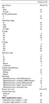
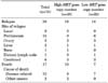
This study analyzed 70 tumor tissues samples from 74 patients enrolled in the pilot study. The patient characteristics are shown in Table 1. The median patient age was 56 years (range, 22-71 years), and 43 patients were male. The stages after surgical resection were as follows: stage II (n=22, 31.4%), stage IIIA (n=15, 21.4%), stage IIIB (n=13, 18.6%), and stage IV (n=20, 28.6%). The predominant histology was poorly differentiated adenocarcinoma. Most patients were classified as diffuse type by the Lauren classification. Among the 70 patients, 30 relapses (42.9%) were documented and 17 patients (24.3%) died (Table 2). The most common site of relapse was the peritoneum (50.0%). With a median follow-up duration of 26.4 months (range, 2.6-73.2 months), the median RFS time was 39.9 months (95% CI: 14.7-65.2), whereas OS time cannot yet be calculated. The estimated 3-year RFS and OS rates were 54.3% and 77.4%, respectively.
2. Relationship between MET gene copy number and clinicopathologic factors
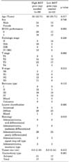
When using the cutoff MET gene copy number of ≥2 copies, 38 patients (54.3%) were in the high MET gene copy number group, and 32 patients (45.7%) were in the low MET gene copy number group. The relationships between the MET gene copy number and the clinicopathologic factors are shown in Table 3. No significant correlations were observed between the MET gene copy number and the clinicopathologic features.
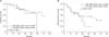
3. Survival analysis
In the univariate and multivariate analyses including age, sex, tumor size, and stage, no significant association was observed between the MET gene copy number and survival (Table 4 and Fig. 1). Stage, tumor size, and age were found to be independent prognostic factors of survival for the patients with resected gastric cancer.
DISCUSSION
When investigating the MET gene copy number and its impact in patients with surgically resected gastric cancer, no significant association was observed between the MET gene copy number and patient survival.
Activation of the MET pathway is mainly accompanied by amplification of the MET gene, which leads to subsequent protein overexpression and kinase activation.2 Thus, alteration of the MET gene can be detected by overexpression, gene mutations, amplification, or rearrangements.19 In general, the frequency of overexpression is high (40-80%) and amplification is present in 5% to 10% of gastric cancer.2021 In contrast, 38 patients (54.3%) were in the high MET gene copy number group (median, 2.0; range, 1.14-6.27). As such, there may have been different sensitivities and specificities between the methods used to detect the MET gene copy number.
Clinical studies have already demonstrated that activation of the MET pathway produces a higher grade and worse survival outcome for solid tumors including gastric cancer.5678910 For example, overexpression of MET has been connected to the risk and progression of gastric cancer.13 In a study by Graziano et al. that evaluated the effects of MET gene copy number and sequencing for HGF on survival in 230 patients with gastric cancer, the survival outcome was worse among the patients with five or more copies.16 Recently, Lee et al. also showed that an increased MET gene copy number was associated with poorer survival.15 Furthermore, MET activation has been more specifically associated with liver and peritoneal metastases,1214 whereas activation of the MET gene has been strongly linked to uncontrolled cell proliferation, oncogenesis, and aggressive cellular invasiveness, leading to poor survival outcome.
In the present study, the prognostic impact of the MET gene copy number in gastric cancer patients was not statistically significant. There are several reasons for this finding. One reason is that the sample size was too small to make conclusions about the prognostic significance of MET gene copy number. Second, although qPCR is considered a standard method for MET testing, various concordance rates have been reported between fluorescence in situ hybridization and qPCR.22 Plus, the methodology of interpreting copy number and the cutoff values will need to be standardized. Third, the present findings need to be confirmed to determine whether the adjuvant chemotherapy may have contributed to the survival outcomes.
In conclusion, the MET gene copy number investigated in this study was not found to be a prognostic marker for patients with curatively resected gastric cancer. However, further investigation is needed to clarify the role of the MET gene as a biomarker and new target in gastric cancer.

 XML Download