

 PDF
PDF ePub
ePub Citation
 Print
Print


INTRODUCTION
An anomalous aortic origin of a coronary artery is a rare congenital anomaly with an incidence of about 0.17%.1 Such anomalies are reported as an important cause of sudden death in specific populations such as athletes or military recruits.2,3 Generally, surgical interventions like coronary artery bypass grafting (CABG), reimplantation, or unroofing are recommended when the patient has symptoms of ischemia or arrhythmia.4 Here we report a case of failed CABG of an anomalously originating right coronary artery (RCA) in a young and symptomatic patient.
CASE REPORT
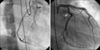
A 21-year-old male soldier visited the division of cardiology of our medical center with the complaint of effort angina. He had no history of smoking and no family history of heart disease. His vital signs were stable, his electrocardiogram showed a normal sinus rhythm, and the results of baseline laboratory findings were nonspecific. However, owing to the typical nature of his chest pain, which suggested an ischemic origin, we decided to perform cardiac computed tomography angiography (CTA) to rule out coronary artery diseases. The cardiac CTA images showed normal left coronary arteries but an RCA anomalously originating from the left sinus of Valsalva. Also, the proximal RCA had an interarterial course, which meant that the RCA was trapped between the aorta and the pulmonary artery (Fig. 1).
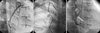
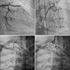
We performed coronary angiography (CAG) to verify the anomaly. CAG from the left sinus of Valsalva showed all 3 coronary arteries, suggesting the anomalously originating RCA (Fig. 2). CAG from the right coronary sinus showed no significant stenosis in the left anterior oblique view. In the right anterior oblique view, however, significant stenosis was noted in the proximal RCA as the result of external compression (Fig. 3). Also during CAG, an intravascular ultrasonography (IVUS) study of the RCA showed that the minimum lumen area in the proximal RCA was 3.8 mm2, which suggested significant stenosis. After CAG, we could not proceed to additional diagnostic tests to verify the evidence of ischemia in the RCA territory, such as myocardial single photon emission computed tomography (M-SPECT) or fractional flow reserve (FFR), because the patient was on a mandatory military service and did not have enough time for many diagnostic tests. However, given the findings from the CAG and IVUS, we regarded the chest pain to be originating from ischemia of the RCA territory. We therefore decided to perform surgical intervention. Owing to a slit-like stenosis of the RCA ostium, we could not choose reimplantation of the RCA ostium, and surgical unroofing was not favored because of a concern for persistent myocardial ischemia in such cases.5 Finally, CABG [right internal mammary artery (RIMA) to RCA] was selected as the surgical intervention. After the CABG, the patient's symptom was relieved, which suggested that the effort angina was caused by myocardial ischemia. A follow-up CAG done 1 week after the operation showed a patent RIMA to RCA graft (Fig. 4). The patient was discharged from our hospital with medications including aspirin.
The patient had no chest pain for more than 10 months after the operation. However, nearly 1 year after the operation, he visited our center and reported suffering from chest pain for about 2 weeks. We recommended that he undergo CAG again. The follow-up CAG revealed no interval changes of the 3 native coronary arteries, but the RIMA to RCA graft was totally occluded (Fig. 5). The right subclavian artery was intact and the patient had no other risk factors for atherosclerosis. Thus, the total occlusion seemed to have developed as the result of complete inhibition by good antegrade flow of the RCA. At this point, we decided to control the patient's symptom with medical treatment, and we recommended that he undergo reimplantation of the right coronary ostium if his symptom persisted after optimal medical treatment.
DISCUSSION
Graft patency of the internal mammary artery is generally excellent, with a 5-year patency rate of more than 90% and a 10-year patency rate of about 80-95%.6 It is well known that the degree of stenosis in the native vessel is a major predictor of internal mammary artery bypass graft patency.7 In the present case, however, the patient's RCA was significantly stenosed, but the graft became occluded. In fact, the degree of stenosis in the anomalously originating RCA may have changed, because the diameter of the 2 major vessels may vary according to sympathetic and parasympathetic tone. Thus, the degree of antegrade flow through the native RCA, and, consequently, the degree of competitive flow, can also vary. This suggests that we should be particularly careful when choosing a surgical intervention for an anomalously originating RCA with an interarterial course. If we could measure flow rates or intraluminal pressures of native arteries and estimate the intensity of possible competitive antegrade flow through the native coronary arteries, we could avoid CABG surgeries in patients who have higher risks of graft occlusion by competitive flows. The use of methods such as FFR for such purposes should be investigated.
One question arises from this case. Did this patient really have to undergo a surgical intervention? Despite the lack of other evidence of myocardial ischemia, such as results from a treadmill test, M-SPECT, or FFR, the patient's chest pain was present with a significantly stenosed RCA, which was relieved by CABG and unfortunately redeveloped with occlusion of the graft. This suggests that the symptom was caused by myocardial ischemia. However, in one report, no significant difference in 10-year survival was seen in patients with an anomalously originating coronary artery from the opposite sinus of Valsalva, which had an interarterial course, after a surgical intervention.8 Of course, this does not mean that a surgical intervention is useless for such cases. The efficacy of surgical intervention versus medical treatment for reducing symptoms is not yet validated in such cases, and this remains a question that should be investigated.
In summary, physicians should think twice before performing a surgical intervention in patients who have an anomalously originating coronary artery with an interarterial course, and methods to evaluate flow rates or the intraluminal pressures of native arteries could be helpful in making a decision. It should be investigated whether such methods would really be helpful, and the treatment efficacy of surgical intervention should also be evaluated.


 XML Download