

 PDF
PDF ePub
ePub Citation
 Print
Print


INTRODUCTION
Tricuspid regurgitation (TR) more often develops as a secondary consequence, rather than as a primary valve disease. The main causes of secondary TR include marked annular dilatation and increased tricuspid leaflet tethering, secondary to right ventricular pressure overload, volume overload, and right ventricular diseases.1
Although a previous study described that atrial fibrillation contributes to the occurrence of TR, previous reports on tachycardia-induced cardiomyopathy primarily addressed left ventricular dysfunction and associated mitral valve regurgitation.234 In the present report, we describe a case of tachycardia-induced severe TR that was completely resolved without surgery after the tachycardia was controlled.
CASE REPORT
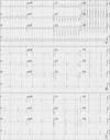
A 48-year-old man presented to our hospital with intermittent palpitations that he had experienced for the past several years. An initial electrocardiography indicated narrow QRS tachycardia, with a ventricular rate of 231 beats per minute (bpm) (Fig. 1A). Chest radiography showed cardiomegaly without pulmonary edema. However, the sinus rhythm was restored after amiodarone infusion, with a rate of 50-70 bpm (Fig. 1B). Transthoracic echocardiography showed a moderately decreased left ventricular systolic function (ejection fraction, 37%), enlargement of the right atrium and ventricle, and dilation of the tricuspid valve annulus.
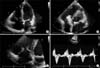
A parasternal long axis image showed a right ventricular end diastolic dimension of 3.22 cm, whereas an apical four-chamber image showed a tricuspid annular diameter of 4.3 cm. Severe TR was detected on Doppler examination with a proximal isovelocity surface area radius of 0.99 mm, effective regurgitant orifice area of 0.77 cm2, and regurgitation volume of 41 mL (Fig. 2).
The tricuspid annulus dilation and right ventricular dilation were believed to be irreversible because the patient's tachycardia was long-standing, probably for several years. Hence, surgery was recommended to prevent further right ventricular remodeling. However, the surgery had to be delayed for personal reasons. Hence, medical treatment consisting of amiodarone, digoxin, carvedilol, ramipril, and warfarin was continued. The palpitations were considerably reduced during the 6 months of medical treatment.
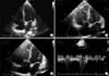
Echocardiography after 6 months of treatment surprisingly showed complete resolution of the TR. The tricuspid annulus diameter had decreased (tricuspid annular diameter, 3.23 cm) and the right atrial dimension had decreased as well (Fig. 3). Coronary angiography, which was performed to exclude ischemic etiology, showed no significant stenosis. Moreover, no pulmonary hypertension was noted on cardiac catheterization.
He underwent electrophysiological examination, and radiofrequency ablation for atrioventricular re-entry was successfully performed. After 5 months of follow-up, his general condition is good, he has not experienced palpitations, and the sinus rhythm has been maintained (Fig. 1C).
DISCUSSION
In the present report, we describe a case of severe TR that was completely resolved without the need for surgery. Tachycardia-induced cardiomyopathy is a well-described disorder associated with various types of arrhythmias. Jeong et al. reported the presence of a dilated atrium and ventricle, reduced left ventricular contractility, and mitral tricuspid regurgitation in patients with tachycardia-induced cardiomyopathy.3 The pathophysiology of left ventricular dysfunction in patients with tachyarrhythmias is unclear. However, a number of mechanisms have been proposed, including myocardial energy depletion, abnormal calcium handling, myocardial ischemia, and extracellular matrix remodeling.4
The right ventricular and left ventricular responses to tachycardia differ. In the left ventricle, chamber dilation develops without an increase in mass, whereas in the right ventricle, hypertrophy of both chamber and myocytes occurs.4 Control of the underlying tachyarrhythmias may resolve the cardiomyopathy. A previous study reported that left ventricular dysfunction and mitral regurgitation could be reversed by controlling the arrhythmias.4
Secondary TR primarily occurs due to tricuspid annular dilation, which can result from right ventricular volume and pressure overload, or dilation of the cardiac chambers.5 The disorders causing secondary right ventricular dilatation include left-sided heart failure, mitral stenosis or regurgitation, primary pulmonary disease, left to right shunt, and hyperthyroidism.6
The management of secondary TR remains controversial. Until recently, surgical treatment for TV repair was not recommended in patients with secondary TR because of the presumption that the TR would recover after treatment of the primary left heart disease.7 Over the past few years, however, investigators have presented evidence in favor of an aggressive surgical approach for secondary TR.789 These investigators believe that once the tricuspid annulus is dilated, its size cannot spontaneously return to normal, and it may continue to dilate.8
However, secondary severe TR with tricuspid annulus dilatation can be completely reversed once the underlying disease is successfully treated. For example, a few cases of reversible right heart failure and severe TR associated with hyperthyroidism have been reported worldwide.6
Yamasaki et al. reported that atrial fibrillation-related severe TR is caused by tricuspid valve systolic coaptation loss due to tricuspid annular dilatation, and that this condition is associated with an atrial abnormality.2 In the present case, the mechanism of severe TR involves coaptation failure due to tricuspid annular dilatation associated with long-standing supra-ventricular tachycardia. The appropriate treatment of the underlying tachycardia includes the prevention of right atrial remodeling and tricuspid annular dilatation. The dilated right atrium and tricuspid annulus can be subsequently resolved to maintain an appropriate sinus rhythm in the long term.
Functional TR and mitral regurgitation (MR) often present simultaneously in patients. In an echocardiographic study of 1,421 consecutive patients with left ventricular ejection fraction of ≤35%, nearly 50% of patients with severe TR also had severe MR.10 However, the patient in the present case had severe TR but did not have severe MR. Moreover, although the right ventricle was dilated, the size of the left ventricle was normal. Considering these points, we believe that the findings of the present case suggest that tachyarrhythmias affect predominantly the right side of the heart.
Here, we describe the case of a patient with tachycardia-induced severe TR that was resolved completely without the need for surgery. Recognizing the reversibility of severe TR with tricuspid annulus dilation is important because these conditions can be completely resolved without the need for surgery.
 XML Download