

 PDF
PDF ePub
ePub Citation
 Print
Print


INTRODUCTION
Cervical radiculopathy is typically caused by lateral disc herniation or osteophytes in the intervertebral foramen. Surgical management for radiculopathy of the cervical spine includes anterior cervical discectomy and fusion (ACDF), cervical foraminotomy via an anterior or posterior approach, and cervical arthroplasty with decompression. Surgeons tend to choose the surgical method that is appropriate to the patient's needs, the pathologic characteristics of the case, and the surgeon's skill.
ACDF has been considered the standard access for cervical degenerative disease owing to the usefulness of exposure, wide exposure of the lesion, and reduced patient discomfort.1 However, additional complications, such as graft-site complications and pseudoarthroses, must be considered. Moreover, loss of motion and long-term consequences of cervical fusion on treated segments have been associated with increased pressure in the disc space and adjacent segment degeneration.23 Artificial cervical disc replacement is advantageous for preserving segment mobility. However, long-term follow-up is needed to establish whether arthroplasty can reduce adjacent level degeneration.
Posterior cervical foraminotomy is an appropriate alternative to anterior-approach surgery. Posterior cervical foraminotomy can maintain the range of motion of the treated cervical segment and minimize adjacent segment degeneration. The posterior approach is especially feasible when soft disc herniation irritating the nerve root originates from the posterolateral location. It may also be feasible for patients with osteophytes originating from the facet joint and for patients who complain of more serious radicular symptoms than neck symptoms.45 However, posterior cervical foraminotomy has a relatively narrow operative window compared with anterior-approach cervical surgery. To preserve the anatomical integrity after spinal surgery, CO2 laser-assisted microscopic discectomy or endoscopic discectomy has been used in a narrow surgical field in the lumbar spine.6 In this study, we report 12 cases of unilateral cervical radiculopathy that were managed by a posterior approach with a CO2 laser for disc removal or foraminal decompression and discuss the clinical and radiological outcomes and efficacy of the CO2 laser.
MATERIALS AND METHODS
We retrospectively reviewed the clinical and radiological data of 12 consecutive patients with unilateral cervical radiculopathy who underwent posterior foraminal decompression or discectomy by use of a CO2 laser between January 2006 and December 2008. The inclusion criteria for this study were as follows: unilateral cervical foraminal stenosis and unilateral posterolateral soft disc herniation as demonstrated by computed tomography or magnetic resonance imaging (MRI), unilateral radicular symptoms and/or neck pain consistent with radiologic findings, and unsuccessful outcome of conservative treatment for at least 6 weeks. Institutional review board/ethics committee approval was obtained from the Institutional Review Board of the Chonnam National University Hospital (IRB No. CNUH-2015-187).
Preoperative MRI was performed to demonstrate either posterolateral disc herniation or foraminal stenosis caused by spondylotic osteophytes. Computed tomography was also performed preoperatively to evaluate the calcified disc. Within 1 day after surgery, the degree of spinal canal and nerve root compression was evaluated by postoperative MRI in all cases. Clinical outcomes were evaluated by using visual analogue scale (VAS) scores for radicular pain and Odom's criteria. Plain cervical radiographs were obtained before the operation, immediately after the operation, and at the final follow-up for assessment of spinal instability and kyphotic deformity. We defined spinal instability as newly developed translation of more than 3.5 mm or angulation of more than 11 degrees in the index level. To evaluate the development of kyphotic deformity, the Cobb angle of the entire segment from C2 to C7 was measured by neutral plain radiograph.
The surgical procedure was as follows. With the patient in the prone position, a midline skin incision centered on the disc space was performed to expose the appropriate space. The level was rechecked intraoperatively by using fluoroscopy. All procedures were performed under the microscopic view. A high-speed drill was used to drill the bone. First, the inferior part of the upper-level lamina and the superior part of the lower-level lamina were drilled away in the lateral third of the lamina, and then the medial half of the facet joint was drilled away. When performing facetectomy, we always tried to preserve more than half of the facet joint. Both the upper and lower pedicle could be palpated. Bleeding from the epidural vein and radicular plexus were controlled by bipolar coagulation, Aviten, and thrombin-soaked gel foam. After carefully retracting the root in the upward direction, annulotomy was performed with a CO2 laser (Lumenis CO, Israel) connected to a microscope. When using the CO2 laser, we used about 300 joules of laser energy. Subsequently, the disc fragment was removed by use of the microprobe and CO2 laser. During CO2 laser treatment, heating injury was prevented by frequent cooling with saline irrigation. After we confirmed under the microscopic view that the root was properly decompressed , the wound was closed layer by layer.
RESULTS
The patients included eight men and four women with a mean age of 53.0 years (range, 41-75 years) and a mean follow-up duration of 33.3 months (range, 19-44 months). All patients had posterior neck pain and radiating pain to the shoulder or arm that was refractory to conservative therapy. The affected levels were as follows: C5-6 in four patients, C6-7 in six patients, and C5-6-7 in two patients. Single-level foraminotomy was performed in 10 patients and two-level foraminotomies were performed in 2 patients. Radicular symptoms were more common on the left side (10 cases) than on the right side (2 cases).
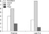
Postoperative VAS scores for radicular symptoms improved or resolved in all patients compared with preoperative states (Table 1). For Odom's criteria, excellent (33.3%) or good (50%) results were obtained at discharge, and patients returned to their preoperative employment and physical activity. At the last follow-up, 11 patients (91.7%) showed excellent or good clinical outcomes with respect to Odom's criteria, and 1 patient (8.3%) was fair at the last follow-up (Fig. 1). His follow-up MRI at 40 months after surgery showed recurrence of disc herniation. Although we recommended ACDF, he refused it and wanted conservative treatment. The postoperative MRI confirmed extensive decompression of the disc protrusion and widening of the cervical foraminal space. In serial follow-up with plain radiographs, the development of significant cervical kyphosis was not detected at the last follow-up. The mean preoperative segmental angulation was 12.4 degrees, and the mean postoperative segmental angulation was 12.0 degrees in our series. At the last follow-up, it was 17.6 degrees. Although two patients had complained of axial neck pain postoperatively, this resolved within 3 months. Furthermore, there were no surgery-related complications.
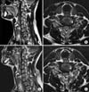
As an illustrative case, we discuss a 46-year-old man who had persistent neck pain and left arm radiating pain for 3 months. The preoperative MRI revealed a herniated disc to the intervertebral foramen of the C6-7 on the left side (Fig. 2A and B). He had been treated conservatively in other hospitals for 2 months; however, he had difficulty in everyday life owing to his neck and left arm pain. The patient underwent a left-sided posterior foraminotomy on C6-7, and we initially confirmed dural sac, nerve root, and a protruding disc in an operative microscopic view (Fig. 2C). After confirmation of the neural structure and disc space, disc fragments were removed via a small operative corridor by microprobe and CO2 laser. Last, we confirmed a freely decompressed cervical nerve root (Fig. 2D). Immediately after surgery, the VAS score of the arm decreased from 8 to 3. Postoperative MRI demonstrated removal of the herniated disc and widening of the intervertebral foramen (Fig. 3A and B).
DISCUSSION
Posterior laminectomy to treat cervical disc herniation was first reported by Mixter and Barr.7 The technique subsequently evolved to a small keyhole foraminotomy. However, the posterior procedure is considered an indirect decompression, because it leaves the anteriorly compressed lesion on the root owing to the difficult approach.8 For direct decompression of lesions such as bony spurs and disc fragments compressing the root, Robison and Smith in 1955 and Cloward in 1958 reported the anterior approach for discectomy and fusion. The anterior approach is recommended for central compressive lesions, especially in the clinical cases of myelopathy or bilateral symptoms. However, complications associated with additional procedures such as graft harvesting must be considered. The risk of graft-site complications has been reported to be up to 18%.9 Problems associated with ACDF have included loss of intervertebral height, pseudoarthroses, complications related to access, and adjacent segment degenerations caused by the loss of mobility.10 The evolution of arthrodeses minimizes the progression of pseudoarthroses; however, the problem of stresses in adjacent levels and adjacent segment degeneration with symptoms remains.1011 Hilibrand et al.2 reported that among patients who experienced ACDF, 2.9% of patients per year had symptomatic adjacent segment disease, 25.6% of patients developed adjacent segment disease within 10 years of the operation, and 7.5% of these patients required reoperation.
Anterior foraminotomy is another anterior-approach surgery for unilateral cervical radiculopathy. Anterior foraminotomy allows a direct approach to the anterior foraminal lesion and avoids fusion. This procedure eliminates some of the risks and complications associated with plating and fusion. However, concerns remain regarding vertebral artery injury, the development of spinal instability, and recurrence.12 An excessive resection of an uncovertebral joint may cause instability of motion of the segment and lead to a second operation.13 A 2% risk of permanent superior laryngeal and recurrent laryngeal nerve injury and a 0.25% risk of esophageal perforation has been reported. Risk of Horner's syndrome has also been reported.71415
The posterior approach is especially appropriate for unilateral radiculopathy caused by lateral or foraminal stenosis. Posterior foraminotomy allows decompression of the nerve root and avoids fusion and several visceral and soft tissue structures on the anterior neck. Furthermore, it can expose the involved nerve root directly and offer better visualization of the exiting nerve root.16 Because of decompression without the fusion, the associated complications such as graft dislodgement, graft site morbidity, plate and implant complications, and pseudoarthroses can be avoided. This approach can avoid several complications caused by manipulation of visceral structures on the anterior neck, such as damage to the trachea and esophagus.11 There are also no risks of cerebrovascular complications caused by manipulation of the vascular structures, and there is a low risk of vertebral artery injury after posterior foraminotomy compared with anterior cervical foraminotomy. Posterior foraminotomy can be performed unilaterally or bilaterally, at a variable number of levels, or in combination with another posterior-approach surgery such as laminectomy or laminoplasty. Bilateral foraminal disease at a single level is treated by the fenestration that includes bilateral foraminotomy while preserving spinous processes and intraspinous and supraspinous ligaments. However, with a posterior foraminotomy, it is difficult to remove the ventral lesion adequately owing to the relatively narrow operative field. Therefore, indirect decompression of the nerve root in some cases, especially on the calcified lesion, could be another potential disadvantage.
The primary concern should be to avoid or minimize manipulation of the root and spinal cord.1718 The surgical goal should be exact decompression under continuous visualization with concurrent minimization of surgery-related trauma and its possible consequences. The goal of posterior foraminotomy is to move the nerve root away from the ventral compressive lesions such as osteophytes. In this study, we used a CO2 laser to overcome this limitation. Since the first trial of a neodymium:yttrium-aluminum-garnet (Nd: YAG) laser during disc surgery of the lumbar spine, many reports about the effectiveness and usefulness of several kinds of lasers for disc surgery have been published.192021 Nerubay et al.21 reported that 50 patients who complained of unilateral radiating leg pain due to lumbar disc disease were successfully managed by percutaneous laser nucleolysis with a CO2 laser. Lee and Lee.6 reported that a CO2 laser enabled sufficient removal of extraforaminal or foraminal lumbar disc herniation via a narrow surgical window without excessive loss of the facet joint or the pars interarticularis.
In cervical disc disease, disc decompression with a percutaneous laser showed significant clinical benefits such as improvement of several symptoms in over 51% of patients observed during a mean period of 43 months.22 These studies strongly support that a CO2 laser can be useful for removing cervical discs. Additionally, when the CO2 laser is applied to a microscopic operation, it enables disc cysts to be readily removed and disc material to be easily vaporized. In this study, the lateral osteophyte of the uncinate process was decompressed adequately by using Kerrison rongeurs and a high-speed drill, and the ventrally protruded disc was removed by using a CO2 laser in cases of posterolateral or foraminal disc protrusion. After annulotomy with the CO2 laser, the protruded disc was carefully removed by microprobe and CO2 laser. Soft disc materials or even calcified components can be removed by laser. This technical advance can achieve minimal manipulation of the nerve root and spinal cord during direct satisfactory decompression despite the narrow working space.
The primary postoperative problem has been access-induced neck pain secondary to the subperiosteal detachment of muscle from bony structures. Some studies have reported that excessive removal of facet (more than 50%) or bilateral procedures at the same level could cause instability.2324 Postoperative kyphosis has been a major concern in some reports with the presence of cervical deformity, which has been a risk factor for kyphosis on the cervical spine. In those cases, the extent of facetectomy played a major role in causing postoperative kyphosis.12526 However, the extent of facet resection required during posterior foraminotomy is typically 25% and rarely exceeds 50%.2728 Particularly, the extent of laminotomy or facetectomy for decompression or disc removal can decrease if a CO2 laser is adequately used through the small space. Therefore, the incidence of segmental instability after surgery can be decreased by minimizing removal of the cervical facet joint.29 In our series, we could preserve the cervical facet joint more than 50% by using a CO2 laser for posterior cervical foraminotomy and discectomy, and segmental instability did not develop during the follow-up period. Therefore, minimal and unilateral paraspinal dissection and facetectomy for one- or two-level radiculopathy may not influence the development of postoperative cervical kyphosis because contralateral paraspinal muscles and midline ligamentous structures are preserved.
If segmental instability is suggested in the evaluation of preoperative flexion and extension views, posterior foraminotomy should be clearly excluded from the surgical options. On imaging studies, the evidence of a central compressive lesion, preexisting kyphosis, or myelopathy could be potential contraindications. In our studies, two patients complained of transient axial neck pain without the development of postoperative segmental instability. We thought that postoperative transient axial neck pain may have developed from an approach-related problem, such as paraspinal dissection or injury to the cervical medial branch during operation. In addition to the advantage of preserving the motion of the segment, adjacent segment disc degeneration is unlikely to occur in patients undergoing posterior foraminotomy. A large-scale, well-designed, randomized clinical trial for patients in this clinical scenario will be necessary to resolve this question.
In conclusion, posterior microscopic foraminotomy and discectomy using a CO2 laser must be considered within the surgical methods for degenerative cervical disc diseases. This technique is a good method in patients with appropriate alignment who do not have any instability. In particular, by using a microscope and CO2 laser, ventral lesions (protruding disc or osteophyte) can be decompressed with minimal manipulation of the nerve root. We consider this technique to be a sufficient and safe procedure in carefully selected cases for unilateral cervical radiculopathy. The limitation of this study was the small number of cases in a single institution. A study with a larger number and longer duration of follow-up will be required to clarify the effectiveness of this technique.


 XML Download