Journal List > Chonnam Med J > v.51(3) > 1074932

Yang, Jung, Ahn, Kim, Min, Bom, Lee, and Kim: Predictive Efficacy of Interim Positron Emission Tomography/Computed Tomography (PET/CT) for the Treatment of Aggressive Lymphoma

Abstract

The prognostic value of whole-body positron emission tomography/computed tomography (PET/CT) with 18F-fluoro-2-deoxy-D-glucose (FDG) shortly after the onset of induction chemotherapy or mid treatment could help to predict long-term clinical outcomes in patients with Hodgkin's or Non-Hodgkin's lymphoma. However, FDG is not a tumor-specific substance, and it may accumulate to the point of being detected in a variety of benign conditions or at physiologic anatomical sites, which may give rise to false-positive interpretation. In an attempt to standardize the reporting criteria for interim PET/CT, the First International Workshop on Interim PET in Lymphoma suggested visual response criteria with the Deauville five-point scale, and the standardized uptake value (SUV) has been investigated in comparison with this visual system. A quantitative approach using the measurement of maximal SUV (SUVmax) or the reduction rate of SUVmax (ΔSUVmax) might be more appropriate in early-response PET/CT for reducing false-positive rates or for decreasing interobserver variability in interpretation. In this review, the predictive efficacy of PET/CT is discussed for the treatment of aggressive lymphoma, especially in terms of an interim PET/CT-based prognostic model.

INTRODUCTION

Whole-body positron emission tomography (PET) with 18F-fluoro-2-deoxy-D-glucose (FDG) is a functional imaging modality used for staging and monitoring in the treatment of malignant lymphoma and has a higher sensitivity and specificity than conventional imaging. Residual abnormalities following chemotherapy, which result from the development of fibrosis or tumor necrosis, are seen in up to 64% of lymphoma patients.1,2,3 Conventional imaging, especially computerized tomography (CT), cannot reliably help in the differentiation between active tumors and fibrosis or necrosis.4,5,6,7 These limitations have restricted the predictive value of conventional CT concerning the clinical outcome of non-Hodgkin's lymphoma (NHL) and Hodgkin's lymphoma (HL).
PET/CT may be a more accurate tool than conventional imaging for assessing treatment effects, correctly identifying patients with residual disease, and predicting therapeutic outcomes. Furthermore, FDG uptake is a tool used to predict the therapeutic response during or after the course of treatment. Several studies have demonstrated the prognostic value of post-therapeutic FDG-PET or PET/CT in malignant lymphoma.8,9 Moreover, higher relapse rates and lower rates of event-free survival are observed in PET-positive than in PET-negative patients.10,11,12 FDGPET or PET/CT images can predict increased risk of treatment failure during or after primary chemotherapy. However, because FDG is not a tumor-specific substance, it may accumulate to the point of being detected in a variety of benign conditions, which may give rise to false-positive results. A correlation with findings on anatomical imaging such as CT is important for identifying changes resulting from primary chemotherapy. Thus, interim FDG-PET/CT has emerged as a powerful predictive method of assessing HL and NHL.
The predictive value of interim PET scans appears to be positively correlated with the International Prognostic Index (IPI), which is used to predict treatment outcome for NHL, and the International Prognostic Score, which is used for HL. Recent studies have demonstrated that FDGPET/CT, shortly after the initiation of induction chemotherapy or mid treatment, can predict long-term clinical outcomes in patients with HD or NHL.9,13,14,15,16 These studies categorized patients by PET-positive or PET-negative results on the basis of visual analysis, and subsequently compared the rates of relapse and progression-free or failurefree survival. The present review describes the role of PET/CT, especially interim PET or a PET/CT-based prognostic model, in the treatment of aggressive lymphoma.

STANDARD CRITERIA FOR INTERIM PET/CT

In an attempt to standardize the reporting criteria for interim PET/CT, the First International Workshop on Interim PET in Lymphoma suggested visual response criteria with use of the Deauville five-point scale (5-PS), and the standardized uptake value (SUV) has been investigated in comparison with this visual system.17 The use of a quantitative approach with the measurement of maximal SUV (SUVmax) or the reduction in SUVmax (ΔSUVmax) might be more appropriate in early-response PET/CT for reducing false-positive rates and for decreasing the interobserver variability in interpretation.18,19 However, ΔSUV max only partially reports on the tumor response because it reflects the changes in metabolic activity per representative tumor slice and not overall tumor activity. Metabolic tumor volume (MTV), which is defined as the volume of tumor tissue with increased FDG uptake, has been reported as an important independent prognostic factor that compensates for the deficiencies of SUV-based assessment in maliagnancies.20,21,22 SUV can only represent metabolic extent with the dependence of SUVmax on the whole dimension of target lesions, whereas MTV represents the amount of high metabolic tumor cells with the volumetric estimation of active tumor burden.

1. Visual assessment by deauville 5-point scores

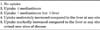
Patients were classified with 5-PS by interim PET/CT analysis based on the Deauville criteria as follows17: 1, no uptake; 2, uptake ≤mediastinum; 3, uptake >mediastinum but ≤liver; 4, uptake moderately increased compared to the liver uptake at any site; 5, markedly increased uptake compared to the liver at any site and/or new sites of disease (Table 1). Interim PET/CT images were graded as negative or positive by comparison with initial PET/CT. Grades 1 through 3 were considered as negative and grades 4 and 5 as positive.23 This grading process is independent of the size of the residual tumor.

2. Quantitative assessment based on SUVmax

Patients were classified by the quantitative analysis of 18F-FDG uptake changes based on the percentage reduction of SUVmax between the initial and interim PET/CT. On axial, coronal, or sagittal coregistered PET/CT slices, simple circular regions of interest (ROIs) were placed so as to cover the lesion or background. SUV measurements were corrected for body weight according to the following standard formula24:
Mean ROI activity (MBq/mL)/[injected dose (MBq)/body weight (kg)]
For each PET dataset, the maximum SUV (SUVmax) was defined as the highest SUV among all hypermetabolic tumor foci. The SUVmax reduction rate (ΔSUVmax) was calculated as follows:
ΔSUVmax (%)=100×[SUVmax (initial)-SUVmax (interim)] /SUVmax (initial)
If all lesions had disappeared on interim PET, ROIs were drawn in the same area on the interim PET as on the baseline PET.

3. Quantitative assessment based on MTV2.5

Patients were classified by the quantitative analysis of metabolic volume changes based on the percentage of MTV reduction (ΔMTV) between initial and interim PET/CT. To define the exact tumor margins around the target lesions, SUV2.5 was used as in previous reports, which means that the tumor volume area in PET/CT was delineated by a circle encompassing regions with an SUV cutoff value of 2.5.21,25 MTV2.5 was measured by use of the AW Volume Share™ workstation on the fused PET/CT images.22 The active MTV2.5 was measured in a 3-D manner by selecting volume of interest (VOI) on the axial image, and the size of the VOI was manually regulated on the corresponding coronal and sagittal images to include entire active tumors. The SUVmax and the sum of the tumor volumes in all hypermetabolic tumor foci were computed automatically by the program. The MTV2.5 reduction rate (ΔMTV 2.5) was calculated by use of the same formula as for the SUVmax reduction rate.

INTERIM PET/CT FOR THE TREATMENT OF DIFFUSE LARGE B CELL LYMPHOMA

Patients with diffuse large B cell lymphoma (DLBCL) are stratified into prognostic groups according to the IPI or molecular profiling.26,27 These therapeutic measures make it possible to predict survival after chemotherapy and to alter therapeutic strategies in groups with a poor risk classification, referred to as "risk-adapted therapy." Since the addition of the anti-CD20 monoclonal antibody, rituximab, to CHOP, the therapeutic outcomes of these patients have improved. The use of rituximab for the treatment of DLBCL has changed the prognostic risk groups based on the IPI, which is referred to as the revised IPI.28 However, there are still 20% to 40% of patients who will not be cured with R-CHOP, and the failure of R-CHOP chemotherapy might be associated with a less effective response to salvage chemotherapy.29 Therefore, there is a continuous effort to improve immune-chemotherapy and to determine which patients have a poor prognosis based on their response to treatment.
Despite the prognostic value of interim PET/CT response, which has important implications for responseadapted therapy in DLBCL, an optimal extension of the use and standardized definition of interim PET/CT is still being investigated (Table 2). The pitfalls of interim PET/CT interpretation may be related to the definition of positivity without concern for tumor physiology or anatomical variations. Aggressive NHLs have different clinical features with multifocal, noncontinuous involvement at diagnosis and involve both nodal and extranodal sites simultaneously compared with other solid malignancies. The prognostic significance of early PET scans after the first or second cycle of chemotherapy may be associated with false determination owing to tracer uptakes by inflammatory or infectious lesions.30,31,32 Quantitative SUV-based assessment at the time of early treatment response has been shown to increase the predictive value of interim PET over visual assessment or to be equivalent to visual assessment for determining midtherapy response.18,33,34,35 However, the quantitative assessment of SUVmax in the early response period has several drawbacks for defining positivity in cases of interobserver interpretation, for considering volumetric changes in the tumor during chemotherapy, and for interpreting minimal residual uptakes or physiologic anatomical FDG uptakes.
Although only a fraction of tumor cells are eradicated during the initial cycles of R-CHOP chemotherapy, it is at this point that the greatest rate of killing occurs.13,31,36 This is therefore an appealing time point at which to assess changes in metabolic activity as a surrogate marker for tumor sensitivity to treatment. It is intuitive that this could allow meaningful prognostic information to direct changes in or escalation of therapy. However, immunochemotherapy may lead to inflammatory changes within the tumor bed, leading to frequent false-positive PET assessments and thereby significantly decreasing the positive predictive value.37,38 Recent studies using the definition of visual or SUV-based assessments reported a low positive predictive value and a high negative predictive value in the treatment of DLBCL with immunochemotherapy. A low positive predictive value for visual or SUV-based assessments could make it difficult to intensify the treatment strategy because of concerns regarding over-treatment of a substantial portion of poor responders.4,32,36,39 In addition, interobserver mismatches in the interpretation of interim scans according to different visual criteria should caution us regarding the use of interim PET in response-adapted treatment.40 To compensate for these discrepancies and to better predict clinical outcomes, the combined evaluation of interim PET/CT response using visual, SUV-based, and MTV-based assessment may allow more differentiated prediction of individual prognosis in patients with DLBCL after the rituximab era.41 However, no definitive information is currently available on the role of interim PET/CT regarding which assessment or which standardized criteria should be applied in DLBCL.

INTERIM PET/CT FOR THE TREATMENT OF PERIPHERAL T CELL LYMPHOMAS

Peripheral T cell lymphoma (PTCL) is a heterogeneous group of aggressive lymphomas. The T-cell phenotype itself is associated with unfavorable prognosis compared to B-cell phenotype lymphomas.42 Although PTCL is chemosensitive to conventional regimens, the clinical outcomes have been uniformly disappointing. In particular, elderly patients generally have impaired bone marrow function, altered drug metabolism, comorbidities, and functional status impairment.43 They frequently show intolerance and treatment-related complications with full-dose salvage chemotherapy44,45,46 and may need to be frequently hospitalized during full courses of salvage chemotherapy. Although the prognostic role of interim PET/CT in PTCL is less well established than in DLBCL, some studies have suggested that the interim PET response may also be useful for predicting the outcome in PTCL. A retrospective study of mature T-cell and NK/T cell lymphoma reported that patients achieving interim PET/CT negativity showed improved 2-year progression-free survival and overall survival compared with those with interim PET/CT positivity.47 Another retrospective study yielded similar results regarding the prognostic role of interim PET/CT.48 In addition, the reviewer also reported that an analysis of 59 patients with PTCLs using interim PET/CT response based on 5-PS, ΔSUVmax, and ΔMTV2.5 after three and four courses of induction treatment had predictive value for progression-free survival; no significant difference was observed between the visual and quantitative assessments for predicting progression.49 However, the major drawback of these reports was the lack of uniform and reliable criteria for interim PET interpretation. Data concerning the role of PET/CT in patients with PTCLs using interim assessment with FDG uptake are limited. T/NK cell lymphomas are mostly FDG avid, with higher uptake in more aggressive subtypes but lower uptake in cutaneous disease.50,51 The advantage of using the baseline metabolic tumor parameters is the availability of prognostic information before treatment and thus allocation to the most optimal treatment intensity from the start. Song et al. measured MTV in extranodal NK/T-cell lymphoma patients and found that high MTV was correlated to poorer overall survival.52 If confirmed in more studies including in other PTCL patients, quantitative PET/CT studies could be an alternative to visual PET/CT analysis for patients with PTCL. Therefore, prospective studies are needed to determine the optimal use of interim PET/CT in PTCLs, including the role of visual or quantitative assessment.

CONCLUSION

Interim PET/CT analysis has significant predictive value for disease progression and survival in the treatment of aggressive lymphoma, and poor responders according to interim PET/CT should be considered for an alternative therapeutic plan or intensification of treatment. However, the visual assessment of interim PET/CT may make it hard to identify patients with poor prognosis as early as possible or to switch therapeutic modalities with intensification or stem cell transplantation for overcoming adverse clinical outcomes. Larger and prospective studies and harmonization of the criteria for interpreting interim PET/CT are needed to confirm the predictive efficacy in future clinical trials.

Figures and Tables

TABLE 1

Visual assessment based on a five-point scale

cmj-51-109-i001
TABLE 2

Predictive power of interim PET/CT in DLBCL

cmj-51-109-i002

DLBCL: diffuse large B cell lymphoma, EFS: event-free survival, PET/CT: positron emission tomography/computed tomography, PFS: progression-free survival, ΔSUVmax: reduction rate in the maximal standardized uptake value.

*SUV-based quantitative analysis.

ACKNOWLEDGEMENTS

This work was supported by a research grant from the Research Institute of Medical Sciences, Chonnam National University (2013-CURIMS-DR007).

Notes

CONFLICT OF INTEREST STATEMENT None declared.

References

1. Spaepen K, Stroobants S, Dupont P, Van Steenweghen S, Thomas J, Vandenberghe P, et al. Prognostic value of positron emission tomography (PET) with fluorine-18 fluorodeoxyglucose ([18F] FDG) after first-line chemotherapy in non-Hodgkin's lymphoma: is [18F]FDG-PET a valid alternative to conventional diagnostic methods? J Clin Oncol. 2001; 19:414–419.
crossref
2. Jerusalem G, Beguin Y, Fassotte MF, Najjar F, Paulus P, Rigo P, et al. Whole-body positron emission tomography using 18F-fluorodeoxyglucose for posttreatment evaluation in Hodgkin's disease and non-Hodgkin's lymphoma has higher diagnostic and prognostic value than classical computed tomography scan imaging. Blood. 1999; 94:429–433.
crossref
3. Mikhaeel NG, Timothy AR, O'Doherty MJ, Hain S, Maisey MN. 18-FDG-PET as a prognostic indicator in the treatment of aggressive Non-Hodgkin's Lymphoma-comparison with CT. Leuk Lymphoma. 2000; 39:543–553.
crossref
4. Cheson BD, Horning SJ, Coiffier B, Shipp MA, Fisher RI, Connors JM, et al. Report of an international workshop to standardize response criteria for non-Hodgkin's lymphomas. NCI Sponsored International Working Group. J Clin Oncol. 1999; 17:1244.
crossref
5. Surbone A, Longo DL, DeVita VT Jr, Ihde DC, Duffey PL, Jaffe ES, et al. Residual abdominal masses in aggressive non-Hodgkin's lymphoma after combination chemotherapy: significance and management. J Clin Oncol. 1988; 6:1832–1837.
crossref
6. Coiffier B, Gisselbrecht C, Herbrecht R, Tilly H, Bosly A, Brousse N. LNH-84 regimen: a multicenter study of intensive chemotherapy in 737 patients with aggressive malignant lymphoma. J Clin Oncol. 1989; 7:1018–1026.
crossref
7. Canellos GP. Residual mass in lymphoma may not be residual disease. J Clin Oncol. 1988; 6:931–933.
crossref
8. Kostakoglu L, Coleman M, Leonard JP, Kuji I, Zoe H, Goldsmith SJ. PET predicts prognosis after 1 cycle of chemotherapy in aggressive lymphoma and Hodgkin's disease. J Nucl Med. 2002; 43:1018–1027.
9. Querellou S, Valette F, Bodet-Milin C, Oudoux A, Carlier T, Harousseau JL, et al. FDG-PET/CT predicts outcome in patients with aggressive non-Hodgkin's lymphoma and Hodgkin's disease. Ann Hematol. 2006; 85:759–767.
crossref
10. Picardi M, De Renzo A, Pane F, Nicolai E, Pacelli R, Salvatore M, et al. Randomized comparison of consolidation radiation versus observation in bulky Hodgkin's lymphoma with post-chemotherapy negative positron emission tomography scans. Leuk Lymphoma. 2007; 48:1721–1727.
crossref
11. Zhao J, Qiao W, Wang C, Wang T, Xing Y. Therapeutic evaluation and prognostic value of interim hybrid PET/CT with (18)F-FDG after three to four cycles of chemotherapy in non-Hodgkin's lymphoma. Hematology. 2007; 12:423–430.
crossref
12. Fields PA, Mikhaeel G, Hutchings M, van der Walt J, Nunan T, Schey SA. The prognostic value of interim positron emission tomography scans combined with immunohistochemical data in diffuse large B-cell lymphoma. Haematologica. 2005; 90:1711–1713.
13. Haioun C, Itti E, Rahmouni A, Brice P, Rain JD, Belhadj K, et al. [18F]fluoro-2-deoxy-D-glucose positron emission tomography (FDG-PET) in aggressive lymphoma: an early prognostic tool for predicting patient outcome. Blood. 2005; 106:1376–1381.
crossref
14. Mikhaeel NG, Hutchings M, Fields PA, O'Doherty MJ, Timothy AR. FDG-PET after two to three cycles of chemotherapy predicts progression-free and overall survival in high-grade non-Hodgkin lymphoma. Ann Oncol. 2005; 16:1514–1523.
crossref
15. Hutchings M, Mikhaeel NG, Fields PA, Nunan T, Timothy AR. Prognostic value of interim FDG-PET after two or three cycles of chemotherapy in Hodgkin lymphoma. Ann Oncol. 2005; 16:1160–1168.
crossref
16. Hutchings M, Loft A, Hansen M, Pedersen LM, Buhl T, Jurlander J, et al. FDG-PET after two cycles of chemotherapy predicts treatment failure and progression-free survival in Hodgkin lymphoma. Blood. 2006; 107:52–59.
crossref
17. Meignan M, Gallamini A, Meignan M, Gallamini A, Haioun C. Report on the first international workshop on interim-PET- scan in lymphoma. Leuk Lymphoma. 2009; 50:1257–1260.
crossref
18. Itti E, Juweid ME, Haioun C, Yeddes I, Hamza-Maaloul F, El Bez I, et al. Improvement of early 18F-FDG PET interpretation in diffuse large B-cell lymphoma: importance of the reference background. J Nucl Med. 2010; 51:1857–1862.
crossref
19. Michallet AS, Trotman J, Tychyj-Pinel C. Role of early PET in the management of diffuse large B-cell lymphoma. Curr Opin Oncol. 2010; 22:414–418.
crossref
20. Cazaentre T, Morschhauser F, Vermandel M, Betrouni N, Prangère T, Steinling M, et al. Pre-therapy 18F-FDG PET quantitative parameters help in predicting the response to radioimmunotherapy in non-Hodgkin lymphoma. Eur J Nucl Med Mol Imaging. 2010; 37:494–504.
crossref
21. Zhu D, Ma T, Niu Z, Zheng J, Han A, Zhao S, et al. Prognostic significance of metabolic parameters measured by (18)F-fluorodeoxyglucose positron emission tomography/computed tomography in patients with small cell lung cancer. Lung Cancer. 2011; 73:332–337.
crossref
22. Wu X, Dastidar P, Pertovaara H, Korkola P, Järvenpää R, Rossi M, et al. Early treatment response evaluation in patients with diffuse large B-cell lymphoma--a pilot study comparing volumetric MRI and PET/CT. Mol Imaging Biol. 2011; 13:785–792.
crossref
23. Meignan M, Gallamini A, Haioun C, Polliack A. Report on the Second International Workshop on interim positron emission tomography in lymphoma held in Menton, France, 8-9 April 2010. Leuk Lymphoma. 2010; 51:2171–2180.
crossref
24. Rijbroek A, Boellaard R, Vriens EM, Lammertsma AA, Rauwerda JA. Comparison of transcranial Doppler ultrasonography and positron emission tomography using a three-dimensional template of the middle cerebral artery. Neurol Res. 2009; 31:52–59.
crossref
25. Hong R, Halama J, Bova D, Sethi A, Emami B. Correlation of PET standard uptake value and CT window-level thresholds for target delineation in CT-based radiation treatment planning. Int J Radiat Oncol Biol Phys. 2007; 67:720–726.
crossref
26. The International Non-Hodgkin's Lymphoma Prognostic Factors Project. A predictive model for aggressive non-Hodgkin's lymphoma. N Engl J Med. 1993; 329:987–994.
27. Rosenwald A, Wright G, Chan WC, Connors JM, Campo E, Fisher RI, et al. Leukemia Molecular Profiling Project. The use of molecular profiling to predict survival after chemotherapy for diffuse large-B-cell lymphoma. N Engl J Med. 2002; 346:1937–1947.
crossref
28. Sehn LH, Berry B, Chhanabhai M, Fitzgerald C, Gill K, Hoskins P, et al. The revised International Prognostic Index (R-IPI) is a better predictor of outcome than the standard IPI for patients with diffuse large B-cell lymphoma treated with R-CHOP. Blood. 2007; 109:1857–1861.
crossref
29. Mikhaeel NG. Interim fluorodeoxyglucose positron emission tomography for early response assessment in diffuse large B cell lymphoma: where are we now? Leuk Lymphoma. 2009; 50:1931–1936.
crossref
30. Castellucci P, Nanni C, Farsad M, Alinari L, Zinzani P, Stefoni V, et al. Potential pitfalls of 18F-FDG PET in a large series of patients treated for malignant lymphoma: prevalence and scan interpretation. Nucl Med Commun. 2005; 26:689–694.
crossref
31. Moskowitz CH, Schöder H, Teruya-Feldstein J, Sima C, Iasonos A, Portlock CS, et al. Risk-adapted dose-dense immunochemotherapy determined by interim FDG-PET in Advanced-stage diffuse large B-Cell lymphoma. J Clin Oncol. 2010; 28:1896–1903.
crossref
32. Han HS, Escalón MP, Hsiao B, Serafini A, Lossos IS. High incidence of false-positive PET scans in patients with aggressive non-Hodgkin's lymphoma treated with rituximab-containing regimens. Ann Oncol. 2009; 20:309–318.
crossref
33. Itti E, Lin C, Dupuis J, Paone G, Capacchione D, Rahmouni A, et al. Prognostic value of interim 18F-FDG PET in patients with diffuse large B-Cell lymphoma: SUV-based assessment at 4 cycles of chemotherapy. J Nucl Med. 2009; 50:527–533.
crossref
34. Lin C, Itti E, Haioun C, Petegnief Y, Luciani A, Dupuis J, et al. Early 18F-FDG PET for prediction of prognosis in patients with diffuse large B-cell lymphoma: SUV-based assessment versus visual analysis. J Nucl Med. 2007; 48:1626–1632.
crossref
35. Yang DH, Min JJ, Song HC, Jeong YY, Chung WK, Bae SY, et al. Prognostic significance of interim 18F-FDG PET/CT after three or four cycles of R-CHOP chemotherapy in the treatment of diffuse large B-cell lymphoma. Eur J Cancer. 2011; 47:1312–1318.
crossref
36. Casasnovas RO, Meignan M, Berriolo-Riedinger A, Bardet S, Julian A, Thieblemont C, et al. Groupe d'étude des lymphomes de l'adulte (GELA).SUVmax reduction improves early prognosis value of interim positron emission tomography scans in diffuse large B-cell lymphoma. Blood. 2011; 118:37–43.
crossref
37. Spaepen K, Stroobants S, Dupont P, Bormans G, Balzarini J, Verhoef G, et al. [(18)F]FDG PET monitoring of tumour response to chemotherapy: does [(18)F]FDG uptake correlate with the viable tumour cell fraction? Eur J Nucl Med Mol Imaging. 2003; 30:682–688.
crossref
38. Juweid ME, Smith B, Itti E, Meignan M. Can the interim fluorodeoxyglucose-positron emission tomography standardized uptake value be used to determine the need for residual mass biopsy after dose-dense immunochemotherapy for advanced diffuse large B-cell lymphoma? J Clin Oncol. 2010; 28:e719–e720. author reply e721-2.
crossref
39. Cashen AF, Dehdashti F, Luo J, Homb A, Siegel BA, Bartlett NL. 18F-FDG PET/CT for early response assessment in diffuse large B-cell lymphoma: poor predictive value of international harmonization project interpretation. J Nucl Med. 2011; 52:386–392.
crossref
40. Horning SJ, Juweid ME, Schöder H, Wiseman G, McMillan A, Swinnen LJ, et al. Interim positron emission tomography scans in diffuse large B-cell lymphoma: an independent expert nuclear medicine evaluation of the Eastern Cooperative Oncology Group E3404 study. Blood. 2010; 115:775–777. quiz 918.
crossref
41. Yang DH, Ahn JS, Byun BH, Min JJ, Kweon SS, Chae YS, et al. Interim PET/CT-based prognostic model for the treatment of diffuse large B cell lymphoma in the post-rituximab era. Ann Hematol. 2013; 92:471–479.
crossref
42. Armitage JO. The aggressive peripheral T-cell lymphomas: 2012 update on diagnosis, risk stratification, and management. Am J Hematol. 2012; 87:511–519.
crossref
43. Fields PA, Linch DC. Treatment of the elderly patient with diffuse large B cell lymphoma. Br J Haematol. 2012; 157:159–170.
crossref
44. Vose JM, Armitage JO, Weisenburger DD, Bierman PJ, Sorensen S, Hutchins M, et al. The importance of age in survival of patients treated with chemotherapy for aggressive non-Hodgkin's lymphoma. J Clin Oncol. 1988; 6:1838–1844.
crossref
45. Wunderlich A, Kloess M, Reiser M, Rudolph C, Truemper L, Bittner S, et al. German High-Grade Non-Hodgkin's Lymphoma Study Group (DSHNHL). Practicability and acute haematological toxicity of 2- and 3-weekly CHOP and CHOEP chemotherapy for aggressive non-Hodgkin's lymphoma: results from the NHL-B trial of the German High-Grade Non-Hodgkin's Lymphoma Study Group (DSHNHL). Ann Oncol. 2003; 14:881–893.
crossref
46. Limat S, Demesmay K, Voillat L, Bernard Y, Deconinck E, Brion A, et al. Early cardiotoxicity of the CHOP regimen in aggressive non-Hodgkin's lymphoma. Ann Oncol. 2003; 14:277–281.
crossref
47. Li YJ, Li ZM, Xia XY, Huang HQ, Xia ZJ, Lin TY, et al. Prognostic value of interim and posttherapy 18F-FDG PET/CT in patients with mature T-cell and natural killer cell lymphomas. J Nucl Med. 2013; 54:507–515.
crossref
48. Casulo C, Schöder H, Feeney J, Lim R, Maragulia J, Zelenetz AD, et al. 18F-fluorodeoxyglucose positron emission tomography in the staging and prognosis of T cell lymphoma. Leuk Lymphoma. 2013; 54:2163–2167.
crossref
49. Jung SH, Ahn JS, Kim YK, Kweon SS, Min JJ, Bom HS, et al. Prognostic significance of interim PET/CT based on visual, SUVbased, and MTV-based assessment in the treatment of peripheral T-cell lymphoma. BMC Cancer. 2015; 15:198.
crossref
50. Zinzani PL. PET in T-Cell Lymphoma. Curr Hematol Malig Rep. 2011; 6:241–244.
crossref
51. Kako S, Izutsu K, Ota Y, Minatani Y, Sugaya M, Momose T, et al. FDG-PET in T-cell and NK-cell neoplasms. Ann Oncol. 2007; 18:1685–1690.
crossref
52. Song MK, Chung JS, Lee JJ, Jeong SY, Lee SM, Hong JS, et al. Metabolic tumor volume by positron emission tomography/computed tomography as a clinical parameter to determine therapeutic modality for early stage Hodgkin's lymphoma. Cancer Sci. 2013; 104:1656–1661.
crossref
TOOLS
Similar articles