

 PDF
PDF ePub
ePub Citation
 Print
Print


INTRODUCTION
Transcatheter closure of patent ductus arteriosus (PDA) by use of the Amplatzer duct occluder shows good results in properly selected patients.1 The advantages of the Amplatzer duct occluder device include a user-friendly delivery system, the ability to retrieve or reposition the device when required, and a high complete closure rate (>99% complete occlusion of PDA within 6 months of implant).2 Although embolization of an Amplatzer duct occluder is rare, it can occur. We present a case of retrieval of an embolized Amplatzer duct occluder by use of a percutaneous method in a young man.
CASE REPORT
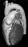
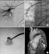
A 33-year-old man presented with exertional dyspnea. He had no specific medical history. Transthoracic two-dimensional echocardiography and computed tomography (CT) confirmed PDA (Fig. 1). For transcatheter closure, we deployed the Amplatzer duct occluder II (9-PDA2-05-06, AGA Med. Corp., USA) after confirmation of the proper position by repeated angiogram. However, 5 min after successful deployment, the Amplatzer duct occluder device was suddenly embolized into the branch of the right pulmonary artery (RPA) (Fig. 2A). Before surgical removal, we decided to remove the embolized device by use of a percutaneous method after obtaining consent from the patient's relatives. Multiple attempts at removal by use of guidewire twisting, a goose-neck snare, and a radiofrequency ablation catheter were unsuccessful. Because the embolized device was floating in the branch of the RPA, it moved into the distal portion of the RPA after several attempts of retrieval with the snare and ablation catheter. In order to stabilize the embolized device, a 5.0×20 mm balloon catheter was placed just distal to the embolized device and inflated. Endomyocardial biopsy forceps (Cordis Corp., USA) were introduced from the left femoral vein through an 8-Fr sheath. After multiple attempts, we caught the screw attachment tip of the embolized device and removed it into the 8-Fr shuttle sheath (Fig. 2B). The entire device was then easily removed (Fig. 2C, attached video file). Subsequently, an Amplatzer duct occluder 8-6 (9-PDA-005) was carefully deployed via a venous route. The device was confirmed to be properly positioned across the defect by echocardiography and the CT angiogram (Fig. 2D). Neither clinical complications nor a residual shunt was observed during the 6-month follow-up.
DISCUSSION
Transcatheter PDA closure is currently the standard of therapy and is performed with high success rates and few complications.3 Although the rate of device embolization is rare (<0.5%), this remains a major complication requiring urgent surgical management.4 The most common cause of embolization is the use of an undersized device. Therefore, exact PDA sizing and the use of the proper size of device is essential to avoid this potential complication. Careful attention must be paid to imperfect alignment or malposition of the device or excessive tension on the delivery cable; performing the wiggle procedure may reduce the acute failure rate of the procedure.5 Excessive tension on the delivery cable was the main cause of embolization in our patient. In this case, stabilizing the floating device by balloon inflation played a pivotal role in success. The use of a larger 8-Fr sheath during retrieval could make easier device entry into the sheath after capture of the embolized device. In general, the following techniques are recommended to increase the chances of successful retrieval: the use of a sheath 2 Fr sizes larger than the delivery sheath, the use of stiff sheaths to avoid kinking, and cutting a bevel or notch at the end of the sheath to facilitate the entrance of the embolized device.6
Our case illustrates a practical transcatheter removal technique for an embolized Amplatzer duct occluder. Even in procedures performed by the most experienced operators, device embolization can occur. Thus, physicians have to keep in mind both the possibility and the treatment of this severe complication during PDA closure.
 XML Download