

 PDF
PDF ePub
ePub Citation
 Print
Print


INTRODUCTION
Rice is a staple food in many countries. In addition, rice, particularly glutinous rice, has been used for the treatment of gastrointestinal disorders in folk medicines in Korea and other countries. Recently, rice extract was reported to protect the gastric mucosa from toxic agents. These reports included the following findings: i) Rice extract protected the gastric mucosa from ethanol-induced injury in rats.1 ii) Rice extract prevented NaCl-induced gastric mucosal damage and cell proliferation in rats.2 iii) Rice extract attenuated the inflammatory changes of the gastric mucosa induced by Helicobacter pylori infection in Mongolian gerbils.3 The rice extract used in the above studies was prepared by saccharization of rice with Aspergillus oryzae followed by fermentation with Saccharomyces cerevisiae. The extract contained high concentrations of saccharides but no protein and no lipid.
Peptic ulcers are believed to result from the imbalance between protective and aggressive factors.4,5 The gastroprotective factors include mucus, mucosal antioxidants, prostaglandins and growth factors, and gastric blood flow. The aggressive factors, such as increased gastric secretion, increased generation of reactive oxygen species and pro-inflammatory cytokines, and infection with Helicobacter pylori, can cause mucosal damage. Researchers have developed many in vivo gastric ulcer models with different gastropathic mechanisms.6 Among them, the ethanol-, indomethacin-, and water immersion restraint stress (WIRS)-induced models have been used in many studies to screen antiulcer substances,7,8,9 because alcohol, nonsteroidal anti-inflammatory drugs, and stress are major risk factors in human life for peptic ulcers and other gastrointestinal diseases.
We have observed that the aqueous extract of glutinous rice (GRE) very effectively protects the gastric mucosa from injury induced by ethanol, indomethacin, and WIRS treatments in rats. Also, we have found that the active substances of GRE responsible for the protection differ from those of the rice extract used in earlier studies.1,2,3 The present study assessed the gastroprotective activity of GRE as exerted in the above-mentioned ulcer models and presents some characteristics of the gastroprotective activity and related GRE substances.
MATERIALS AND METHODS
1. Materials
Three cultivars each of glutinous (Dongjinchal, Baikokchal, and Sinsunchal) and nonglutinous (Hopum, Sindongjin, and Saenuri) rice were supplied from National Agricultural Products Quality Management Service (NAQS) of Korea. Tea extracts were prepared by extracting green and black teas (Amorepacific, Seoul, Korea) with 100 volumes of boiling distilled water for 10 min and filtering and freeze-drying the resultant solutions. Ethanol was purchased from Merck KGaA (Darmstadt, Germany) and indomethacin was from Sigma-Aldrich Co. (St. Louis, MO, USA). All other reagents were of the analytical grade commercially available.
2. Animals
Male Sprague-Dawley rats (230-250 g) were housed in cages with mesh bottoms to prevent coprophagia in an air-conditioned room with a 12-h dark-light cycle and were allowed free access to water and food. Prior to the experiments, the animals were fasted, but allowed water, for 36 h. This study was performed in accordance with the Institutional Guidelines for Experimental Animal Care and Use of Chonnam National University Medical School.
3. Preparation of rice extracts and administration to animals
Rice was ground to pass through a 0.18-mm screen (Daehwa Precision, Cheonan, Korea). Rice flour was added with 10 volumes of distilled water and agitated vigorously for 30 s with a vortex mixer 3 times. After agitation, the mixture was centrifuged at 1,000×g for 10 min to obtain a supernatant. The supernatant was then freeze-dried and stored at 4℃. When administered to animals, the freeze-dried rice extract was suspended in distilled water and administered to 36-h fasted rats by intragastric gavage in a single dose of 0, 10, 30, 100, or 300 mg/kg in 4 mL/kg.
4. Induction and determination of gastric mucosal lesions
1) Ethanol-induced lesion
Thirty minutes after administration of the rice extract, rats were orally administered 4 mL/kg ethanol to induce gastric lesions.10 One hour after the ethanol treatment, the animals were killed and their stomachs were removed. The stomachs were cut along the greater curvature and rinsed gently with saline. Then, the gastric mucosal surface was photographed with a digital camera and the area of hemorrhagic lesions was measured with a software (Image Tool) by a rater who was unaware of the experimental protocol. The lesion index was expressed as the percentage of the hemorrhagic lesions of the whole corpus mucosa. The inhibition rate of gastric lesion formation (IR) in the rice extract-treated group was calculated as follows: IR (%)=(lesion index of control group-lesion index of sample-treated group)/lesion index of control group ×100.
2) Indomethacin-induced lesion
Thirty minutes after administration of the rice extract, rats were orally administered 30 mg/kg indomethacin dissolved in 3% NaHCO3 to induce gastric lesions.6 Five hours after the indomethacin treatment, the rats were killed and gastric mucosal lesions were determined as described above.
3) WIRS-induced lesion
Thirty minutes after administration of the rice extract, the rats were placed in a restraint device and immersed up to their xiphoid process in a water bath at 22℃ for 4 h to induce gastric lesions.11 After the WIRS treatment, the rats were killed and gastric mucosal lesions were determined as described above.
5. Determination of gastric mucin content
1) Total mucin content in gastric mucosa
One hour after the GRE treatment, the rats were killed and their stomachs were removed and opened. Mucosa in the glandular stomach was removed by scraping with a blunt knife. Mucosal preparations from three animals were pooled, lyophilized, and powdered for extraction of mucin as described by Ichikawa et al.12 The sample was homogenized in 50 mM Tris-HCl buffer, pH 7.2, containing 2% Triton X-100 and incubated at 37℃ for 1 h. After centrifugation at 8,000×g for 30 min at 4℃, an aliquot of the supernatant was applied to a Bio-Gel A-1.5m column (Bio-Rad, Hercules, CA, USA) and eluted with the above Tris-Triton buffer. The hexose content in the eluates was measured by the phenol-sulfuric acid method13 with galactose as the standard, and the void volume fraction was collected as mucin. Mucin content was expressed as hexose content per stomach.
2) Gastric mucus content
After the rats were treated with GRE and ethanol, they were sacrificed and their gastric mucus content was measured by the Alcian blue method.14 The glandular portion of the stomach was immersed in 10 mL of 0.1% Alcian blue containing 0.16 M sucrose and 50 mM sodium acetate, pH 5.8, for 2 h and rinsed twice with 10 mL of 0.25 M sucrose for 20 min. The dye complexed with the gastric mucus was then extracted with 0.5 M MgCl2 for 2 h. An aliquot of the blue extract was vortex-mixed vigorously with an equal volume of diethyl ether and the mixture was centrifuged at 1,500×g for 10 min. The absorbance of the aqueous layer was read at 580 nm.
6. Determination of nonprotein sulfhydryl (NP-SH) concentration in gastric mucosa
This was done as described by Szabo et al.15 One hour after the GRE treatment, gastric mucosa was obtained from rats as described above. The mucosa was homogenized in 40 volumes of 20 mM EDTA and the NP-SH concentration in the homogenate was measured with Ellman's reagent as described by Sedlak and Lindsay.16
7. Separation of the particulate (GREP) and soluble (GRES) fractions of GRE
GRE was allowed to stand overnight at room temperature until it became aggregated and formed a precipitate. After centrifugation at 1,000×g for 10 min, the precipitate was used as GREP after freeze-drying and the soluble part was referred to as GRES.
8. Isolation of proteins from glutinous rice flour by the Osbourne method
Different kinds of proteins were extracted from glutinous rice flour on the basis of their solubility at room temperature in water, 5% NaCl, 1% lactic acid, and 70% ethanol by the procedure described by Ju et al.17 with minor modifications. Glutinous rice flour (50 g) was defatted with 200 mL hexane and dried in a fume hood at room temperature for 24 h. The defatted flour was then extracted by shaking with 200 mL distilled water for 4 h and centrifuged at 3,000×g for 30 min to isolate the soluble albumin fraction. After water extraction, the flour was similarly extracted with 200 mL of 5% NaCl to isolate the globulin fraction. The flour was then extracted with 200 mL of 1% lactic acid for 1 h to isolate the glutelin fraction, followed by extraction with 200 mL of 70% ethanol for 4 h to isolate the prolamin fraction. Each extraction step was repeated twice. The protein fractions were then dialyzed against distilled water, lyophilized, and stored at 4℃.
RESULTS
1. Gastroprotective effects of GRE in different gastric ulcer models
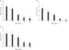
Hemorrhagic lesions of a linear type were observed in gastric mucosa after rats were treated with ethanol, indomethacin, or WIRS. The lesion indices were much higher in the ethanol-treated rats than in the indomethacin- and WIRS-treated rats (controls in Fig. 1). When the rats were pretreated with GRE prepared from the cultivar Dongjinchal, the gastric lesions were obviously reduced in all the ulcer models. As shown in Fig. 1, GRE dose-dependently blocked the gastric lesions induced by ethanol, indomethacin, and WIRS treatments up to more than 70%. The efficacy of GRE, however, differed among the ulcer models. GRE could significantly block the ethanol-induced gastric lesion at the dose of 30 mg/kg, whereas it could significantly block the lesions induced by indomethacin and WIRS at the dose of 100 mg/kg.
2. Comparison of gastroprotective effects of glutinous (GRE) and nonglutinous (NGRE) rice extracts
Rice extracts were prepared from three cultivars of glutinous and nonglutinous rice, respectively, and their gastroprotective effects were compared at 100 mg/kg in the ethanol and indomethacin ulcer models. As shown in Table 1, all GREs exerted much stronger protective effects against ethanol- and indomethacin-induced gastric lesions than did the nonglutinous rice extracts (NGREs) tested, although the NGREs also showed considerable degrees of protection against both lesions. In the following experiments of this study, the cultivar Dongjinchal was used for the preparation of GREs.
3. Mechanisms for the gastroprotective action of GRE
Because GRE showed effective protection against ethanol-, indomethacin-, and WIRS-induced gastric ulcers, we examined the effects of GRE on the levels of two important protective factors in gastric mucosa: mucin and nonprotein sulfhydryls (NP-SHs).
1) Effect of GRE on the total mucin content in gastric mucosa
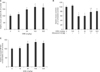
To study the effect of GRE on the synthesis of gastric mucin, the GRE-induced change in the total gastric mucin content was determined. The dose-related changes in the gastric mucin content following GRE administration to rats are shown in Fig. 2A. The average mucin content of the control was 0.86 mg hexose per stomach. The mucin content significantly increased to 148%, 167%, and 163% of the control with GRE treatment at 30, 100, and 300 mg/kg, respectively.
2) Effect of GRE on the gastric mucus content
To study the effect of GRE on the gastric mucin release, GRE- and ethanol-induced changes in the content of the mucus layer lining the surface of the gastric mucosa were determined on the basis of Alcian blue binding ability (Fig. 2B). GRE administration at 100 mg/kg alone did not induce a significant change in the mucus content. Ethanol treatment (4 mL/kg) reduced the mucus content to 58.9% of the control, which is consistent with earlier observations.18,19 When GRE was administered before ethanol treatment, the ethanol-induced reduction in the mucus content was partially blocked.
3) Effect of GRE on the NP-SH concentration in gastric mucosa
The dose-related changes in the gastric mucosal NP-SH concentration following GRE administration to rats are shown in Fig. 2C. Average NP-SH concentration of the control was 0.68 µmol/g tissue. The NP-SH concentration significantly increased to 132% and 137% of the control with GRE treatment at 100 and 300 mg/kg, respectively.
4. Identification of gastroprotective substances in GRE
1) Distribution of gastroprotective activity in the particulate (GREP) and soluble (GRES) fractions of GRE
For this study, GRE of 25 mg/mL was allowed to stand overnight at room temperature and centrifuged at 1,000×g for 10 min. The resultant soluble fraction was used as GRES, and the precipitate was suspended in the starting volume of distilled water to use as GREP. To make the GREP plus GRES sample, the precipitate was suspended in the starting volume of GRES. The GRE, GREP, GRES, and GREP plus GRES samples thus obtained were administered to rats at a dose of 4 mL/kg and their protective effects against ethanol-induced ulcers were determined. The resulting lesion indices for the different experimental groups were as follows (N=8): distilled water control, 23.7±3.9%; GRE, 5.1±2.1%; GREP, 12.3±3.0%; GRES, 21.9±3.5%; and GREP plus GRES, 5.9±1.8%. GREP showed a significant gastroprotective activity, although the activity was far weaker than that of GRE. GRES did not show a significant gastroprotective activity. However, it could enhance the activity of GREP when the two fractions were combined. This result suggests that active substances responsible for the gastroprotective effect of GRE are present in GREP and that the active substances may need some soluble activators for full activity.
2) Enhancement of the gastroprotective activities of GRE and GREP by tea extracts
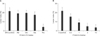
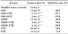
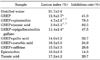
We have observed that tea extracts can enhance the gastroprotective activities of GRE and GREP at such low doses that alone they do not exert a significant gastroprotective effect. As shown in Table 2, the activities of GRE and GREP were markedly enhanced by co-treatment with 8 mg/kg green tea or black teaextract. As shown in Fig. 3, the black tea extract dose-dependently increased the gastroprotective activities of GRE and GREP at doses of 0 to 8 mg/kg. Green tea extract also dose-dependently increased the activities of GRE and GREP under the condition of Fig. 3 (data not shown). Of the tea constituents, epicatechin and tannic acid showed significant enhancing effects for the gastroprotective activity of GREP at 40 µmol/kg, whereas epigallocatechin gallate, gallic acid, ascorbic acid, and caffeine did not (Table 3).
3) Effects of heat and pepsin treatments and solvent extraction on the gastroprotective activity of GREP
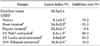
The gastroprotective activity of GREP was found to be very labile to heat or pepsin treatment (Table 4). When GREP was heat-treated at 80℃ for 3 min, its activity was totally lost. GREP also lost its gastroprotective activity by incubation with 0.01% pepsin at 37℃ for 1 h. These results strongly suggest that the active substances in GREP are proteins. In an effort to explore which proteins, if any, are involved in the gastroprotective activity of GREP, GREP was extracted with different solvents to remove different proteins: 5% NaCl, 1% lactic acid, and 70% ethanol were used as solvents to remove globulins, glutelins, and prolamins, respectively. As shown in Table 4, extraction with 5% NaCl and 1% lactic acid did not cause a significant change in the remaining gastroprotective activity of GREP. With 70% ethanol extraction, however, GREP markedly lost its gastroprotective activity. These results suggest that prolamins are involved in the gastroprotective activity of GREP.
4) Gastroprotective activity of the prolamin fraction isolated from glutinous rice flour
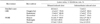
To study further the gastroprotective activities of glutinous rice proteins, the albumin, globulin, glutelin, and prolamin fractions were isolated sequentially from glutinous rice flour by the Osbourne method and their gastroprotective activities were determined (Fig. 4). From 50 g of glutinous rice flour, 0.28, 0.32, 1.09, and 0.24 g of albumin, globulin, glutelin, and prolamin fractions were isolated, respectively. Among the isolated fractions, only the prolamin fraction showed a significant gastroprotective activity at 1 to 10 mg/kg in the presence of 8 mg/kg black tea extract. These results support the above assumption that prolamins are involved in the gastroprotective activity of GREP.
DISCUSSION
In this study, GRE effectively protected the gastric mucosa from injury induced by ethanol, indomethacin, and WIRS. GRE exerted significant effects dose-dependently at doses of 30 to 300 mg/kg against ethanol-induced ulcers and at doses of 100 to 300 mg/kg against indomethacin- and WIRS-induced ulcers (Fig. 1). When rice extracts prepared from different cultivars of glutinous and nonglutinous rice were compared for their gastroprotective effects in the ethanol and indomethacin ulcer models, GREs showed much stronger effects than NGREs, although NGREs also showed considerable effects (Table 1). Considering that alcohol, nonsteroidal anti-inflammatory drugs, and stress are major risk factors for peptic ulcer and other gastrointestinal diseases, our results indicate that GRE may be useful for the prevention and treatment of gastritis and peptic ulcer.
It is well known that gastric mucin is depleted in animals treated with ethanol,18,19 indomethacin,20 and WIRS.21 Because the gastric mucin is important to protect the mucosa from physical and chemical stimuli and back diffusion of hydrogen ions and pepsin, depletion of mucin may result in gastropathy.18,22 Our results (Fig. 2A and B) showed that the GRE treatment increased the total gastric mucin content but did not alone cause a significant change in the content of the mucus layer lining the gastric mucosal surface. These results suggest that GRE stimulates mucin synthesis in the gastric mucosa but not its secretion into gastric lumen. However, pretreatment with GRE before ethanol treatment could partially block the ethanol-induced depletion of gastric mucus (Fig. 2B). This effect might be due to the fact that the intramucosal mucin content was increased by GRE treatment. Accordingly, more mucin might be secreted from the mucosa by the subsequent ethanol stimulation in the GRE-pretreated rats than in the control rats without GRE pretreatment. The exact mechanisms for the changes in gastric mucin content by GRE treatment remain to be clarified.
Free radicals are also involved as a major aggressive factor in the pathogenesis of gastric ulcers induced by ethanol, 23 indomethacin,24 and WIRS,25 which is supported by the facts that lipid peroxide levels are increased26,27,28 and the major antioxidants (including reduced glutathione; vitamins A, C, and E; and enzymes such as superoxide dismutase, catalase, and glutathione peroxidase) are depleted in the insulted gastric tissues.26,29 Among the antioxidants, NP-SH compounds play a pivotal role in gastroprotection from free radicals and maintenance of gastric mucosal integrity.30,31,32 NP-SHs react with reactive oxygen species to transform them to inert products and also keep sulfhydryl groups of proteins in reduced form.33,34 They also help in recycling endogenous antioxidant vitamins, thereby preventing lipid peroxidation.20 In addition, NP-SHs scavenge superoxide anions, thus preventing their interaction with nitric oxide to form peroxynitrite. Nitric oxide thus preserved can improve tissue perfusion and attenuate neutrophil infiltration and free radical generation.35 The facts that NP-SH concentration in the gastric mucosa is relatively high36 and treatment of animals with sulfhydryl blockers results in induction of gastric mucosal injury37 suggest the importance of NP-SH in gastroprotection and maintenance of gastric mucosal integrity. In the present study, GRE treatment significantly increased the NP-SH concentration in gastric mucosa, which suggests that the gastroprotective effect of GRE may involve the mechanisms by which it enhances the NP-SH concentration in gastric mucosa.
In this study, the gastroprotective activity of GREP was totally destroyed by heat treatment at 80℃ for 3 min or treatment with 0.01% pepsin at 37℃ for 1 h, and the activity of GREP was decreased markedly by extraction with 70% ethanol (Table 4). In addition, the prolamin fraction isolated from glutinous rice flour by the Osbourne method exhibited a remarkable gastroprotective activity in the presence of black tea extract as an activator (Fig. 4). These results suggest that prolamins are responsible, at least in part, for the gastroprotective activity of GRE. This article is the first to describe the gastroprotective activity of prolamin. According to Jayaraj et al,38 fresh rice bran and its oil showed a gastroprotective effect but stored rice bran and its oil were ulcerogenic in pylorus ligated rats. Those authors suggested that unsaturated fatty acids play a critical role in the above actions of rice bran and its oil. Matsuhashi et al.1 reported that rice extract, which was prepared through saccharization of rice with Aspergillus oryzae and fermentation with Saccharomyces cerevisiae, protected rat gastric mucosa from ethanol-induced injury, and the protective activity was stable to heat treatment at 80℃ for 3 min. The rice extract was reported to contain mainly saccharides but no protein and no lipid.3 Therefore, it seems that our GRE preparation is a novel rice extract with gastroprotective activity whose active compounds differ from those of the rice preparations used by the above investigators.
The gastroprotective action of GRE was markedly enhanced by co-treatment with tea extracts. It is uncertain how the tea extracts enhance the GRE action. We observed that tea constituents such as epicatechin and tannic acid showed significant enhancing effects for the GREP action at 40 µmol/kg, whereas epigallocatechin gallate, gallic acid, ascorbic acid, and caffeine did not (Table 3). Some polyphenol compounds present in tea extracts may keep the functional groups of GRE prolamins in reduced forms to make the proteins active. Another possibility is that tea extracts may increase, synergistically with GRE, the levels of protective factors including gastric mucosal NP-SH and mucin. To apply GRE clinically for the prevention and treatment of gastritis and peptic ulcer, it is recommended to use a combination therapy with GRE and tea extracts. Clinical studies are required for evaluation of this point. Also, further studies are needed to clarify the mechanisms of the gastroprotective actions of GRE and tea extracts.

 XML Download