

 PDF
PDF ePub
ePub Citation
 Print
Print


WHAT CAUSES FREQUENT URINARY TRACT INFECTION AND GROSS HEMATURIA?
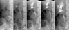
A 31-year-old woman suffered from frequent urinary tract infection and intermittent gross hematuria. Ultrasound disclosed unilateral hydronephrosis and intravenous urography (IVU) showed delayed visualization of right kidney. Therefore, retrograde pyelography (RP) was performed. We found that dynamic imaging exhibited "firework" appearances and static imaging showed "bouquet of flower" appearance (Fig. 1).
DIAGNOSIS: MEDULLARY SPONGE KIDNEY
These images are typical characteristic of medullary sponge kidney (MSK) on IVU. Firework appearance is a description given to the appearances of medullary sponge kidney on consecutively dynamic retrograde pyelography (RP) images. From a fireball to a glamour firework of explosion of the series of pictures is what we want to define as a trait of MSK on RP. The static RP images also showed bouquet of flowers appearance similar to those from a traditional IVU. Most MSK patients are asymptomatic and will not be diagnosed until they are complicated by renal colic, urinary tract infections, or hematuria. They are liable to have kidney stone formation due to hypercalciuria, urine stasis in the dilated collecting tubules and impaired distal tubular acidification. Treatment, if any, depends on the underlying conditions such as recurrent stone formation and urinary tract infection.1 Ultrasound, IVU, CT urogram and magnetic resonance imaging (MRI) might be useful to detect MSK.2 RP provides an extraordinary efficient tool to diagnose MSK or other etiologies of obstructive uropathy.
 XML Download