

 PDF
PDF ePub
ePub Citation
 Print
Print


INTRODUCTION
Radiotherapy or chemical therapy in cancer patients causes xerostomia and reduces saliva flow rates1 and may induce highly specific changes in microbiota.2 In addition, low saliva flow rates may be associated with markedly increased susceptibility to dental caries.3 The major organisms responsible for caries are mutans streptococci and lactobacilli4; of these, Streptococcus mutans and Streptococcus sobrinus are generally considered to be the primary etiological bacteria of human dental caries.5,6 Furthermore, Lactobacillus species have been reported to occur in high numbers in caries sites.7,8 In particular, Lactobacillus salivarius and Lactobacillus acidophilus are predominant species in the mouth.9
Investigations of bacterial profiles in saliva for the identification of caries risk groups are popular and produce reliable results.10 A variety of methods, such as conventional culture methods, direct enzyme tests, enzyme-linked immunosorbent assays, and conventional end-point polymerase chain reaction (PCR) have been used to detect and identify oral microbes.11-13 However, these methods do not allow for accurate quantification; thus, reliance on them means that important diagnostic aspects are overlooked. However, quantitative real-time PCR (qRT-PCR) is a highly specific, relatively fast, and sensitive means of detecting and quantifying bacteria as compared with conventional culture methods and end-point PCR.14
Study of the prevalences of oral microbes in cancer patients is important because it provides basic data that aid in control of the oral complications of cancer therapies. Furthermore, the availability of an accurate quantitative assay for the detection of cariogenic bacteria could facilitate the monitoring of therapies and enable more accurate epidemiological studies on the progression of caries. Recently, our research group showed that PCR can be used to compare the frequencies of oral microbes in the saliva of oncological patients and healthy controls.15 However, relatively few data are available on the quantification of salivary caries-associated bacteria in oncological patients by qRT-PCR.
The aim of this study was to determine and compare the physiologic values of salivary flow, pH, and the levels of S. mutans, S. sobrinus, L. salivarius, and L. acidophilus in saliva samples from oncological patients and healthy controls by use of qRT-PCR.
MATERIALS AND METHODS
1. Subjects and saliva collection
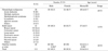
The study population consisted of 40 systemically healthy control subjects and 71 cancer patients, which included 30 patients with a hematologic malignancy (HE) and 41 patients with a solid tumor (SO) who visited Chonnam National University Hwasun Hospital for an oral examination. The 111 study subjects comprised 55 men and 56 women ranging in age from 13 to 78 years (mean, 53.6±14.4 years); their characteristics are shown in Table 1. None of the patients had received antibiotics or irradiation therapy during the preceding 3 months. Etiological factors including smoking and alcohol were not analyzed. All subjects signed an informed consent form approved by the Ethics Committee of Chonnam National University Hwasun Hospital (HCRI 09 032-3).
The subjects were asked to refrain from eating, drinking, and dental hygiene control for a minimum of 1 h before sialometry. Subjects were asked to hold their head slightly forward and to expectorate accumulated saliva into a collection tube (SPL, Pocheon, South Korea) and to take care not to swallow during the 5-min collection period. Saliva was expectorated into the tube at 1-min intervals. Amounts of saliva collected were measured in milliliters to gauge salivary flow, and salivary pH was measured with a pH meter (pH-200L; iStek, Seoul, South Korea). Saliva samples were stored immediately at -20℃ before genomic DNA extraction.
2. Bacterial strains
S. mutans Ingbritt, S. sobrinus KCTC 3308, L. salivarius KCTC 3157, and L. acidophilus KCTC 3164 were used as reference strains. S. mutans and S. sobrinus were grown in brain heart infusion broth (BHI broth; Difco, Detroit, MI, USA), and L. salivarius and L. acidophilus were grown in De Man, Rogosa, Sharpe broth (MRS broth; Difco). Bacteria were incubated at 37℃ for 16 h under aerobic conditions.
3. Quantitative real-time PCR amplification
Bacterial DNAs were extracted from bacterial pellets obtained by centrifuging (12,000×g at 4℃ for 2 min) 1-ml aliquots of overnight culture and clinical saliva samples by using a G-spin™ Genomic DNA Extraction Kit (iNtRON Biotechnology Inc., Sungnam, South Korea).
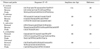
Real-time PCR was performed by using the Rotor-Gene Q system (QIAGEN, Strasse, Hilden, Germany). Each reaction tube contained 10 µl of a mixture containing 2 µl of DNA template, 5 µl of 2×Rotor Gene™ Probe PCR Kit (QIAGEN), 2 µl of each primer (10 pmol), and 1 µl of probe (10 pmol). The primers and probes used for qRT-PCR targeted the gtfB, gtfT, and 16S-23S intergenic spacer regions of S. mutans, S. sobrinus, and Lactobacillus species, respectively. Another universal primer targeting the 16S rRNA gene was used to quantify the total bacterial load in samples. Primer and probe sequences are listed in Table 2. Probes were labeled with 6-carboxyfluorescein (FAM) at their 5'-ends and with Black Hole Quencher (BHQ) at their 3'-ends. Primers and probes were synthesized by Applied Biosystems (Foster City, CA, USA).
The PCR cycling conditions used were as follows: 1 cycle of denaturation at 95℃ for 3 min, followed by 50 amplification cycles of 95℃ for 3 s and 58℃ for 10 s. Fluorescence was automatically measured during PCR and all amplifications were performed in duplicate. The data were analyzed by using complementary software from QIAGEN. Bacterial cell numbers were determined by using DNA from known amounts of the four different bacterial strains. Proportions of target bacteria were expressed as percentages of total bacteria.
4. Statistics
Statistical analysis was performed by using SPSS version 19.0 (SPSS Inc., Chicago, IL, USA). The chi-square and Kruskal-Wallis tests were used to determine statistical significance, which was accepted for p values of <0.05. Fisher's least significant difference test was used for non-parametric post-hoc multiple comparisons.
RESULTS
1. Salivary flows and pH values
Salivary flow and pH values are presented in Table 3. Salivary flow was nonsignificantly lower in patients than in controls. Salivary pH, however, was significantly lower in patients (p<0.05).
2. Detection range of the real-time PCR system for cariogenic bacteria
A standard curve was plotted by using 10-fold serial dilutions of positive controls for all four target bacteria. Standard curves were generated from the amplification plots in the insets (correlation coefficients were 0.9955 for S. mutans, 0.9968 for S. sobrinus, 0.9912 for L. salivarius, and 0.9899 for L. acidophilus). The real-time PCR assay was found to have a detection range of 101 to 108 bacterial cells. To generate a standard curve, CT values of standard dilutions were plotted against number of bacteria. Bacterial species used in this study had CT values in the range of 12 to 37, which corresponded to bacterial counts of 108 to 101, respectively.
3. Quantitative analysis of cariogenic bacteria levels in saliva samples
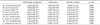
Saliva samples were evaluated by real-time PCR for the four cariogenic bacteria (Table 4). S. mutans was detected in 40.0% of HE samples, in 78.0% of SO samples, and in 60% of control samples. Mean numbers of S. mutans in cancer patients were not significantly higher than in healthy controls. However, mean numbers of S. mutans in the SO group were significantly higher than in the HE group (p<0.05). S. sobrinus was detected less frequently than S. mutans; S. sobrinus was detected in 3.3%, 22.0%, and 7.5% of subjects in the HE, SO, and control groups, respectively. The mean S. sobrinus number in the SO group was significantly higher than in both the HE group and the controls (p<0.05). L. salivarius was detected in 70% of the HE samples, in 85.4% of the SO samples, and in 70.0% of the control samples. The mean number of L. salivarius was significantly higher in the SO group than in the control group (p<0.05). L. acidophilus was detected less frequently than L. salivarius; L. acidophilus was detected in 40.0%, 48.8%, and 37.5% of the HE, SO, and control samples, respectively. However, the mean number of L. acidophilus in saliva was nonsignificantly lower in patients than in controls.
The proportions of the four cariogenic bacteria versus total bacteria by real-time PCR were also determined (Table 4). Proportions of S. mutans, S. sobrinus, and L. salivarius were significantly higher in the SO group than in the control group. Proportions of S. mutans and S. sobrinus were also significantly higher in the SO group than in the HE group. However, no significant intergroup differences were found in proportions of L. acidophilus among total bacteria.
DISCUSSION
The determination of accurate salivary flow rates is important in managing the altered oral environment of patients who have undergone radiotherapy or chemotherapy. Because saliva has a protective effect, clinically significant changes in salivary functions can contribute to the development or prevention of dental caries. In addition, it has been established that radiotherapy and chemical therapy in cancer patients causes marked reductions in the saliva flow rate and may cause clinical xerostomia.1 In the present study, no significant intergroup differences were found for salivary flow rates, but salivary pH was significantly lower in cancer patients. This finding is consistent with published data.15,16
Many researchers have reported that S. mutans levels in the oral cavity are correlated with the presence of dental caries.17,18 Blay et al.19 reported that the S. mutans levels in the oral cavity are correlated with a history of caries and caries susceptibility. In another study, the prevalence of S. sobrinus was found to be closely associated with high caries activity.20 In the present study, consistent with that previously reported,15 the frequency of S. mutans among all study subjects was higher than that of S. sobrinus. Furthermore, the prevalences of S. mutans and S. sobrinus and the levels and proportions of S. mutans and S. sobrinus, as determined by qRT-PCR, were higher in patients than in controls. This result suggests that high proportions and levels of cariogenic bacteria in the saliva of cancer patients are sufficient to predict the occurrence of dental caries. On the other hand, the frequencies of S. sobrinus in the saliva were lowest in patients and controls. That finding concurs with a report issued by Homer et al.,21 in which the presence of N-acetylglucosamine in the oral cavity was found to inhibit the growth of S. sobrinus and to reduce the frequency of its isolation.
Lactobacilli constitute approximately 1% of cultivable oral microflora, and they are well known to play opportunistic roles during the development of dental caries by producing lactic acid and extracellular polysaccharides. Several research groups have reported that lactobacilli including L. salivarius and L. acidophilus are numerically dominant in caries-susceptible sites.8,22,23 The results of the present study show that L. salivarius is detected most frequently in patients with a solid tumor, although it is probably not a major cariogenic pathogen. In addition, the levels and proportions of L. salivarius in saliva were higher in patients than in controls. In contrast, higher levels of L. acidophilus were present in controls, although its levels were lower than any other bacteria tested, which suggests that L. acidophilus levels are not correlated with dental caries. This result is not consistent with a study conducted by Martin et al.,8 in which L. acidophilus was found to be numerically dominant in caries samples. This discrepancy suggests that bacterial compositions are dependent on the carious sites included.
In the present study, saliva samples were collected and not plaque samples, because the latter is known to be unreliable for predicting the prevalence of caries-associated bacteria because bacterial counts on different tooth surfaces show large variations.24 Emilson25 reported that saliva better reflects the colonization of cariogenic bacteria on all dentition because it is in continuous contact with all oral surfaces. Thus, the use of saliva samples in the present study is believed to provide an accurate overall view of microbiological loadings.
The detection and quantification of mutans streptococci and lactobacilli in the oral cavity are important because these bacteria are relevant in the context of diagnosis and treatment planning for dental caries. In this study, we chose qRT-PCR for bacterial quantification because it is the most accurate and reliable tool for determining bacterial levels in clinical samples. By use of this technique, the prevalences and the levels of four cariogenic bacteria in the saliva samples of oncological patients and healthy controls were determined. High levels of three cariogenic bacteria were found in cancer patients. Nonetheless, the prevalences of mutans streptococci and lactobacilli were similar in patients and healthy controls. These results agree with our earlier PCR study,15 except that Lactobacillus species were not detected in all subjects. This finding indicates that real-time PCR is more specific and accurate than conventional end-point PCR.
In the present study, we focused on comparing the levels of cariogenic bacteria between cancer patients and a control group. Although the levels of cariogenic bacteria in cancer patients were higher than in controls, the caries status of cancer patients was not determined in this study. We did not find a significant correlation between salivary flow and salivary pH. High levels of cariogens are found to be closely associated with high caries activity. Nevertheless, we can suggest that the quantification of cariogenic bacteria is a valid method for evaluating caries susceptibility.
In conclusion, this study demonstrated that the saliva of oncological patients has significantly lower pH levels and harbors larger amounts of S. mutans, S. sobrinus, and L. salivarius than does the saliva of healthy individuals. Therefore, periodic follow-up is required for dental caries detection and plaque removal in patients with cancer. Further studies on salivary microbial levels in relation to caries development in cancer patients are needed.

 XML Download