

 PDF
PDF ePub
ePub Citation
 Print
Print


INTRODUCTION
The urachus is a vestigial fibrous cord that lies between the peritoneum and the transversalis fascia. The urachus is the embryological remnant of the allantois, which originally communicates with the apex of the bladder to the umbilicus. During embryonic development, the urachus begins to be obliterated and then becomes completely obliterated shortly after birth, giving rise to the median umbilical ligament.1 Different portions of the urachus may not be fully obliterated, however, which can lead to the formation of a cyst, sinus, diverticulum, or patent urachus. Urachal defects are rare, with urachal cysts being the most common anomaly and occurring in approximately 1/5,000 births. Usually, urachal cysts are asymptomatic and manifest themselves only when infected.1,2
Urachal remnants (most commonly cysts) occasionally require intervention when they become infected and symptomatic. Intervention is recommended over drainage of the abscess cavity and antibiotic therapy to prevent the risk of recurrence and the potential for malignant change of the urachal remnant. The traditional approach for removing a urachal remnant has been open surgery with a hypogastric transverse or midline infraumbilical incision, which is associated with increased morbidity and longer convalescence.3,4
Herein, we report our experience with complete laparoscopic removal of symptomatic urachal remnants with or without a cuff of bladder tissue as a minimally invasive surgery and evaluate the perioperative records and pathologic results.
MATERIALS AND METHODS
Seven men and one woman (mean age, 36.5 years; range, 5 to 78 years) visited our institutions with symptomatic urachal remnants between July 2004 and July 2012. The patients' medical records were reviewed retrospectively. The patients were interviewed by telephone to evaluate their long-term outcomes. All patients presented with a low abdominal infraumbilical mass, with or without fever, pain, and umbilical discharge. Preoperative evaluations included ultrasonography, computerized tomography (CT), and/or magnetic resonance imaging (MRI) of the abdomen. Initial treatment consisted of antibiotics and drainage with bacterial cultures and sensitivity tests. After the patients' acute symptoms subsided, each patient underwent complete excision of the urachal remnant laparoscopically. We evaluated communication or adhesion with the bladder by urine analysis, cystography, ultrasonography, CT, and/or MRI. We reviewed perioperative and postoperative records to assess perioperative data, operation time, blood loss, complications, pathological evaluation, and follow-up.
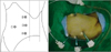
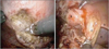
A Foley catheter was inserted with the patient under general anesthesia in a supine position. The visualization port was accessed by using the Endopath Xcel 120-mm trocar (Ethicon, USA) via the umbilicus and was insufflated by using CO2 with intraabdominal pressure maintained at 12 mmHg. Another three 5-mm working camera ports were inserted under direct vision on the right side of the abdomen (Fig. 1). The patient was then placed in the 60° left oblique position. A 0° laparoscope was used in all procedures. First, any bowel or omental adhesions from prior surgeries or inflammatory reactions to the infected urachal remnant were lysed off by using monopolar scissors, and the median and lateral umbilical ligaments were identified (Fig. 2). The umbilicus was not excised; the cephalic side of the urachus was dissected from the umbilicus, and the caudal stump of the median umbilical ligament was transected just above the bladder dome with ultrasonic scissors. If preoperative or intraoperative investigation revealed communication or adhesion between the urachus and the bladder, we performed a bladder cuff resection and then sutured transversely with a continuous intracorporeal 3-0 Vicryl suture. A Barovac® closed wound drainage evacuator system was positioned in the pelvic cavity, and the excised specimen was exteriorized with a laparoscopic retrieval bag via the umbilicus port and sent for histopathological examination.
RESULTS
Patient demographic and perioperative data are shown in Table 1. The mean surgery time was 2.7 hours (range, 1.2 to 3.5). The mean hospital stay was 14.6 days (range, 7 to 22 days). All patients started to eat an oral diet and began ambulating on postoperative day 1. The period of postoperative Foley catheterization was a mean of 7.2 days (range, 0.5 to 14 days). Bladder cuff resection followed by transverse suturing with a continuous intracorporeal 3-0 Vicryl suture was performed in 5 patients. The patients with bladder cuff resection had a long admission and Foley catheterization period (mean, 14.4, 11 days). Postoperative cystography was done to find any urine leakage in patients undergoing bladder cuff resection. In our study, there were no intraoperative complications. In addition, we found no postoperative complications including any symptom recurrence with or without infection suggestive of incomplete excision during a mean follow-up of 46.3 months (range, 3 to 96 months). The results of the pathological evaluations were as follows: 6 cases of infected urachal cysts, 1 case of infected urachal sinus, and 1 case of urachal adenocarcinoma.
DISCUSSION
The urachus is a vestigial structure arising from the anterior bladder wall and extending cranially to the umbilicus within the extraperitoneal fat between the peritoneum and the transversalis fascia. Embryologically, the urachus is said to arise as a result of the separation of the allantois from the ventral cloaca during the midtrimester.5,6
Before birth, approximately 98% of the urachus is obliterated. Urachal anomalies can be divided into congenital and acquired.2 Congenital anomalies manifest themselves as loss of urine through the navel and cause a persistence of the permeability of the urachus or the bladder's inability to descend to the pelvis, with an incidence of 1:300,000 in infants and 1:5,000 in adults, and with a male to female ratio of 3:1. The acquired anomalies appear when the urachus closes after birth and reopens later owing to pathological factors. Various types of remnants have been described, including cysts, sinus, diverticulum, and patent urachus. These anomalies are extremely rare in adults; most are detected in childhood as a result of infection.1,2,7
History and physical examination can be diagnostic, but several radiographic tests and cystoscopic investigation are needed to confirm a urachal anomaly. However, it is difficult to find an obvious connection between the bladder and the urachus. We used various imaging studies including cystography, ultrasound, CT, and MRI. Adult patients with urachal anomalies should especially undergo an abdominal CT and/or MRI scan because of the high risk of malignancy of up to 25% and the increased risk of malignancy of a cystic urachal mass with increasing age. Furthermore, 20% of patients with urachal cancer present with metastatic disease. Therefore, early surgical treatment is mandatory in aged patients with urachal diseases.8,9
Complicated urachal cysts usually warrant surgical treatment, but initially conservative management can be attempted. A 25% to 38% rate of recurrence is seen when complicated urachal cysts are managed conservatively by means of drainage and antibiotic therapy. Therefore, the basic scheme for urachal remnants is the complete excision of the complicated lesion with or without a partial cystectomy of the bladder apex.2,10 In the cases of communication or difficult separation by significant fibrotic attachments of the urachal cyst to the bladder, we resected a segment of bladder cuff with the specimen. Bladder cuff resection was associated with longer Foley catheter placement, more complications, and a delayed recovery.11-13
In 1993 Trondsen et al.14 reported the first laparoscopic excision of a urachal remnant. Since then, there have been several other case reports of laparoscopic excision of various types of urachal remnants in patients of various ages.15-18 There have also been two very short series of laparoscopic management of urachal remnants, one in four adults19 and one in four children.20 Both of these series used basically the same technique and showed that a laparoscopic technique can be used safely and effectively to remove all urachal remnant tissue with minimal morbidity. In the present series, we showed that a laparoscopic technique is feasible for excising urachal remnants whether the patent abnormal section is at the umbilicus or at the bladder. We also showed that the procedure is possible in a wide age range of patients. Our method had some distinct features, such as patient position and trocar sites, compared with other laparoscopic urachal surgeries. First, after the Endopath Xcel 12-mm trocar (Ethicon, USA) was inserted via the umbilicus, another three 5-mm working camera ports were inserted under direct vision on the right side of the abdomen with the patient in a supine position. Second, we changed the patient's position to the 60° left oblique position to avoid sigmoid colon, and the surgery was done from looking through the 5-mm camera port on the patient's right side. This port provided a good view of the full length of the urachus and allowed adequate access to both the umbilicus and the bladder dome without any hindrance or discomfort. This lateral port placement may reduce the risk of incomplete excision of the urachal remnant.
The largest recent series of open surgery for urachal anomalies was reported by Mesrobian et al.21 They investigated 21 children: 9 with a urachal cyst, 9 with a urachal-umbilical sinus, 2 with a urachal fistula, and 1 with a vesico-urachal diverticulum. In the cases in which no communication with the bladder was found, no bladder was excised. There were no complications and no hospital readmissions or reoperations. The authors did not comment about the hospital stay, analgesic requirements, or time of convalescence. However, the open operation appeared to be safe and extremely effective, even without taking a bladder cuff in all cases.
Postoperative complications including any symptom recurrence with or without infection could indicate an incomplete excision. We did not observe any symptom recurrence suggestive of incomplete excision during the follow-up period in our series, which agrees with most series of other reports using open traditional or robot-assisted techniques.9,10,19,22-27
In our series, 8 of 11 patients with complicated urachal cysts underwent laparoscopic surgery and 3 patients underwent open traditional surgery. Because of the low-priced medical insurance for hospitalization in Korea, most patients want to be hospitalized until full convalescence, defined as a return to normal life without pain. Therefore, the duration of hospitalization with laparoscopic surgery was lengthened. In the case of patient no. 1 (Table 1), antibiotics therapy was applied for a week initially; thus, the duration of hospitalization has increased to 22 days. In a case of urachal adenocarcinoma, the patient did not want any additional treatments; thus, we performed only laparoscopic removal of the urachal cyst with bladder cuff resection and did not find any cancer recurrence in follow-up computed tomography.
The primary limitation of this study was the small number of patients and the retrospective analysis. Another limitation was the lack of comparison among patients of group 2 between those with and without the bladder cuff resection.
Laparoscopic excision seems to be a safe and less invasive method for the treatment of complicated urachal anomalies, but a prospective, large, multi-institutional randomized study is needed.

 XML Download