

 PDF
PDF ePub
ePub Citation
 Print
Print


INTRODUCTION
Fibroblasts maintain a key role in extracellular matrix metabolism, and their diverse cellular interactions in the dermal inflammatory reaction are important in various cutaneous disorders. Fibroblasts are essential in producing many connective tissue matrix proteins (collagen, mucopolysaccharides, etc) as well as in the secretion of various soluble factors (cytokines, growth factors, etc). In the skin, dermal fibroblasts are known to process pathologic fibrous changes via unknown pathways when inflammation is overexpressed in some conditions.1
Histamine is also a well-known mediator of allergic inflammation in which it performs various functions including vascular dilation, activation of inflammatory cells, gastric secretion regulation, and nerve signal transmission. Four types of histamine receptors have been discovered (H1, H2, H3, H4), all of which are in the G-protein-coupled receptor family.2 Many dermal inflammatory disorders are related to the interaction between fibroblasts and histamine, yet the exact mechanism of this relation is not fully explained.
Toll-like receptors (TLRs) are pattern recognition receptors that were first recognized as a functional component of the innate immune system. They interact with various endogenous and exogenous ligands and antigens. Also, TLRs influence acquired immune responses as well as various intracellular signaling processes.3 Until now, a total of 11 TLRs have been discovered, and research is ongoing on the ligands and some functional interactions of these receptors. Only a few studies on TLRs in fibroblasts have been presented in the literature, and those studies were mainly focused on certain groups of TLRs such as TLRs 2 and 4 and their co-expression pattern with keratinocyte.
Previous studies that focused on the role of TLRs 2 and 4 reported an enhanced expression of TLRs by histamine treatment, which resulted in stronger inflammatory reactions.4-6 Moreover, Proost et al.7 observed Th1 cytokine profile increment when TLRs 2, 3, 4, 5, and 9 were activated in human fibroblasts. These previous studies suggest a potential role of TLRs in fibroblasts under diverse inflammatory reactions.
The purpose of the present study was to detect whether TLR expression in skin fibroblasts is detectable and then to see how TLR expression could be modulated by histamine, because histamine-associated chronic inflammatory reactions in the dermis have been seen in fibroblast-associated disorders such as scleroderma and keloid.
MATERIALS AND METHODS
1. Cell culture
Fibroblasts were collected from a 12-year-old boy during circumcision. The collected cells were cultured in Dulbecco's modified Eagle's medium (Hyclone, Logan, UT, USA) with 1% penicillin-streptomycin (Hyclone, Logan, UT, USA) and 10% fetal bovine serum (Hyclone, Logan UT, USA). An approximate amount of 1×10⁵ cells was initially injected in a petri dish containing 10 ml of culture medium. The cells were cultured in a humidified incubator (MCO-18AIC, Sanyo, Sakata, Japan) with 5% CO2 and at 37℃. Cells were subcultured when they reached 80% confluence. Subculture was done by the following procedures. The culture medium was removed from the dish and the cells were washed with phosphate-buffered saline (PBS, Hyclone, Logan, UT, USA). PBS was removed and the plate was placed in the incubator with 2 ml of trypsin (Hyclone, Logan, UT, USA). After 2 minutes, trypsin was deactivated with 6 ml of horse serum (Hyclone, Logan, UT, USA) and all the contents were centrifuged at 800 rpm for 3 minutes. After removing the supernatants, 30 ml of culture medium was added and the samples were stirred well before being divided into three petri dishes. The cells were used in experiments when they reached 90% confluence at cell passages between 4 and 5.
2. Histamine treatment
Before treatment, cells were divided into a 6-well plate with approximately 5×10⁵ cells per well. The MTT assay was carried out before treatment to check for cytotoxicity of histamine (Sigma Chemical Co., St. Louis, MO, USA). Histamine was treated for 1, 2, 3, and 6 hours at a concentration of 10 µM. The cells were harvested with EDTA (Hyclone, Logan, UT, USA) and trypsin to analyze TLR gene expression. A separate experiment was done to see the antagonism of antihistamine by using fibroblasts that were pre-treated with 10 µM diphenhydramine (DPH, Sigma Chemical Co., St. Louis, MO, USA) for 1 hour. These cells were then further treated with 10 µM histamine for 1, 2, and 6 hours.
3. Real-time PCR for TLRs
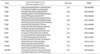
TriReagent (Molecular Research Center Inc., Cincinnati, OH, USA) was used to extract total RNA from experimental cells. Approximately 1 µg of total RNA per group was treated with M-MLV (Gibco BRL, Grand Island, NY, USA) according to the manufacturer's instructions to synthesize cDNA. cDNA was quantified by using Qubit™ (Invitrogen, Carlsbad, CA, USA) and 15 pg of cDNA entered 50 cycles of real-time PCR (Takara Bio Inc., Shiga, Japan) with SYBR Green (Takara Bio Inc., Shiga, Japan) tags. TLR primers (Bioneer Co., Daejon, Korea) were specified by using basic BLAST (Basic logical alignment search tool) tools and the standard material was GAPDH (Bioneer Co., Daejon, Korea) (Table 1).
4. Statistical analysis
Data obtained from triplicate sets of experiments were analyzed under paired-t tests between control and experimental groups by using SPSS (SPSS Inc., Chicago, IL, USA). A p value derived from the average comparison less than 0.05 was regarded as being statistically significant.
RESULTS
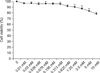
1. Effect of histamine on cell viability
Cell viability tended to decrease as the histamine concentration was gradually increased. However, there was no significant cell toxicity under the concentration level of 0.313 mM (Fig. 1).
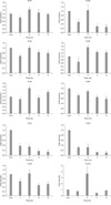
2. TLR mRNA expression in human dermal fibroblasts and effects of histamine treatment
TLRs 1 to 10 were all expressed in human dermal fibroblasts, and the level of expression did not differ significantly. The expression of each TLR was measured as a ratio of the standard expression of GAPDH via real-time PCR. The expression of all TLRs except TLRs 7 and 8 reached a peak value after 2 hours of histamine treatment and then showed a tendency to decrease. Among the increased TLRs, TLR 4 was the only receptor with a significant increase even after 6 hours of treatment, but TLRs 5 and 10 showed statistically significant increases only after 2 hours of treatment. The expression of TLRs 2, 3, 6, 7, 8, and 9 decreased significantly compared with the control after 6 hours of treatment. Among the TLRs showing decreased expression, TLRs 7 and 8 showed a continuously significant decreasing pattern throughout the treatment period (Fig. 2).
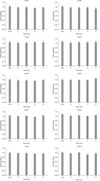
3. Changes in TLR mRNA expression with diphenhydramine
The mRNA expression of TLRs 3, 4, 5, and 8 of DPH-treated fibroblasts decreased instantly with statistical significance. The increments of TLRs 4, 5, and 10 were abrogated, but the changes in TLRs 2, 6, 7, 8, and 9 were insignificant compared with the results for treatment with the same concentration of histamine in the previous experiment. Interestingly, the expression of TLRs 3 and 10 was decreased compared with the control after alternative histamine treatment of 6 hours (Fig. 3).
DISCUSSION
Fibroblasts are the most abundant cells forming connective tissue and are known to interact with various proteins and mediators from surrounding tissues. Abnormal metabolic stimulation of the formation of extracellular matrix in the skin causes keloid or hypertrophic scars in which the over-activated chronic inflammatory process is involved.8,9 The signaling pathway of abnormal fibrosis caused by cellular proliferation and inflammation is now being studied from a different point of view, and recent reports have shown some proof that TLR activation can increase inflammatory cytokines in fibroblasts.7
It was shown in the present study by real-time PCR that the mRNAs of all 10 TLRs were well expressed in human dermal fibroblasts even though some results were rather inconsistent and the pattern was insufficient to verify the detail of activity of each receptor. However, the present data are enough to suggest that TLR research in fibroblasts could give us a clue to understanding the dermal inflammatory reaction, which seems to be closely related to TLR function. Histamine has been widely studied as an inflammatory mediator that acts in vascular dilation, gastric acid secretion, and neuronal signal transmitting.10,11 It is also known to control immune cells such as mononuclear cells, macrophages, T-cells, neutrophils, and endothelial cells related to inflammation.12-18 Besides the above known findings for histamine, a new functional class of its kind is also being explored. Recent study has shown an interesting role of histamine in enhancing the sensitivity and activity of TLRs via H1 receptor activation. Talreja et al.4 treated human umbilical endothelial cells with histamine and lipopolysaccharide (LPS) in which increased TLR 2 and 4 expression and secretion of IL-6 were found compared with LPS treatment alone. Other experiments that were performed with exposure to peptidoglycan and lipoteichoic acid showed similar results. The effect of histamine was detected from 0.1 µM and was strongest at a concentration of 10 µM. This enhanced expression of TLRs 2 and 4 was abrogated by 10 µM DPH but was not affected by H2 receptor antagonist, thus demonstrating the exclusive role of H1 receptor activation. The expression of TLR mRNA was most increased after 2 hours of treatment, and protein expression was highest after 16 hours.4 Hou et al.5 treated A549 and H292 cells with 1, 10, or 100 µM histamine for 2, 12, and 24 hours. This study reported a histamine-dependent increase of TLR 3 mRNA expression that was antagonized by DPH treatment.5 Another study conducted by Kobayashi et al.6 reported an increase in TLR 2 receptor and cytokine secretion with 1 µg/ml histamine treatment.
In the present study, we first focused on the expression of TLRs in fibroblasts from human skin. All TLRs seem to be well expressed. Next, we explored whether the TLR expression was influenced by histamine treatment and found increases in the mRNA expression of TLRs 4, 5, and 10 in contrast with decreases in the expression of TLRs 2, 3, 6, 7, 8, and 9 after treatment with 10 µM histamine. The increment of TLR 4 was similar to results from other studies, which suggests that TLR 4 responds to histamine similarly even in different cells. However, the mRNA expression of TLRs 7 and 8 was significantly decreased by histamine in this experiment. Known ligands of TLRs 7 and 8 are imidaziquinoline and virus-oriented ssRNA. Thus, we can speculate that this finding may be an explanation for the common clinical finding that patients with allergic disease are frequently more susceptible to low-infectivity viral infections than are normal patients. Indeed, there is a report concerning the increase of histamine-releasing factor along with allergic reaction mediators as a result of ssRNA virus treatment on low-airway endothelial cells.19 All these findings suggest the possibility of a negative feedback between TLRs 7 and 8 and tissue histamine concentrations, which may be a useful means of approach to prevent excessive allergic reactions.
Another interesting aspect of this study was the increasing pattern of TLR 4 mRNA expression with exposure time of histamine. This increased pattern was sustained for more than 6 hours of treatment, which was similar to TLRs 7 and 8 but in the opposite direction. According to Petricevic et al.20 TLR 4 expression decreases in response to TLRs 7 and 8 ligand-activating treatment. Collectively, these results suggest a complex interaction between these ligands. It is well known that TLRs 4, 7, and 8 are essential in pathogenic antigen recognition in the response to bacterial and viral antigens, respectively, which have some common characteristics and differences. Their ligand-binding activation sites in the cell are different: TLR 4 is located on the cell membrane but TLRs 7 and 8 are located in the cytoplasm. TLR 4 ligand activation shares the MyD88 signaling pathway with TLRs 7 and 8 but it uses an additional TRIF pathway that is MyD88 independent. Because TLRs 4, 7, and 8 function as a gateway of pathogenic inflammation, excessive inflammatory reaction may happen in the case of synergistic infections. On the contrary, an inhibitory interaction between TLR 4 and TLRs 7 and 8 can reduce the severity of abnormal inflammatory activation, such as sepsis.
Thus, the present results suggest that an interesting interactive mechanism may exist in skin disorders in which TLRs 4, 7, and 8 are activated by histamine. However, it is also speculated that the histamine-induced TLR expression pattern and its kinetic interaction may be dependent on cell type or dominant inflammatory signals, because our data also show some inconsistent mRNA expression patterns. Thus, the post-translational process and protein expression during chronic inflammatory processes may vary if the histamine exposure pattern is modulated under specific conditions, as seen in previous reports.
It is well known that the chronic dermal inflammatory process in scleroderma and keloid results from the stimulation of fibroblasts and the overproduction of extracellular matrix such as collagen, in which histamine can act as an inflammatory signal. Our study indicated the complicated role of TLRs 4, 7, and 8 in dermal inflammatory conditions, and successive studies will re-evaluate these receptor-ligand interactions.
Another interesting finding in this experiment was the higher TLR 10 expression pattern in fibroblasts with histamine. Until now, the role of TLR 10 was not clear.
In conclusion, we found that human dermal fibroblasts express all TLRs 1 through 10, and among them, TLRs 4, 5, and 10 increase with histamine treatment but TLRs 2, 6, 7, 8, and 9 decrease. These effects were antagonized by DPH, which is known to inhibit the action of the H1 receptor. We believe that the functional interaction and signaling pathways of these TLR receptors in fibroblasts are worthy of being explored in various dermal inflammatory disorders.
 XML Download