

 PDF
PDF ePub
ePub Citation
 Print
Print


INTRODUCTION
Orbital wall reconstruction by the transconjunctival approach involves reduction of the protruded tissue from the fracture site and insertion of a prosthesis to recover both the functional and anatomical roles of the orbital wall.1 Diplopia, enophthalmos, ocular deviation, lower lid retraction, infra-orbital nerve damage, occlusion of the lacrimal passage, dacryocystitis, pupil dilation, and lymph edema are well-known complications that can occur after orbital wall reconstruction. Among these, lower lid retraction can result in severe problems associated with cosmesis and tear film function and is known to be caused by contracture between the orbicularis oculi muscle and orbital septum.2
Recently, many studies have been performed on the use of various anti-adhesive agents after orbital wall reconstruction by the transconjunctival approach.3-5 Anti-adhesive agents form a physical barrier around the surgical area to prevent adhesion and are absorbed by the surrounding tissue after the wound healing process. In addition, such agents do not require an additional removal process after application because the absorption of the adhesive material into the body is harmless. In ophthalmologic surgery, anti-adhesives have been used in strabismus surgery to prevent adhesion between the extraocular muscles and sclera as well as in dacryocystorhinostomy to prevent adhesion at the rhinostomy site.6-9 In addition, a mixture of sodium hyaluronate (HA) and carboxymethylcellulose (CMC), also known as Seprafilm® (Genzyme Corp., Cambridge, MA, USA), has been used in orbital wall reconstruction.10
In the present study, we evaluated the clinical anti-adhesive effects of HACMC, a mixture of HA and CMC, by applying it between the orbicularis oculi muscle and orbital septum during a transconjunctival surgical procedure for orbital wall fracture.
MATERIALS AND METHODS
A prospective study was performed on 87 eyes of 87 patients who visited our hospital, underwent orbital wall reconstruction by the transconjunctival approach, and were followed up for at least 6 months from July 2009 to August 2010. No patients had a history of surgery, damages, or abnormality of the lower lid. Anti-adhesive solution was applied between the orbicularis oculi muscle and orbital septum in 47 eyes of 47 patients (the HACMC group), whereas no anti-adhesive solution was administered to the other 40 eyes of 40 patients (the control group) during the operation. The anti-adhesive solution, Guardix-sol® (Hanmi Pharm Co., Seoul, Korea), is a colorless clear liquid that uses a 1:1 mixture ratio of HA and CMC. External ocular muscle movement, forced duction, diplopia, and Hertel exophthalmometer test were evaluated along with routine ophthalmologic examinations. In addition, computed tomography scanning was performed to determine the size and location of the orbital wall fracture and the involvement of orbital soft tissues.
Surgery was performed through the transconjunctival approach under general anesthesia by a single surgeon (K.C.Y.). The lower lid was retracted downward and an incision was made on the conjunctiva along the border of the tarsal plate by unipolar electrocautery. The orbicularis oculi muscle was removed from the orbital septum, and a DesMarres retractor was used to retract the lower lid and orbicularis muscle downward. A malleable retractor was inserted behind the orbital septum to expose the periosteum of the inferior orbital border. A #15 Bard-Parker knife was used to make an incision on the periosteum, and a rugine was used to expose the fracture site. Then, the malleable retractor, suction tip, and rugine were used to repair the muscles and soft tissues. The damage site was measured, and Medpor® (Stryker, Newnan, GA, USA) was inserted on the fracture site after applying anti-bacterial solution. The periosteum was sutured with 5-0 Vicryl sutures, and Guardix-sol® was applied between the orbicularis oculi muscle and the orbital septum. Finally, the conjunctiva was sutured with 6-0 Vicryl sutures.
To prevent lower lid retraction, a traction suture was applied to the lower lid to pull it upward. The suture was removed 3 days after the surgery, and dexamethasone was injected intravenously after the surgery to reduce edema, which was tapered over 3 days. Ofloxacin (Ocuflox, Samil, Seoul, Korea) and 0.1% fluorometholone (Ocumetholone, Samil, Seoul, Korea) eyedrops were applied three times a day.
Follow-up examination was performed at 1 week and 1, 3, and 6 months after surgery. Ocular movement was classified into five grades as follows: grade 0, no ocular movement impairment; grade 1, 75% of ocular movement compared with the other eye; grade 2, 50-75% of ocular movement compared with the other eye; grade 3, 25-50% of ocular movement compared with the other eye; grade 4, less than 25% of ocular movement compared with the other eye.11,12 Diplopia was also classified into five grades as follows: grade 0, no diplopia; grade 1, diplopia above 60 degrees; grade 2, diplopia above 45 degrees; grade 3, diplopia above 30 degrees; grade 4, diplopia on central gaze. Lower lid retraction was followed up during the period by using marginal reflex distance2 (MRD2) for quantitative analysis. Lower lid retraction was defined as the sclera shown over the lower lid or MRD2 over 6 mm. The decision of 6 mm was based on the average MRD2 value of 4.6±0.7 mm in the Korean population.13 Because MRD2 was not measured before the surgery owing to edema and bleeding, it was measured only after the surgery and during the follow-up period. Lower lid ectropion was defined as visible retraction of the lid margin.
Chi-square test and independent t-test using SPSS version 17.0 (SPSS Inc., Chicago, IL, USA) were performed to compare the results between groups. p values less than 0.05 were considered statistically significant.
RESULTS
In the HACMC group, 36 of 47 patients were male and 11 were female, and the mean patients' age was 28.87±14.26 years. In the control group, 32 of 40 patients were male and 8 were female, and the mean age was 28.05±10.85 years. The average time between initial damage and surgery was 15.37±8.01 days in the HACMC group and 12.76±7.57 days in the control group. The distribution of fracture location and size is demonstrated in Table 1. There were no significant differences in gender, age, fracture location, fracture size, or period between initial fracture and surgery between the two groups (p>0.05).
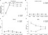
There were no significant differences in diplopia, enophthalmos, or ocular movement impairment between the two groups at 1 week and 1, 3, and 6 months after surgery. MRD2 values for quantitative analysis of lower lid retraction were 4.53±0.55 mm at 1 week, 4.48±0.75 mm at 1 month, 4.68±0.83 mm at 3 months, and 4.73±0.86 mm at 6 months in the HACMC group. The respective values were 5.19±0.95 mm (p=0.04), 4.98±0.84 mm (p=0.04), 4.90±0.83 mm (p=0.78), and 4.86±0.81 mm (p=0.73) in the control group. MRD2 values were significantly lower in the HACMC group than in the control group at postoperative 1 week and 1 month (Fig. 1). Two patients showed lower lid ectropion (5.0%) in the control group from 1 month after surgery, which persisted until 6 months. However, no patients showed lower lid ectropion in the HACMC group (p=0.08).
DISCUSSION
Transconjunctival, subtarsal, and subciliary approaches are the major approach methods for orbital fracture surgery. Because the transconjunctival approach does not involve making an incision on the skin, lower lid ectropion and edema occur less frequently. The transconjunctival approach is also commonly being used for its advantage of a wide surgical view by performing canthotomy during the surgery.14
Diplopia, enophthalmos, ocular deviation, infraorbital nerve damage, occlusion of the lacrimal passage, dacryocystitis, pupil dilation, lymph edema, and lower lid retraction are well-known complications that occur after orbital wall fracture surgery. Lower lid ectropion or lower lid retraction is caused by contracture around the orbital septum and orbicularis oculi muscle. Also, it may occur after combined sutures between the periosteum and orbital septum. It also occurs when the lower lid is sutured on a lower position than its original position after canthotomy. Traction suture of the lower lid to pull it upward helps to prevent lower lid complications. Also, steroid injection and lower lid massages may be applied on the lesion after lower lid retraction and ectropion occur.2
Compared with subciliary and subtarsal approaches, for which the risk of lower lid ectropion exists owing to adhesion between the conjunctiva and fat as well as damage to the orbicularis oculi muscles, the transconjunctival approach has a lower risk for lower lid complications because only part of the inferior tarsal muscle and capsulopalpebral fascia are involved.15 According to Mullins et al.16 in 2,086 patients who underwent orbital fracture repair, lower lid ectropion developed in 4.7%, lower lid entropion in 0.5%, lower lid edema in 1.1%, and hypertrophic scarring in 0.6%. In the subciliary approach group, 14.0% of patients showed lower lid ectropion, whereas only 1.5% of patients showed lower lid ectropion in the transconjunctival approach group. In the lower lid transdermal approach group, 1.5% showed lower lid entropion, whereas 0.7% of patients showed lower lid entropion in the transconjunctival approach group. Novelli et al.17 reported that 4 cases (7.1%) out of 56 patients who underwent orbital fracture repair showed complications after the surgery. Two patients (3.6%) showed lower lid entropion and two patients (3.6%) showed trichiasis.
Adhesion after surgery causes the damage site to release vasoactive factors and causes accumulation of inflammatory cells, which thereby results in the formation of collagen-rich vessels and infiltration of fibroblasts, macrophages, and giant cells. The tissues are replaced with angiogranular tissues and the adhesion becomes permanent.18 These facts suggest that HACMC can be used to create a physical barrier to prevent the adhesion process. HA is an anionic polysaccharide that is composed of D-glucuronic acid and N-acetyl-D-glucosamine. It has high viscosity and elasticity that prevent adhesion by preventing fibrosis of the tissue when sprayed on the tissue.19 However, HA is limited as a physical barrier because the physiological half-life of HA is only 1 to 3 days and it is easily decomposed by hyaluronidase. On the other hand, CMC uses cellulose from plants and is not easily disintegrated within the body. Because of its ability to maintain a physical barrier within the body tissues, a mixture of the two ingredients allows adequate prevention of postoperative adhesion.20,21 Several reports have indicated that the use of HACMC solution (Guardix-sol®) in strabismus surgery can prevent adhesion after the surgery.6,8 The use of HACMC solution (Guardix-sol®) during dacryocystorhinostomy was reported to effectively reduce occlusion and adhesion of the rhinostomy site.9 Furthermore, the use of Seprafilm®, a film structure of HACMC, significantly reduced the occurrence of adhesion between the conjunctiva, periosteum, and muscles after surgery and reduced the occurrence of adhesion and ocular movement disorder after orbital fracture repair surgery.7,10 However, Seprafilm® was difficult to manipulate and reposition once it contacted the tissue.7 In contrast, Guardix-sol®, a mixture solution of HA and CMC, is easy to handle and does not cause problems such as displacement and distortion after injection in an area that requires flexibility and movability.8
To our knowledge, no study has been performed on the use of anti-adhesives during wound repair of orbital wall reconstruction. In the present study, we used Guardix-sol® during orbital wall reconstruction by the transconjunctival approach to evaluate the anti-adhesive effects of the solution after the surgery. There were no significant differences in enophthalmos, ocular movement impairment, or diplopia between the HACMC and control groups. No lower lid ectropion was found in the HACMC group, whereas 2 cases of lower lid ectropion (5.0%) were found in the control group after the first month. Although there were no statistically significant differences in MRD2 values between the two groups between 3 and 6 months after the operation, MRD2 values were significantly lower in the HACMC group than in the control group at postoperative 1 week and 1 month. This may have been due to the anti-adhesive effect of HACMC, which can form a temporary physical barrier and hence decrease adhesion during the early postoperative period.8 The limitations of this study include a small sample size and limited follow-up period.
In conclusion, HACMC can be used as a supplementary agent during orbital wall reconstruction by the transconjunctival approach to reduce early postoperative adhesion. The use of HACMC seems to reduce the complications related to eyelids such as lower lid ectropion when contracture around the orbicularis oculi muscle and orbital septum is expected.

 XML Download