Journal List > Chonnam Med J > v.47(2) > 1074840

Lee: Vascular Dementia

Abstract

Cerebrovascular disease is the second leading cause of cognitive impairment in the elderly, either alone or in combination with Alzheimer's disease (AD). Vascular dementia (VaD) is heterogeneous in terms of both clinical phenotype and pathogenetic mechanisms. It may result from multiple cortical infarctions due to cerebral large vessel pathologies or to subcortical ischemic changes such as leukoaraiosis or lacunar infarction due to cerebral small artery disease. Clinical symptoms and signs vary depending on the location and size of the stroke lesion, and no single neuropsychological profile characteristic of VaD has been defined, although dysexecutive function is common. A slightly higher mortality rate and slower progression are reported in VaD compared with AD. VaD is potentially preventable by rigorous identification and treatment of cardiovascular disease risk factors, and modest symptomatic improvement with cholinesterase inhibitors has been reported.

INTRODUCTION

Vascular dementia (VaD) is the impairment of memory and cognitive functioning resulting from cerebrovascular disease (CVD) such as infarcts and leukoaraiosis. Although VaD is considered to be the second most common cause of cognitive impairment after Alzheimer's disease (AD) in the elderly, controversy remains concerning the terminology, classification, and diagnostic criteria of VaD. Cognitive status after a stroke is unstable and wide variability in the rates of cognitive impairment and dementia after stroke is reported, regardless of study duration or methods.1

EPIDEMIOLOGY AND RISK FACTORS

It is estimated that 6% to 10% of people over the age of 65 have dementia and up to 60% of patients with AD likely have an overlap with VaD. VaD alone is presumed to account for nearly 20% to 40% of dementia cases.2 Seventy-five percent of all strokes occur in individuals older than 65 years.3 In poststroke patients, age is the greatest risk factor for the development of VaD.4 Although the incidence rate of VaD varies considerably depending on the study methodology and criteria used, in one study, the risk of dementia with stroke was higher for those older than 80 years of age (odds ratio [OR]=12.7) or aged 70-79 years (OR=3.0) than for those aged 60-69 years, for those with 8 or fewer years of education versus those with 13 or more years (OR=4.1), for major hemispheric stroke versus posterior fossa (OR=3.0), for those with diabetes mellitus (OR=1.8), for anterior/posterior cerebral artery distribution versus other vascular territories (OR=1.7), and for those with prior stroke (OR=1.7).5
Research has established a strong relationship between age, low education level, and poststroke dementia as well as vascular risk factors (e.g., hypertension, diabetes, hyperlipidemia, smoking).3,5
Ethnic comparisons in the Cardiovascular Health Study showed nearly twofold higher incidence rates among African Americans than in whites.6 Men, those of Asian descent, and older people have a tendency for a higher risk of VaD.5,6
VaD is considered to result from interactions between vascular etiologies, changes in the brain, host factors, and cognition.7 Some studies have found that infarcts may precipitate dementia through an additive or synergistic relationship with AD neuropathology.8-10

CLASSIFICATION AND CLINICAL FEATURES OF VaD

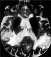
Post-stroke dementia, which includes multi-infarct dementia (MID), strategic infarct dementia, subcortical vascular dementia (SVD), and hypoperfusion dementia, is defined as dementia occurring in close temporal relation to thromboembolic or hemodynamic events.11 Large-artery infarctions are responsible for most cases of poststroke dementia. MID is characterized by transient ischemic attack (TIA) and stroke episodes in close temporal relation to the onset or development of dementia. Neuroimaging (brain computed tomography [CT] or magnetic resonance imaging [MRI]) shows multiple cortico-subcortical infarcts (Fig. 1). MID is related to atherothrombotic strokes, cardiac embolic strokes, and major hemodynamic events. Typical clinical features of MID are focal neurological signs such as hemiparesis or sensory deficits and stepwise progression with cognitive impairment. The presence of 'patchy' or unequal cognitive deficits are only to be expected in MID where there are only very few (two or three) cortical infarcts.
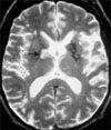
Strategic infarct dementia is caused by isolated infarcts in regions that are important for cognition of the brain, such as the thalamus, hippocampus, caudate, or genu of the internal capsule (Fig. 2). It is characterized by the abrupt onset of cognitive or behavioral changes, which vary depending on the infarct location.
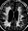
Most VaD results from multi-infarcts where cortical damage is responsible for cognitive impairment. However, this is now known to be a relatively rare pattern of disease in VaD and in the far more common subcortical form of VaD (SVD), a history of stroke may be absent in up to 40%.12 The primary types of brain lesions in SVD are lacunar infarcts and ischemic white matter lesions (WMLs) (Fig. 3). Therefore, accumulation of small infarcts in the deep white matter and gray matter may lead to SVD. Lacunar infarcts represent approximately 25% of symptomatic ischemic strokes.12 WMLs have been associated with age, hypertension, diabetes, metabolic syndrome, microvascular retinopathy, elevated homocysteine levels, and ischemic heart diseases.13
The onset of SVD is insidious in over half of patients and the course is usually continuous and slowly progressive.13,14 Neurological symptoms and signs related to SVD include cognitive deterioration, gait disturbance (imbalance, fall, short-stepped gait), urinary dysfunction, and dysarthria. Behavioral and cognitive changes include mental slowness (bradyphrenia), emotional lability, personality changes, and depression.15 Depression is common in VaD, occurring in up to 20% of cases, and is disproportionately prominent in those cases with small amounts of infarction. It may be particularly related to frontal deep white matter lesions.16
Cognitive and behavioral changes in SVD are due to disruption of the frontal cortico-subcortical circuit, resulting in 'frontal-subcortical syndrome' or 'dysexecutive syndrome.'17
The primary pathophysiological mechanism underlying SVD seems to be vessel wall damage in the long penetrating arterioles that drain into the white matter, with subsequent white matter degeneration and occurrence of lacune.18
Orthostatic hypotension and blood pressure variability may play a role in hypoperfusion dementia or WMLs.19 Proposed mechanisms for this role include nocturnal hypotension superimposed upon limited perfusion reserve and impaired vasoreactivity producing partial ischemia leading to incomplete infarction.20
Neuropsychological tests used in the assessment of AD such as the Mini-Mental State Examination (MMSE) are commonly used to evaluate cognition in VaD but are not appropriate tools in patients with VaD because neuropsychological characteristics in both subtypes of dementia are not similar. VaD predominantly involves deficits in executive functioning, whereas AD involves earlier and more significant memory impairment.12,15-17
Instruments that include assessment of frontal, executive, and subcortical function are required for the diagnosis of VaD.
Because the pathogenesis, risk factors, and comorbidities for VaD are similar to those for AD, it is sometimes difficult to differentiate the two.13 The major clinical differences between VaD and AD are summarized in Table 1.

DIAGNOSIS

The diagnosis of VaD is developed by considering clinical, imaging, and pathological findings. Given the heterogeneity of the VaD phenotype, large, uniform data sets are necessary to unravel the links between CVD and cognitive impairment. Three basic elements for a diagnosis of VaD are 1) evidence of dementia or cognitive impairment, 2) evidence of CVD, and 3) evidence of a casual relationship between cognitive impairment and CVD. Although there are no uniform diagnostic criteria for VaD, guidelines for interpreting neuroimaging findings have been incorporated into recent criteria for the clinical diagnosis of VaD.
The main set of criteria for the clinical diagnosis of VaD are the Diagnostic and Statistical Manual of Mental Disorders, Fourth Edition (DSM-IV)21; the International Classification of Diseases 10th version (ICD-10)22; the State of California Alzheimer's Disease Diagnostic and Treatment Centers (ADDTC) criteria23; and the National Institute of Neurological Disorders and Stroke and Association Internationale pour la Recherché et l'Enseignement en Neurosciences (NINDS-AIREN) criteria.24 All the currently used clinical criteria are 'consensus criteria' derived from expert opinion based on prevailing knowledge and pathogenetic hypotheses of dementia. All are based on the ischemic infarct concept, with the NINDS-AIREN, DSM-IV, and ICD-10 also including hemorrhages. The DSM-IV criteria for VaD require focal neurological signs and symptoms or laboratory evidence of focal neurological damage clinically judged to be etiologically related to the disturbance, but the criteria lack neuroimaging guidelines. The ICD-10 criteria require unequal, patchy distribution of cognitive deficits, focal neurological signs, and significant CVD judged to be etiologically related to the dementia, but the shortcomings of these criteria include a lack of detailed etiological and neuroimaging guidelines. The ADDTC criteria are exclusive for ischemic VaD and require two or more ischemic strokes by history, neurological signs, or neuroimaging. The NINDS-AIREN criteria include definitions for the dementia syndrome, CVD, and their relationship. A relationship between dementia and CVD is inferred from the onset of dementia within 3 months following recognized stroke, or abrupt deterioration in cognitive functions, or fluctuating, stepwise progression of cognitive deficits. The NINDS-AIREN criteria had moderate to substantial inter-rater reliability and were more specific (80%), although less sensitive (58%).25 The various diagnostic criteria are neither perfect nor interchangeable and none of these criteria have the required sensitivity and specificity to identify patients with mixed VaD reliably.26,27 This accounts for the variations in published prevalence and incidence rates and in treatment outcomes. The heterogeneity in the clinical presentation of VaD is compounded by the lack of consensus regarding the pathologic diagnosis of VaD. The main features of the most commonly used clinical criteria for VaD are summarized in Table 2.

NEUROIMAGING

Although neuroimaging is not required for diagnosis by all clinical references, it is recommended in patients with suspected VaD owing to a stroke history, vascular risk factors, or abnormal neurological examination. If CT shows significant evidence of vascular pathology, MRI is not necessary. However, MRI is more sensitive than CT for ischemic damage and vascular lesions. The absence of cerebral vascular lesions on CT or MRI excludes the diagnosis of VaD. Features on CT or MRI that are suggestive of VaD include cortical or subcortical infarctions, multiple lacunar strokes, and WMLs.28

PREVENTION AND TREATMENT

Because stroke is common and its risk factors are well defined and modifiable, it is reasonable to intensify efforts to control cardiovascular risk factors to reduce the population of stroke and VaD. Rigorous control of vascular risk factors is important in the primary and secondary prevention of VaD. In terms of specific treatment, the use of cholinesterase inhibitors and N-methyl-d-aspartate receptor antagonists for VaD has been evaluated.29 Donepezil (Aricept) 5 mg to 10 mg per day showed improvement in AD Assessment Scale-Cognitive (ADAS-Cog) subscale scores.30-32 Galantamine (Reminyl) has been studied for the treatment of VaD in two randomized, double-blind, placebo-controlled trials of 592 patients and 788 patients, respectively. ADAS-Cog score was a primary efficacy measure. At 26 weeks, there were improvements in galantamine-treated patients at doses of 16 mg to 24 mg per day. However, a significant percentage of patients dropped out owing to adverse events, particularly gastrointestinal effects.33,34
Rivastigmine (Exelon) was studied in a randomized, placebo-controlled trial of 710 patients and it was superior to placebo on the ADAS-Cog score as a primary efficacy measure, except in younger (age 50-75 years) patients.35
Memantine (Ebixa) was studied for the treatment of VaD in two large trials and both trials concluded that memantine 20 mg per day improved ADAS-Cog scores.36,37
The therapeutic approach to VaD is summarized as 1) primary prevention with cardiovascular risk factor control, 2) correct diagnosis of VaD, 3) secondary stroke prevention with anti-platelet therapy, and 4) cholinesterase inhibitors or NMDA receptor antagonists.
The principles of anti-dementia drugs for VaD are summarized below38:
  • Make an accurate diagnosis of the dementia syndrome

  • Characterize the severity of the dementia (mild, moderate, or severe)

  • Inform the patient and caregiver of reasonable treatment expectations (small symptomatic improvements and delay in decline are typical responses)

  • Optimize the dose of each medication

  • Slow the titration of the medication if side effects emerge during the titration period

  • Monitor multiple domains for beneficial responses (e.g., cognition, behavior, function)

  • Monitor side effects (especially gastrointestinal side effects with cholinesterase inhibitors)

  • Switch from one cholinesterase inhibitor to another if the patient is intolerant or shows no response to the current treatment

  • Continue therapy until the severe phases of dementia are reached

  • Monitor for rapid emergence of new cognitive, behavioral, or functional symptoms if the medications are discontinued

  • Combine a cholinesterase inhibitor and memantine for optimal therapeutic response

PROGNOSIS AND COMPLICATIONS

The prognosis of VaD varies considerably according to the criteria used to make the diagnosis. MID shortens life expectancy to about 50% of normal at 4 years from the initial evaluation.39 About one-third of the most severely affected elderly die from complications of the dementia itself, one-third from cerebrovascular disease, 8% from other cardiovascular disease, and the rest from miscellaneous causes.40 Overall, the effect of VaD on mortality is similar to or mildly worse than that of AD.5

CONCLUSIONS

VaD is characterized most commonly by subcortical frontal and executive dysfunction of cognitive impairment as opposed to the concept of amnesia-predominant AD. It may begin with very subtle deficits arising on the basis of leukoaraiosis or WMLs and proceeds through infarction to a more advanced state. Early identification of vascular cognitive impairment due to vascular brain damage as an early stage of VaD and proper management through modification of vascular risk factors especially in midlife are important for the prevention of VaD.

Figures and Tables

FIG. 1
Multi-infarc dementia type of vascular dementia on T2-weighted brain MRI.
cmj-47-66-g001
FIG. 2
Strategic infarct dementia type of vascular dementia on T2-weighted brain MRI.
cmj-47-66-g002
FIG. 3
Subcortical vascular dementia type of vascular dementia on fluid attenuated inversion recovery (FLAIR) brain MRI.
cmj-47-66-g003
TABLE 1
Comparison of vascular dementia and Alzheimer disease
cmj-47-66-i001

VaD: vascular dementia, AD: Alzheimer's disease.

TABLE 2
Comparison of clinical criteria for vascular dementia
cmj-47-66-i002

DSM-IV: the Diagnostic and Statistical Manual of Mental Disorders, Fourth Edition, ICD-10: the International Classification of Diseases 10th version, ADDTC: the State of California Alzheimer's Disease Diagnostic and Treatment criteria, NINDS-AIREN: the National Institute of Neurological Disorders and Stroke and Association Internationale pour la Recherché et l'Enseignement en Neurosciences criteria.

References

1. Knopman DS. Dementia and cerebrovascular disease. Mayo Clin Proc. 2006. 81:223–230.
crossref
2. Fratiglioni L, Rocca WA. Boller F, Cappa SF, editors. Epidemiology of dementia. Aging and Dementia. Handbook of Neu-ropsychology. 2001. Vol. 6:2nd ed. Amsterdam: Elsevier;193–215.
crossref
3. Pinkston JB, Alekseeva N, González Toledo E. Stroke and dementia. Neurol Res. 2009. 31:824–831.
crossref
4. de Leeuw FE, de Groot JC, Achten E, Oudkerk M, Ramos LM, Heijboer R, et al. Prevalence of cerebral white matter lesions in elderly people: a population based magnetic resonance imaging study. The Rotterdam Scan Study. J Neurol Neurosurg Psychiatry. 2001. 70:9–14.
crossref
5. Chui HC, Nielsen-Brown N. Vascular cognitive impairment. Continuum Lifelong Learning Neurol. 2007. 13:109–143.
crossref
6. Fitzpatrick AL, Kuller LH, Ives DG, Lopez OL, Jagust W, Breitner JC, et al. Incidence and prevalence of dementia in the Cardiovascular Health Study. J Am Geriatr Soc. 2004. 52:195–204.
crossref
7. Skoog I. Status of risk factors for vascular dementia. Neuroepidemiology. 1998. 17:2–9.
crossref
8. Neuropathology Group. Medical Research Council Cognitive Function and Aging Study. Pathological correlates of late-onset dementia in a multicentre, community-based population in England and Wales. Neuropathology Group of the Medical Research Council Cognitive Function and Ageing Study (MRC CFAS). Lancet. 2001. 357:169–175.
9. Snowdon DA, Greiner LH, Mortimer JA, Riley KP, Greiner PA, Markesbery WR. Brain infarction and the clinical expression of Alzheimer disease. The Nun Study. JAMA. 1997. 277:813–817.
crossref
10. Bowler JV, Munoz DG, Merskey H, Hachinski V. Fallacies in the pathological confirmation of the diagnosis of Alzheimer's disease. J Neurol Neurosurg Psychiatry. 1998. 64:18–24.
crossref
11. Lopez OL, Kuller LH, Becker JT, Jagust WJ, DeKosky ST, Fitzpatrick A, et al. Classification of vascular dementia in the Cardiovascular Health Study Cognition Study. Neurology. 2005. 64:1539–1547.
crossref
12. Román GC, Erkinjuntti T, Wallin A, Pantoni L, Chui HC. Subcortical ischaemic vascular dementia. Lancet Neurol. 2002. 1:426–436.
crossref
13. Reed BR, Eberling JL, Mungas D, Weiner M, Kramer JH, Jagust WJ. Effects of white matter lesions and lacunes on cortical function. Arch Neurol. 2004. 61:1545–1550.
crossref
14. Knopman DS, Parisi JE, Boeve BF, Cha RH, Apaydin H, Salviati A, et al. Vascular dementia in a population-based autopsy study. Arch Neurol. 2003. 60:569–575.
crossref
15. Scheltens P, Barkhof F, Leys D, Wolters EC, Ravid R, Kamphorst W. Histopathologic correlates of white matter changes on MRI in Alzheimer's disease and normal aging. Neurology. 1995. 45:883–888.
crossref
16. Udaka F, Sawada H, Kameyama M. White matter lesions and dementia: MRI-pathological correlation. Ann N Y Acad Sci. 2002. 977:411–415.
crossref
17. Wallin A, Sjögren M, Edman A, Blennow K, Regland B. Symptoms, vascular risk factors and blood-brain barrier function in relation to CT white-matter changes in dementia. Eur Neurol. 2000. 44:229–235.
crossref
18. Brun A. Pathology and pathophysiology of cerebrovascular dementia: pure subgroups of obstructive and hypoperfusive etiology. Dementia. 1994. 5:145–147.
crossref
19. Goldstein IB, Bartzokis G, Guthrie D, Shapiro D. Ambulatory blood pressure and the brain: a 5-year follow-up. Neurology. 2005. 64:1846–1852.
crossref
20. Yamamoto Y, Akiguchi I, Oiwa K, Hayashi M, Ohara T, Ozasa K. The relationship between 24-hour blood pressure readings, subcortical ischemic lesions and vascular dementia. Cerebrovasc Dis. 2005. 19:302–308.
crossref
21. American Psychiatric Association. Diagnostic and Statistical Manual of Mental Disorders. 1994. Fourth Edition (DSM-IV). Washington, DC: American Psychiatric Association.
22. World Health Organization. The ICD-10 Classification of Mental and Behavioural Disorders: Diagnostic Criteria for Research. 1993. Geneva: WHO.
23. Chui HC, Victoroff JI, Margolin D, Jagust W, Shankle R, Katzman R. Criteria for the diagnosis of ischemic vascular dementia proposed by the State of California Alzheimer's Disease Diagnostic and Treatment Centers. Neurology. 1992. 42:473–480.
crossref
24. Román GC, Tatemichi TK, Erkinjuntti T, Cummings JL, Masdeu JC, Garcia JH, et al. Vascular dementia: diagnostic criteria for research studies. Report of the NINDS-AIREN International Workshop. Neurology. 1993. 43:250–260.
crossref
25. Chui H, Lee AE. Clinical criteria for dementia subtypes. Evidence-based dementia practice. 2002. Oxford, England: Blackwell Science;106–119.
26. Chui HC, Lee AY. Evidence-based diagnosis of Alzheimer disease. Neurologia. 2000. 15:suppl. 8–14.
27. Pohjasvaara T, Mäntylä R, Ylikoski R, Kaste M, Erkinjuntti T. Comparison of different clinical criteria (DSM-III, ADDTC, ICD-10, NINDS-AIREN, DSM-IV) for the diagnosis of vascular dementia. National Institute of Neurological Disorders and Stroke-Association Internationale pour la Recherche et l'Enseignement en Neurosciences. Stroke. 2000. 31:2952–2957.
crossref
28. Pohjasvaara T, Mäntylä R, Salonen O, Aronen HJ, Ylikoski R, Hietanen M, et al. MRI correlates of dementia after first clinical ischemic stroke. J Neurol Sci. 2000. 181:111–117.
crossref
29. Molnar FJ, Man-Son-Hing M, Fergusson D. Systematic review of measures of clinical significance employed in randomized controlled trials of drugs for dementia. J Am Geriatr Soc. 2009. 57:536–546.
crossref
30. Wilkinson D, Doody R, Helme R, Taubman K, Mintzer J, Kertesz A, et al. Donepezil 308 Study Group. Donepezil in vascular dementia: a randomized, placebo-controlled study. Neurology. 2003. 61:479–486.
crossref
31. Black S, Román GC, Geldmacher DS, Salloway S, Hecker J, Burns A, et al. Donepezil 307 Vascular Dementia Study Group. Efficacy and tolerability of donepezil in vascular dementia: positive results of a 24-week, multicenter, international, randomized, placebo-controlled clinical trial. Stroke. 2003. 34:2323–2330.
crossref
32. Wilkinson D, Róman G, Salloway S, Hecker J, Boundy K, Kumar D, et al. The long-term efficacy and tolerability of donepezil in patients with vascular dementia. Int J Geriatr Psychiatry. 2010. 25:305–313.
crossref
33. Auchus AP, Brashear HR, Salloway S, Korczyn AD, De Deyn PP, Gassmann-Mayer C. GAL-INT-26 Study Group. Galantamine treatment of vascular dementia: a randomized trial. Neurology. 2007. 69:448–458.
crossref
34. Craig D, Birks J. Galantamine for vascular cognitive impairment. Cochrane Database Syst Rev. 2006. (1):CD004746.
crossref
35. Ballard C, Sauter M, Scheltens P, He Y, Barkhof F, van Straaten EC, et al. Efficacy, safety and tolerability of rivastigmine capsules in patients with probable vascular dementia: the VantagE study. Curr Med Res Opin. 2008. 24:2561–2574.
crossref
36. Orgogozo JM, Rigaud AS, Stöffler A, Möbius HJ, Forette F. Efficacy and safety of memantine in patients with mild to moderate vascular dementia: a randomized, placebo-controlled trial (MMM 300). Stroke. 2002. 33:1834–1839.
crossref
37. Wilcock G, Möbius HJ, Stöffler A. MMM 500 group. A double-blind, placebo-controlled multicentre study of memantine in mild to moderate vascular dementia (MMM500). Int Clin Psychopharmacol. 2002. 17:297–305.
crossref
38. Cummings JL. The Black Book of Alzheimer's Disease, Part 2. Primary Psychiatry. 2008. 15:69–90.
39. Seshadri S, Wolf PA. Lifetime risk of stroke and dementia: current concepts, and estimates from the Framingham Study. Lancet Neurol. 2007. 6:1106–1114.
crossref
40. Ravaglia G, Forti P, Maioli F, Martelli M, Servadei L, Brunetti N, et al. Incidence and etiology of dementia in a large elderly Italian population. Neurology. 2005. 64:1525–1530.
crossref
TOOLS
Similar articles