

 PDF
PDF ePub
ePub Citation
 Print
Print


INTRODUCTION
A pulmonary thromboembolism (PTE) causes a dramatic pressure overload to the right heart. Previous case reports have shown that elevated right atrial (RA) pressure secondary to a PTE can cause right-to-left shunting in the presence of an atrial septal aneurysm (ASA).1 The clinical significance of interatrial shunting in PTE, however, has not been clearly defined. The interesting case presented here shows that right-to-left shunting in the presence of an ASA can cause normal RA pressure, thus masking the pressure overload of the right heart in a patient with submassive PTE.
CASE REPORT
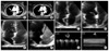
A 57-year-old female with diabetes, hypertension, and an old cerebral infarction was admitted to our hospital with 5 days of progressive dyspnea (NYHA III). Her blood pressure was within normal limits (135 mmHg/85 mmHg) and tachycardia (120/min) and tachypnea (20/min) were detected. No abnormal lung or heart sounds were noted. The chest X-ray was normal, but oxygen saturation was 83%. D-dimer rose up to 10.31 ug/mL and Troponin-I was 0.34 ng/mL. A chest computed tomography angiogram (CTA) confirmed acute PTE involving the right main, left upper, and lower lobar pulmonary arteries (Fig. 1A). Initial transthoracic echocardiography (TTE) showed an ASA swing from the right side to the left side (Fig. 1C), and right-to-left shunting was detected immediately in the agitated saline test (Fig. 1E). However, definite signs of pressure overload of the right heart, such as marked right ventricular (RV) enlargement and RV dysfunction, were not detected in the echocardiography. Also, RV hypertrophy as a sign of chronic pressure overload was not seen. The pulmonary artery systolic pressure calculated by maximal velocity of tricuspid regurgitation (Fig. 1G) and predicted RA pressure (Fig. 1H) was in the normal range. Because of the extensive embolic burden on the CTA and the possibility of a patent foramen ovale (PFO), the patient received thrombolytic therapy with supply of oxygen. Her symptoms gradually improved. An extremity venous Doppler sonography showed venous thrombosis in the left popliteal vein. The results of tests for several tumor markers and imaging studies for hidden malignancy were normal, and anti-nuclear antibody for systemic lupus was within the normal range. We did no further evaluation of other hereditary or hematologic causes, because she had acquired thromboembolic risk factors such as diabetes, old cerebral infarction, and long duration of immobilization. Therefore, we thought that the PTE originated as a deep vein thrombosis rather than an ASA. After 2 weeks, a follow-up chest CTA showed improved PTE (Fig. 1B). The follow-up TTE showed the ASA fixed to the right side (Fig. 1D), and no right-to-left shunting was detected by the agitated saline test despite coughing (Fig. 1F). However, transesophageal echocardiography revealed tiny right-to-left shunting via a PFO. The patient finally improved and was discharged.
DISCUSSION
An ASA is defined as redundant and mobile interatrial septal tissue in the region of the fossa ovalis with phasic excursion of at least 10 to 15 mm.1,2 ASAs have been found in 2% of patients undergoing TTE.1 Although an ASA is considered a clinically benign entity, it has been considered a potential source of cardiogenic embolism.1-3 Another more common complication of ASAs is a right-to-left shunting, which occurs in about 60% of cases, usually via a PFO.4
There are 3 mechanisms of interatrial shunting: an increase in pulmonary vascular resistance leading to a right-to-left shunt, an increase in systemic vascular resistance resulting in a left-to-right shunt, and a decrease in systemic vascular resistance (vasogenic shock) resulting in a right-to-left shunt.5 In patients with PTE, right-to-left shunting has been described in previous case reports as a complication of ASA.1 However, those cases have demonstrated right-to-left shunting only in the presence of elevated RA pressure secondary to PTE. In our case, by contrast, calculated pulmonary artery pressure was within normal limits. The right-to-left shunting of the ASA had concealed the pressure overload of the right heart caused by the PTE. Also, right-to-left shunting through a PFO can result in a paradoxical embolus that originates in the systemic circulation and enters the systemic arterial circulation. A paradoxical embolus can present with cryptogenic stroke.6 As well as pathological states, transient increases in right arterial pressure occur in normal individuals during early ventricular systole, during the Valsalva maneuver, and with repetitive cough.7 In our case, a large quantity of right-to-left shunting via a PFO was observed at the initial presentation because of the acute PTE and, after improvement, tiny right-to-left shunting was also seen during the Valsalva maneuver. Our patient was diagnosed with an old cerebral infarction through brain images from 1 year ago, although with no symptoms or signs. Therefore, we thought that the stroke history of our patient may be associated with a PFO in the ASA.
A PTE causes a dramatic increase in pressure overload to the RV, and an ASA is not an uncommon abnormality. Blood flow from the right to the left through the ASA reduced the pressure of the right heart and, as a result, the signs of pressure overload were not detected in the echocardiogram. This case highlights the possibility of masking of pressure overload of the right heart through the interatrial shunting of an ASA via a PFO in a patient with PTE.
 XML Download